Skip to main page content
U.S. flag

An official website of the United States government

Dot gov

The VSports app下载. gov means it’s official. Federal government websites often end in . gov or . mil. Before sharing sensitive information, make sure you’re on a federal government site. .

Https

The site is secure. The https:// ensures that you are connecting to the official website and that any information you provide is encrypted and transmitted securely V体育官网. .

. 2011 Dec;141(6):2188-2199.e6.
doi: 10.1053/j.gastro.2011.08.005. Epub 2011 Aug 18.

Autophagy suppresses age-dependent ischemia and reperfusion injury in livers of mice

Affiliations

Autophagy suppresses age-dependent ischemia and reperfusion injury in livers of mice

Jin-Hee Wang et al. Gastroenterology. 2011 Dec.

"V体育官网" Abstract

Background & aims: As life expectancy increases, there are greater numbers of patients with liver diseases who require surgery or transplantation. Livers of older patients have significantly less reparative capacity following ischemia and reperfusion (I/R) injury, which occurs during these operations. There are no strategies to reduce the age-dependent I/R injury VSports手机版. We investigated the role of autophagy in the age dependence of sensitivity to I/R injury. .

Methods: Hepatocytes and livers from 3- and 26-month-old mice were subjected to in vitro and in vivo I/R, respectively V体育安卓版. We analyzed changes in autophagy-related proteins (Atg). Mitochondrial dysfunction was visualized using confocal and intravital multi-photon microscopy of isolated hepatocytes and livers from anesthetized mice, respectively. .

Results: Immunoblot, autophagic flux, genetic, and imaging analyses all associated the increase in sensitivity to I/R injury with age with decreased autophagy and subsequent mitochondrial dysfunction due to calpain-mediated loss of Atg4B. Overexpression of either Atg4B or Beclin-1 recovered Atg4B, increased autophagy, blocked the onset of the mitochondrial permeability transition, and suppressed cell death after I/R in old hepatocytes. Coimmunoprecipitation analysis of hepatocytes and Atg3-knockout cells showed an interaction between Beclin-1 and Atg3, a protein required for autophagosome formation. Intravital multi-photon imaging revealed that overexpression of Beclin-1 or Atg4B attenuated autophagic defects and mitochondrial dysfunction in livers of older mice after I/R. V体育ios版.

Conclusions: Loss of Atg4B in livers of old mice increases their sensitivity to I/R injury. Increasing autophagy might ameliorate liver damage and restore mitochondrial function after I/R. VSports最新版本.


Figures

Figure 1
Figure 1
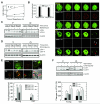
Age-and MPT-dependent cell death after I/R. (A) Hepatocytes from 3(Young)- and 26-month (Old)-old mice were subjected to simulated I/R. Necrosis at 5, 60 and 120 minutes of reperfusion was measured by PI fluorometry. (B) After 2 hours of ischemia, young (top panel) and old hepatocytes (bottom panel) were reperfused and confocal images of calcein, TMRM and PI (arrows) were simultaneously collected. Scale bar: 10 μm. The number of polarized mitochondria per cell was counted at 5 and 20 minutes of reperfusion. To determine autophagic flux, lysates from young (Y) and old (O) hepatocytes were collected at 0 minute of ischemia and at 20 and 60 minutes after reperfusion. LC3 was immunoblotted with 50 nM bafilomycin (C) or 10 μM chloroquine (D). (E) Hepatocytes infected with AdmCherry-GFP-LC3 were subjected to I/R. Confocal images of yellow (autophagosomes) and red (autolysosomes) puncta were collected after reperfusion with and without bafilomycin (Baf). Arrows indicate PI-labeled nuclei (necrosis). (F) After 2 hours of starvation in amino acid- and serum-free KRH, LC3 was immunoblotted with chloroquine (CQ). LC3-II expression was assessed relative to the level in control young hepatocytes and expressed as percent change. *P < 0.05.
Figure 2
Figure 2
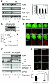
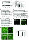
Atg4B loss in reperfused old hepatocytes. (A) Immunoblots at 0 and 120 minutes of ischemia (I) and at 20 and 60 minutes after reperfusion (R). The expression of Atg4B relative to the level at 0 hour of ischemia in young hepatocytes was determined. ** P < 0.01. (B) Old hepatocytes were incubated with different titers of AdAtg4B and overexpression was determined by immunoblotting. Necrosis with AdAtg4B (10 MOI) or AdGFP (viral control) was evaluated by PI fluorometry. (C) Confocal analysis of the MPT. (D) Immunoblots with Atg4B overexpression. (E) Confocal analysis in mCherry-GFP-LC3-labeled old hepatocytes. The number of puncta was counted after 20 minutes of reperfusion. *P < 0.05.
Figure 3
Figure 3
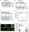
Atg4B loss by calpain 2. Expression of calpain 1(A) and calpain 2(B) was determined by immunoblotting. (C) Old hepatocytes were subjected to I/R with 50 μM ALLM and protein expressions were compared to control. (D) Necrosis in reperfused old cells was assessed with or without ALLM. Autophagic flux in old cells was determined by mCherry-GFP-LC3 analysis (E and F). *P < 0.05.
Figure 4
Figure 4
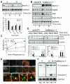
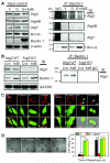
Cytoprotection by Beclin-1. (A) Immunoblots of Beclin-1 after I/R. Beclin-1 expression was expressed relative to the level at 0 hour of ischemia in young hepatocytes. (B) Atg expression in Beclin-1-overexpressed old hepatocytes. (C) Suppression of necrosis by Beclin-1. (D) Changes in autophagic flux by Beclin-1 overexpression were determined by immunoblotting (top panel) and mCherry-GFP-LC3 analysis (bottom panel). *P < 0.05. (E) Old hepatocytes were infected with either AdGFP-LC3/AdLacZ or AdGFP-LC3/AdBeclin-1. Confocal images of GFP-LC3 and TMRM were collected before and after reperfusion. Magnified images represent AdBeclin-1-treated cells after 60 minutes of reperfusion (bottom panel). Scale bar: 3 μm. (F) Hepatocytes at different ages were subjected to either 2 hours of ischemia/12 hours of reperfusion (I/R) or normoxia (Nor). Caspase-3 activation was evaluated by immunoblotting.
Figure 5
Figure 5
Interaction of Beclin-1 with Atg3. (A) Cell lysates at different ages were first immunoprecipitated (IP) with Beclin-1 and subsequently immunoblotted (IB). Bcl-xL and normal rabbit IgG were used for a positive and negative control, respectively. Some old hepatocytes were treated with AdBeclin-1(AdB). (B) Cell lysates from Atg3 WT and KO MEF were immunoblotted with viral control (Con) or AdBeclin-1. Atg3 was immunoblotted after Beclin-1 immunoprecipitation (right panel). (C) Atg3 WT and KO cells were subjected to either 6 hours of normoxia (left panel) or 2 hours of ischemia/4 hours of reperfusion (right panel). Note a lack of the MPT and cell death after normoxia. While WT cells tolerated I/R, mitochondrial dysfunction and cell death (arrows) were evident in KO cells with or without Beclin-1 overexpression. (D) Apoptosis was morphologically evaluated after 4 hours of reperfusion in Atg3 cells (left panel). Cell death was assessed after reperfusion (right panel).
Figure 6
Figure 6
Cathepsin D expression. (A) Immunoblotting of cathepsin D. (B) Immunoblotting in AdBeclin-1-treated old hepatocytes. Cytoprotection by Atg4B or Beclin-1 in in vivo I/R. After in vivo I/R, tissue lysates were collected at given times. Immunoblotting analysis was performed with Beclin-1 (C) and Atg4B (D) overexpression. (E) Old mice were injected with either AdGFP-LC3/AdLacZ or AdGFP-LC3/AdBeclin-1 and intravital multiphoton images of autophagosomes after in vivo I/R were collected at 0.7-μm intervals. (F) Livers were labeled with rhodamine 123 after in vivo I/R and multiphoton images of the mitochondria were collected. Punctate bright green fluorescence of rhodamine 123 represents polarized mitochondria. The number of cells containing polarized mitochondria, indicative of viable cells, was counted after 40 minutes of reperfusion. *P < 0.05.
Figure 7
Figure 7. Scheme of MPT induction and cell death after I/R
Reperfusion of ischemic old hepatocytes increases calpain 2 activity, which in turn hydrolyzes Atg4B. Atg4B loss subsequently impairs autophagy and promotes onset of the MPT and cell death. This chain reaction is prevented by calpain inhibitor (ALLM) and overexpression of Atg4B. Beclin-1 overexpression increases autophagy and blocks MPT onset by its interaction with Atg3.

References

    1. Selzner M, Selzner N, Jochum W, et al. Increased ischemic injury in old mouse liver: an ATP-dependent mechanism. Liver Transpl. 2007;13:382–390. - "VSports" PubMed
    1. Jaeschke H, Lemasters JJ. Apoptosis versus oncotic necrosis in hepatic ischemia/reperfusion injury. Gastroenterology. 2003;125:1246–1257. - PubMed
    1. Schweiger H, Lutjen-Drecoll E, Arnold E, et al. Ischemia-induced alterations of mitochondrial structure and function in brain, liver, and heart muscle of young and senescent rats. Biochem Med Metab Biol. 1988;40:162–185. - PubMed
    1. Clavien PA, Selzner M, Rudiger HA, et al. A prospective randomized study in 100 consecutive patients undergoing major liver resection with versus without ischemic preconditioning. Ann Surg. 2003;238:843–850. - PMC - PubMed
    1. Yorimitsu T, Klionsky DJ. Autophagy: molecular machinery for self-eating. Cell Death Differ. 2005;12(Suppl 2):1542–1552. - PMC (VSports最新版本) - PubMed

Publication types

MeSH terms