Skip to main page content
U.S. flag

An official website of the United States government

Dot gov

The . gov means it’s official. Federal government websites often end in . gov or . mil. Before sharing sensitive information, make sure you’re on a federal government site. VSports app下载.

Https

The site is secure V体育官网. The https:// ensures that you are connecting to the official website and that any information you provide is encrypted and transmitted securely. .

. 2011 Sep 15;187(6):2923-31.
doi: 10.4049/jimmunol.1100698. Epub 2011 Aug 10.

VSports手机版 - The impact of pre-existing memory on differentiation of newly recruited naive CD8 T cells

Affiliations

"VSports在线直播" The impact of pre-existing memory on differentiation of newly recruited naive CD8 T cells

"V体育安卓版" Matthew D Martin et al. J Immunol. .

VSports最新版本 - Abstract

One goal of immunization is to generate memory CD8 T cells of sufficient quality and quantity to confer protection against infection. It has been shown that memory CD8 T cell differentiation in vivo is controlled, at least in part, by the amount and duration of infection, Ag, and inflammatory cytokines present early after the initiation of the response VSports手机版. In this study, we used models of anti-vectorial immunity to investigate the impact of pre-existing immunity on the development and differentiation of vector-induced primary CD8 T cell responses. We showed that existing CD8 T cell memory influences the magnitude of naive CD8 T cell responses. However, the differentiation of newly recruited (either TCR-transgenic or endogenous) primary CD8 T cells into populations with the phenotype (CD62L(hi), CD27(hi), KLRG-1(low)) and function (tissue distribution, Ag-driven proliferation, cytokine production) of long-term memory was facilitated when they were primed in the presence of vector-specific memory CD8 T cells of the same or unrelated specificity. Therefore, these data suggested that the presence of anti-vectorial immunity impacts the rate of differentiation of vector-induced naive CD8 T cells, a notion with important implications for the design of future vaccination strategies. .


Figures
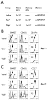
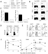
Figure 1
Figure 1. Primary memory OT-I CD8 T cells influence the phenotype of naïve OT-I responses after infection
A) Experimental design. Naïve Thy1.1/1.1 OT-I alone (‘naïve’) or mixed with 3×10⁴ (‘low’) or 5×10⁵ (‘high) primary memory Thy1.1/1.2 OT-1 were transferred into naïve B6 Thy1.2/1.2 recipients one day before infection with Att LM-OVA (1×10⁷ CFU/mouse i.v.). B–C) Blood samples were pooled and the expression of the indicated phenotypic markers was evaluated at B) d12 and C) d41 after infection. Shaded histograms represent isotype control staining and open histograms represent staining on gated primary OT-I (Thy1.1/Thy1.1) CD8 T cells. Numbers indicate the percentage of cells positive for CD127, CD62L, and CD27hi. One out of three experiments with similar results is shown.
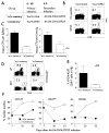
Figure 2
Figure 2. Naïve TCR-Tg CD8 T cells transferred into recipients with endogenous polyclonal memory CD8 T cells of the same specificity progress to a CD27hi/CD62Lhi phenotype faster
A) Experimental design. Naïve B6 mice were infected with either VacV-GP33 (3×10⁶ PFU/mouse i.p.; ‘w/o memory’ group) or VacV-OVA (3×10⁶ PFU/mouse i.p.; ‘w/memory’ group). Naïve OT-I (Thy1.1) were injected 48 days later, and 24 hrs after transfer, mice were infected with Att LM-OVA (5×10⁶ CFU/mouse i.v.). B) Splenocytes from mice infected 47 days previously with VacV-GP33 or VacV-OVA were incubated in the presence or absence of indicated peptides and monitored for IFN-γ production. Numbers represent the percentage of IFN-γ-positive CD8 T cells. C) Bacterial titers in the spleen (CFU/spleen) and liver (CFU/g Liver) were determined three days after Att LM-OVA infection. Data are presented as mean + SD of three mice pergroup. D) Representative plots showing the frequency of tetramer positive (KbOva257) endogenous Ova257-specific cells (Thy1.1 negative) and OT-I cells (Thy1.1 positive) in the blood at day 5 p.i. Numbers indicate the percentage of cells positive for the indicated molecules. E) Blood samples were pooled and the expression of the indicated markers was evaluated at d7 after Att LM-OVA infection. Shaded histograms represent isotype control staining and open histograms represent staining of OT-I (Thy1.1) CD8 T cells. Numbers indicate the percentage of cells positive for the indicated molecules. F) Kinetic analysis of the expression of CD27hi, CD62L, and KLRG-1 molecules on OT-I (Thy1.1) CD8 T cells. Data are presented as the percentage of cells positive for the indicated marker. The experiment was repeated twice with similar results.
Figure 3
Figure 3. Naïve endogenous CD8 T cells primed in the presence of endogenous memory CD8 T cells of unrelated specificity show decreased levels of expansion but progress to a long-term memory phenotype faster
A) Experimental design. Naïve B6 mice were infected with either VacV-β-Gal (3×10⁶ PFU/mouse i.p.; ‘w/o memory’ group) or VacV-GP33 (3×10⁶ PFU/mouse i.p.; ‘w/memory’ group), and 68 days later mice were infected with Att LM-OVA/GP33 (1×10⁷ CFU/mouse i.v.). B) Splenocytes from mice infected 67 days previously with VacV-β-Gal or VacV-GP33 were incubated in the presence or absence of GP33 peptide and monitored for IFN-γ production. Numbers represent the percentage of Ag-specific IFN-γ-positive CD8 T cells. C) Bacterial titers in the spleen (CFU/spleen) and liver (CFU/g Liver) were determined three days after Att LM-OVA/GP33 infection. Data are presented as mean + SD of three mice per group. D) Representative plots showing the frequency of KbOva257-specific CD8 T cells at d7 and d33 after infection. Numbers indicate the percentage of Ova-specific CD8 T cells. E) Percentage of Kb OVA257 positive CD8 T cells in PBL at d33 p.i. presented as mean + SD (n=4 mice per group). F) Kinetic analysis of the expression of the indicated markers for Kb OVA257+ CD8 T cells. Data are presented as the percentage of cells positive for the indicated marker. The experiment was repeated twice with similar results.
Figure 4
Figure 4. Anti-vectorial immunity decreases the magnitude of newly evoked primary CD8 T cell responses after infection
A) Experimental design. Naïve B6 mice were either not infected or infected with Att LM (5×10⁶ CFU/mouse i.v.). Naïve OT-I (Thy1.1, 5×10² cells/mouse) were transferred one day before infection with Att LM-OVA (1×10⁷ CFU/mouse i.v). B) Representative plots showing the expansion of OT-I cells at d7 and d32 after Att LM-OVA infection. Numbers indicate the percentage of OT-I in PBL. C) Kinetic analysis of the primary OT-I CD8 T cell responses in PBL. Data are presented as the percentage (mean + SD for 5 to 15 mice per group per time point) of OT-I cells in PBL. D) The percentage of OT-I (Thy1.1) CD8 T cells in PBL of individual mice on d32 after Att LM-OVA infection. Dots represent individual mice and the line represents the mean. The p values are indicated. E) The expression of CD27 and CD62L was evaluated at d24 after Att LM-OVA infection. Shaded histograms represent isotype control staining and open histograms represent staining of OT-I (Thy1.1) CD8 T cells. Numbers indicate the percentage of cells positive for the indicated molecules. Representative profiles out of 3 per group are shown.
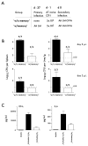
Figure 5
Figure 5. Anti-vectorial immunity decreases the duration of infection and inflammation
A) Experimental design. Naïve OT-I (Thy1.1, 5×10² cells/mouse) were transferred into age matched naïve or previously infected (Att LM) groups of mice one day before infection with Att LM-OVA (1×10⁷ CFU/mouse i.v). B) Bacterial titers in the spleen (CFU/spleen) and liver (CFU/g Liver) were determined two and three days after Att LM-OVA infection. Data are presented as mean + SD of three to four mice per group. Numbers above bars indicate percentage of mice analyzed that showed detectable levels of infection (above LOD -limit of detection). C) The concentrations of IFNγ and IL-6 were determined in serum of naïve and Att LM-Ova infected groups of mice 24 hours post infection. The results are presented as mean + SD of three mice per group.
Figure 6
Figure 6. Anti-vectorial immunity influences the tissue distribution of primary CD8 T cells
A) Representative plots showing the percentage of OT-I (Thy1.1) CD8 T cells in the indicated organs one month after Att LM-OVA infection. B) Ratios of the percentage of OT-I (Thy1.1) CD8 T cells in the indicated organs to the percentage of OT-I CD8 T cells in PBL at 3 to 4 weeks after Att LM-OVA infection. Data are presented asmean + SD of three mice per group. Numbers above bar graphs indicate statistical p value and ‘ns’ indicates not statistically significant. C) Total number of OT-ICD8 T cells in ml of PBL and in the spleen, LN, and lung. Dots represent individual mice analyzed in two separate experiments, and lines indicate the mean. Numbers indicate statistical p value and fold difference in numbers.
Figure 7
Figure 7. Primary CD8 T cells primed in the presence of anti-vectorial immunity show different cytokine profiles after direct ex vivo stimulation with antigen
A) One month post Att LM-OVA infection the cytokine production by OT-I CD8 T cells from the spleen from groups of mice described in Figure 4A were determined after short ex vivo incubation in the presence of OVA257 peptide. Numbers represent the percentage of OT-I (Thy1.1) CD8 T cells that were positive for IFN-γ and IL-2. B) Percentage of IL-2 producing IFN-γ positive OT-I CD8 T cells. Data are presented as mean + SD of three mice per group. Numbers above bar graphs indicate statistical p value.
Figure 8
Figure 8. Primary CD8 T cells primed in the presence of anti-vectorial immunity have a greater proliferative potential after booster challenge
A) Primary memory OT-I cells (Thy1.1) from ‘w/o memory’ and ‘w/memory’ groups of mice (described in Figure 4A) were isolated on day 35 post Att LM-OVA infection by positive selection and transferred in equal numbers (3×10⁴) into naïve recipients one day before Att LM-OVA or VacV-OVA infection. Representative dot plot examples showing the expansion of OT-I cells (Thy1.1) at d7 after Att LM-OVA (5×10⁶ CFU/mouse i.v.) or VacV-OVA (3×10⁶ PFU/mouse i.p.) infections. Numbers indicate the percentage of OT-1 (Thy1.1) CD8 T cells per PBL. B) Kinetic analysis of the OT-I CD8 T cells in PBL after infection. Data are presented as the frequency of OT-I cells in PBL (mean +SD for three mice per group). Numbers above boxes indicate the statistical p value.

"VSports在线直播" References

    1. Badovinac VP, Harty JT. Programming, demarcating, and manipulating CD8+ T-cell memory. Immunol Rev. 2006;211:67–80. - "V体育官网入口" PubMed
    1. Harty JT, V, Badovinac P. Shaping and reshaping CD8+ T-cell memory. Nat Rev Immunol. 2008;8:107–119. - PubMed
    1. Kaech SM, Wherry EJ, Ahmed R. Effector and memory T-cell differentiation: implications for vaccine development. Nat Rev Immunol. 2002;2:251–262. - PubMed
    1. Lefrancois L. Development, trafficking, and function of memory T-cell subsets. Immunol Rev. 2006;211:93–103. - PubMed
    1. Seder RA, Darrah PA, Roederer M. T-cell quality in memory and protection: implications for vaccine design. Nat Rev Immunol. 2008;8:247–258. - PubMed

Publication types