Skip to main page content
U.S. flag

An official website of the United States government

Dot gov

The . gov means it’s official. Federal government websites often end in . gov or . mil. Before sharing sensitive information, make sure you’re on a federal government site. VSports app下载.

Https

The site is secure. The https:// ensures that you are connecting to the official website and that any information you provide is encrypted and transmitted securely. V体育官网.

. 2011 Aug 19;286(33):28749-28760.
doi: 10.1074/jbc.M110.202440. Epub 2011 Jun 28.

VSports最新版本 - Resveratrol induces p53-independent, X-linked inhibitor of apoptosis protein (XIAP)-mediated Bax protein oligomerization on mitochondria to initiate cytochrome c release and caspase activation

Affiliations

Resveratrol induces p53-independent, X-linked inhibitor of apoptosis protein (XIAP)-mediated Bax protein oligomerization on mitochondria to initiate cytochrome c release and caspase activation

Raghu Gogada et al. J Biol Chem. .

Abstract

Resveratrol, a naturally occurring phytoalexin, is known to induce apoptosis in multiple cancer cell types, but the underlying molecular mechanisms remain unclear. Here, we show that resveratrol induced p53-independent, X-linked inhibitor of apoptosis protein (XIAP)-mediated translocation of Bax to mitochondria where it underwent oligomerization to initiate apoptosis. Resveratrol treatment promoted interaction between Bax and XIAP in the cytosol and on mitochondria, suggesting that XIAP plays a critical role in the activation and translocation of Bax to mitochondria VSports手机版. This process did not involve p53 but required accumulation of Bim and t-Bid on mitochondria. Bax primarily underwent homo-oligomerization on mitochondria and played a major role in release of cytochrome c to the cytosol. Bak, another key protein that regulates the mitochondrial membrane permeabilization, did not interact with p53 but continued to associate with Bcl-xL. Thus, the proapoptotic function of Bak remained suppressed during resveratrol-induced apoptosis. Caspase-9 silencing inhibited resveratrol-induced caspase activation, whereas caspase-8 knockdown did not affect caspase activity, suggesting that resveratrol induces caspase-9-dependent apoptosis. Together, our findings characterize the molecular mechanisms of resveratrol-induced caspase activation and subsequent apoptosis in cancer cells. .


Figures

FIGURE 1.
FIGURE 1.
Resveratrol induces caspase-dependent apoptosis. A, MDA-MB231 cells were treated with resveratrol (Resv) for 36 h, and the percentage of cell death (mean ± S.D. from four independent experiments) was determined using trypan blue dye. *, p < 0.01. B, MDA-MB231, GM701 (GM), LNCaP, and PC3 cells were treated with resveratrol for the indicated time intervals, and equal amounts of protein (50 μg) were subjected to caspase-3 activity measurements. Caspase activities are presented as values (mean ± S.D. from at least three independent experiments) relative to those in controls. *, p < 0.01. C, GM701 and MDA-MB231 cells were pretreated with the pan-caspase inhibitor Z-VAD (50 μm) for 1 h and then treated with resveratrol for 36 h. At the end of treatment, equal amounts of protein were used for Western blotting and DEVDase activity assay. The number in DEVDase activity represents -fold change against untreated control (for example, lane 1 is control for GM701 cells and lane 5 is control for MDA-MB231 cells). Data are the representative of four independent experiments. D, MDA-MB231 and GM701 cells were pretreated with the pan-caspase inhibitor Z-VAD (50 μm) for 1 h and then treated with resveratrol for 36 h. At the end of treatment, the percentage of cell death was determined, and data are represented as mean ± S.D. from four independent experiments. *, p < 0.01 as compared with resveratrol-treated cells. Cont, control; Resv, resveratrol; MDA, MDA-MB231; Procasp-3, procaspase-3. p17 is a cleaved caspase-3 fragment.
FIGURE 2.
FIGURE 2.
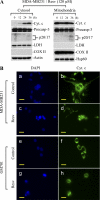
Resveratrol induces cytochrome c release from mitochondria into cytosol and is accompanied by fragmentation of nucleus. A, MDA-MB231 cells were treated with resveratrol (Resv; 120 μm) for the indicated time intervals. At the end of the treatment, cytosolic and mitochondrial fractions were isolated, and equal amounts of protein were subjected to Western blotting for the detection of the indicated molecules. Cyt. c, cytochrome c; Procasp-3, procaspase-3; COX II, cytochrome c oxidase subunit II; LDH, lactate dehydrogenase. p20/17 represents cleaved products of caspase-3. Actin and Hsp60 serve as loading controls. B, cells were treated with resveratrol (Resv; 120 μm) for 24 h. At the end of the treatment, live cells were labeled with DAPI (panels a, c, e, and g) and MitoTracker Orange to detect the nucleus and mitochondria (data not shown), respectively. Cells were then immunolabeled for cytochrome c (Cyt. c; panels b, d, f, and h). Representative micrographs are shown, and magnification bars represent 20 μm. Consistent with the Western blot analysis, the diffuse staining for cytochrome c in individual cells reveals that it was released from mitochondria. Apoptotic cells show fragmented/shiny nucleus with DAPI staining in the panels c and g. Data are representative of at least three independent experiments.
FIGURE 3.
FIGURE 3.
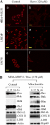
Resveratrol causes loss of mitochondrial membrane potential and induces Bax translocation to mitochondria and up-regulates Bim and t-Bid on mitochondria. A, MDA-MB231, LNCaP, and GM701 cells were treated without (Control; panels a, c, and e) or with resveratrol (Resv; 120 μm for 24 h) (panels b, d, and f) and just prior to fixation were labeled live with MitoTracker Orange, which accumulates in mitochondria in a membrane potential-dependent manner. The images were captured by microscope, representative micrographs are shown, and magnification bars represent 20 μm. B, MDA-MB231 cells were treated with resveratrol (Resv; 120 μm) for the indicated time intervals. Cytosolic and mitochondrial fractions were purified followed by Western blotting for the indicated molecules by loading equal amounts of protein. COX II, cytochrome c oxidase subunit II. Actin and Hsp60 serve as loading controls. Data are representative of three independent experiments.
FIGURE 4.
FIGURE 4.
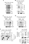
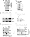
Bax undergoes homo-oligomerization on mitochondria, and Bax oligomerization on mitochondria is accompanied by cytochrome c release. A and B, MDA-MB231 (A) cells were treated with resveratrol (Resv; 120 μm) for the indicated time intervals, and HCT116 WT or HCT116 Bax−/− cells (B) were treated with resveratrol (50 μm). Purified mitochondria were cross-linked with BMH and subjected to Western blotting for Bax to detect its oligomers. Hsp60 serves as a loading control. C and D, MDA-MB231 and HCT116 cells were treated with resveratrol (50 μm) for the indicated time periods. At the end of treatments, cells were cross-linked with BMH or ethylene glycol bis(succinimidylsuccinate) (EGS) followed by Western blotting for Bax or Hsp60. E, mitochondrial lysates isolated from MDA-MB231 cells treated with resveratrol (120 μm for 36 h) or from unstimulated cells were subjected to immunoprecipitation followed by Western blotting for the indicated molecules. F, MDA-MB231 and MDA-MB435 cells were treated with resveratrol (50 μm) followed by mitochondrial cross-linking and cytochrome c release assayed by Western blotting. MDA, MDA-MB231; MB435, MDA-MB435; Cyt. c, cytochrome c; LDH, lactate dehydrogenase. The asterisks in A, B, and F indicate a nonspecific band. Data are representative of at least three independent experiments.
FIGURE 5.
FIGURE 5.
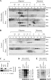
Bax undergoes oligomerization on mitochondria; Bax translocation and activation of Bax/Bak do not involve p53. A and B, mitochondrial (A) and cytosolic (B) fractions isolated from MDA-MB231 cells treated with resveratrol (120 μm for 36 h) or unstimulated were fractionated on a Superdex 200 column. Fractions (0.5 ml) were collected, and a portion (20 μl) of fractions 6–28 was analyzed by Western blotting for Bax, Bak, and p53. Arrows in A indicate Bax oligomers. C, cytosol isolated from MDA-MB231 treated with resveratrol (120 μm for 36 h) was subjected to immunoprecipitation with p53 or Bax followed by Western blotting for the indicated proteins. D and E, MDA-MB231 (D) and MDA-MB435 (E) cells were pretreated with PFT-α (30 μm) followed by resveratrol (50 μm) treatment. Cells were cross-linked and subjected to Western blotting to detect Bax oligomerization. Resv, resveratrol. Data are representative of at least three independent experiments.
FIGURE 6.
FIGURE 6.
Resveratrol induces Bax activation/oligomerization in cytosol, and XIAP interacts with Bax to regulate its activation and translocation to mitochondria. A and B, equal amounts of cytosols obtained from MDA-MB231 cells (A) treated with resveratrol (120 μm for 0, 12, 24, and 36 h) or from HCT116 cells (B) treated with resveratrol (50 μm for 0, 12, and 24 h) were cross-linked with BMH followed by Western blotting for Bax. C, purified mitochondria from resveratrol-treated (50 μm for 24 h) or untreated cells were subjected to Western blotting to detect XIAP and Bax translocation to mitochondria. D, purified cytosol and mitochondria obtained from resveratrol-treated or untreated MDA-MB231 cells were reconstituted to examine XIAP translocation to mitochondria. E and F, IP was performed using mitochondria (E) or cytosol (F) obtained from MDA-MB231 cells treated with resveratrol (50 μm for 18 h) followed by Western blotting for the indicated proteins. MDA, MDA-MB231; MB435, MDA-MB435; HCT, HCT116 WT; Resv, resveratrol; Cont, control; Cyto, cytosol; Mito, mitochondria. The asterisks in A and B indicate a nonspecific band. Data are representative of at least three independent experiments.
FIGURE 7.
FIGURE 7.
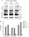
Bax plays prominent role to induce cytochrome c release upon resveratrol treatment. A, HCT116 WT, Bax−/−, and p53−/− cells were treated with resveratrol (120 μm for 12 or 48 h). Cytosolic and mitochondrial fractions were subjected to Western blotting to detect cytochrome c release. Resv, resveratrol; Cyt. c, cytochrome c; COX II, cytochrome c oxidase subunit II. Actin and Hsp60 serve as loading controls. Data are representative of three independent experiments. B, MDA-MB231, MDA-MB435, and MCF-7 cells were pretreated for 2 h with PFT-α (30 μm) or PFT-μ (10 μm) followed by resveratrol treatment (50 μm for 24 h). At the end of treatment, the percentage of cell death was quantified. Data are mean ± S.D. (n = 3). PFT-a, PFT-α; PFT-m, PFT-μ.
FIGURE 8.
FIGURE 8.
Activation of caspase-9 is initiating event in resveratrol-induced apoptosis. A, LNCaP cells were infected with caspase-9, caspase-8, or control shRNA lentiviral particles at a multiplicity of infection of 3. Stable cells were used for Western blotting to detect caspase-9 or caspase-8 silencing. B, caspase-8- and caspase-9-silenced or control (non-targeting) cells were treated with resveratrol (120 μm for 24 h). C, caspase-8 or caspase-9 was silenced in MDA-MB231 cells. D, cells were treated with resveratrol (50 μm for 24 h). At the end of treatment, caspase-3 activity was determined. Resv, resveratrol; Casp-9, caspase-9; Casp-8, caspase-8. Actin and Hsp60 serve as loading controls. Data are mean ± S.D. (n = 3). *, p < 0.01 as compared with resveratrol-treated control cells.

References

    1. Mathew R., White E. (2011) Curr. Opin. Genet. Dev. 21, 113–119 - VSports在线直播 - PMC - PubMed
    1. Zois C. E., Koukourakis M. I. (2009) Autophagy 5, 442–450 - PubMed
    1. Blagosklonny M. V. (2002) Int. J. Cancer 98, 161–166 - PubMed
    1. Lowe S. W., Lin A. W. (2000) Carcinogenesis 21, 485–495 - VSports在线直播 - PubMed
    1. Delmas D., Rébé C., Lacour S., Filomenko R., Athias A., Gambert P., Cherkaoui-Malki M., Jannin B., Dubrez-Daloz L., Latruffe N., Solary E. (2003) J. Biol. Chem. 278, 41482–41490 - PubMed

"VSports" Publication types

MeSH terms

Substances