Skip to main page content
U.S. flag

An official website of the United States government

Dot gov

The . gov means it’s official. Federal government websites often end in . gov or . mil. Before sharing sensitive information, make sure you’re on a federal government site. VSports app下载.

Https

The site is secure. The https:// ensures that you are connecting to the official website and that any information you provide is encrypted and transmitted securely. V体育官网.

. 2011 Sep;43(3):598-608.
doi: 10.1016/j.nbd.2011.05.008. Epub 2011 May 23.

Systemic delivery of NEMO binding domain/IKKγ inhibitory peptide to young mdx mice improves dystrophic skeletal muscle histopathology

Affiliations

Systemic delivery of NEMO binding domain/IKKγ inhibitory peptide to young mdx mice improves dystrophic skeletal muscle histopathology

Daniel P Reay et al. Neurobiol Dis. 2011 Sep.

Abstract

The activation of nuclear factor κB (NF-κB) contributes to muscle degeneration that results from dystrophin deficiency in human Duchenne muscular dystrophy (DMD) and in the mdx mouse VSports手机版. In dystrophic muscle, NF-κB participates in inflammation and failure of muscle regeneration. Peptides containing the NF-κB Essential Modulator (NEMO) binding domain (NBD) disrupt the IκB kinase complex, thus blocking NF-κB activation. The NBD peptide, which is linked to a protein transduction domain to achieve in vivo peptide delivery to muscle tissue, was systemically delivered to mdx mice for 4 or 7 weeks to study NF-κB activation, histological changes in hind limb and diaphragm muscle and ex vivo function of diaphragm muscle. Decreased NF-κB activation, decreased necrosis and increased regeneration were observed in hind limb and diaphragm muscle in mdx mice treated systemically with NBD peptide, as compared to control mdx mice. NBD peptide treatment resulted in improved generation of specific force and greater resistance to lengthening activations in diaphragm muscle ex vivo. Together these data support the potential of NBD peptides for the treatment of DMD by modulating dystrophic pathways in muscle that are downstream of dystrophin deficiency. .


Figures

Figure 1
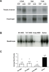
Figure 1. EMSA analysis of NF-κB activation of control and PTD-NBD peptide treated mdx mice
(A) Nuclear extracts from tibialis anterior and diaphragm muscle tissue were assessed for NF-κB activation by electrophoretic mobility shift assay (EMSA) in untreated C57BL/10 and mdx mice at 4.5, 9, and 12 weeks of age. (B) Nuclear extracts from quadriceps muscles of 8–9-week-old 8K-, TAT- and Antp-NBD peptide treated mdx mice (n=3) and saline treated mdx mice (n=3) were assessed for NF-κB activation by EMSA. (C) Densitometric analysis of the blots shown in B represented as mean ± standard error. 8K- and TAT-NBD peptide treated mdx mice received a single injection of 10mg/kg peptide. Antp-NBD peptide treated mdx mice received intraperitoneal injections of 200µg peptide 3 times per week for 4 weeks, beginning at 4–5 weeks of age. * indicates that PTD-NBD peptide treatment group is significantly different from saline mdx control animals (p<0.05). n=number of animals analyzed.
Figure 2
Figure 2. Histological analysis of mdx tibialis anterior muscle histopathology after 4 weeks of PTD-NBD peptide-mediated therapy
10µm tibialis anterior muscle sections from age-matched control mdx mice, PTD-NBD wild type peptide-treated mdx mice, and PTD-NBD mutant peptide treated mdx mice were stained with hematoxylin and eosin for gross morphology (A–F), labeled with mouse IgG for detection of necrotic fibers (G–L), and immunostained for embryonic myosin heavy chain for detection of regenerating fibers (M–R). Representative images for each treatment group are shown. Bar represents 100µm.
Figure 3
Figure 3. Quantification of mdx tibialis anterior and diaphragm histopathology after 4 weeks of PTD-NBD peptide-mediated therapy
10µm muscle sections from age-matched control C57BL/10 and mdx mice, PTD-NBD wild type peptide-treated mdx and PTD-NBD mutant peptide-treated mdx mice were quantified to assess levels of necrosis (A) and regeneration (B) in tibialis anterior muscle and necrosis (C) and regeneration (D) in diaphragm muscle. Data is represented as mean % necrotic or regenerating fibers ± standard error quantified from 2 representative sections per muscle. * indicates that PTD-NBD peptide treatment group is significantly different from untreated mdx control animals (p<0.05). n=number of animals analyzed.
Figure 4
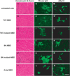
Figure 4. Histological analysis of mdx diaphragm muscle histopathology after 4 weeks of PTD-NBD peptide-mediated therapy
10µm sections of diaphragm muscle from age-matched control mdx mice, 8K-, TAT-, and Antp-NBD wild type peptide-treated mdx mice, and 8K- and TAT-NBD mutant peptide-treated mdx mice were stained with hematoxylin and eosin for gross morphology (A–F), labeled with mouse IgG for detection of necrotic fibers (G–L), and immunostained for embryonic myosin heavy chain for detection of regenerating fibers (M–R). Representative images for each treatment group are shown. Bar represents 100µm.
Figure 5
Figure 5. EMSA analysis of NF-κB activation after 7 weeks of PTD-NBD peptide or saline treatment of mdx mice
Nuclear extracts of muscle tissue collected from mdx mice after treatment for 7 weeks with 8K- or TAT-NBD peptide, or saline were analyzed for NF-κB activation in the tibialis anterior (A,C) and costal diaphragm (B,D) muscles by electrophoretic mobility shift assay (EMSA). Individual lanes in A and B each show the level of NF-κB in nuclear extract from one animal in the study (n=8 mice/group). Mouse numbers indicate corresponding tibialis anterior and diaphragm samples taken from the same mouse. Panels C and D show densitometric analysis of blots shown in A and B, respectively, represented as mean ± standard error. * indicates that PTD-NBD peptide treatment group is significantly different from saline mdx control animals (p<0.05). n=number of animals analyzed.
Figure 6
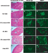
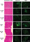
Figure 6. Histological analysis of mdx tibialis anterior and costal diaphragm muscle histopathology after 7 weeks of PTD-NBD peptide-mediated therapy
10µm sections of tibialis anterior and costal diaphragm muscle from age-matched saline treated mdx mice and 8K- and TAT-NBD peptide-treated mdx mice were stained with hematoxylin and eosin for gross morphology (A–F), labeled with mouse IgG for detection of necrotic fibers (G–L), and immunostained for embryonic myosin heavy chain for detection of regenerating fibers (M–R). Representative images for each treatment group are shown. Bar represents 100µm. TA=tibialis anterior. DIA=diaphragm.
Figure 7
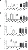
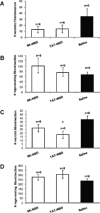
Figure 7. Quantification of mdx tibialis anterior and costal diaphragm muscle histopathology after 7 weeks of PTD-NBD peptide-mediated therapy
10µm muscle sections from age-matched, saline treated mdx mice, and 8K- and TAT-NBD wild type peptide-treated mdx mice were immunostained and quantified to assess levels of necrosis (A) and regeneration (B) in tibialis anterior muscle and necrosis (C) and regeneration (D) in diaphragm muscle. Data is represented as mean total number of necrotic or regenerating fibers ± standard error quantified from 2 representative sections per muscle. * indicates that PTD-NBD peptide treatment group is significantly different from saline treated mdx control animals (p<0.05). n=number of animals analyzed.
Figure 8
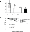
Figure 8. Force generation properties in mdx costal diaphragm after 7 weeks of PTD-NBD peptide therapy
Costal diaphragm muscles were collected after 7 weeks of treatment with 8K- or TAT-NBD peptide or saline. Diaphragms from age-matched control C57BL/10 mice, age-matched saline treated mdx mice, and 8K- and TAT-NBD peptide-treated mdx mice were analyzed for (A) specific force (N/cm2) and (B) residual force following 10 repetitive lengthening activations. Lengthening activation data is expressed as a percentage of the force generated in response to the first lengthening activation. The data is shown as mean ± standard error. * indicates that C57BL/10 group is significantly different from 8K-NBD, TAT-NBD and saline treated mdx mice (p<0.05). ** indicates that PTD-NBD peptide treated groups are significantly different from saline treated mdx mice (p<0.05). ***indicates that C57BL/10 group is significantly different from TAT-NBD and saline treated mdx mice (p<0.05). **** indicates that 8K-NBD peptide group is significantly different from saline treated mdx mice (p<0.05), but not significantly different from C57BL/10 mice. n=number of animals analyzed.

References

    1. Acharyya S, Villalta SA, Bakkar N, Bupha-Intr T, Janssen PML, Carathers M, et al. Interplay of IKK/NF-kappa B signaling in macrophages and myofibers promotes muscle degeneration in Duchenne muscular dystrophy. J Clin Invest. 2007;117:889–901. - "VSports最新版本" PMC - PubMed
    1. Bulfield, Siller WG, Wight PA, Moore KJ. X chromosome-linked muscular dystrophy (mdx) in the mouse. Proc Natl Acad Sci USA. 1984;81:1189–1192. - PMC - PubMed
    1. Dave SH, Tilstra JS, Matsuoka K, Li F, Karrasch T, Uno JK, et al. Amelioration of chronic murine colitis by peptide-mediated transduction of the IkappaB kinase inhibitor NEMO binding domain peptide. J Immunol. 2007;179:7852–7859. - PMC - PubMed
    1. Di MP, Ianaro A, Ghosh S. Amelioration of acute inflammation by systemic administration of a cell-permeable peptide inhibitor of NF-kappaB activation. Arthritis Rheum. 2005;52:951–958. - PubMed
    1. Disatnik MH, Dhawan J, Yu Y, Beal MF, Whirl MM, Franco AA, et al. Evidence of oxidative stress in mdx mouse muscle: studies of the pre-necrotic state. J Neurol Sci. 1998;161:77–84. - PubMed

"V体育官网入口" Publication types

MeSH terms (V体育平台登录)

LinkOut - more resources