Skip to main page content
U.S. flag

An official website of the United States government

Dot gov

The . gov means it’s official. Federal government websites often end in . gov or . mil. Before sharing sensitive information, make sure you’re on a federal government site VSports app下载. .

Https

The site is secure. The https:// ensures that you are connecting to the official website and that any information you provide is encrypted and transmitted securely V体育官网. .

. 2010 Oct;84(20):10533-42.
doi: 10.1128/JVI.01263-10. Epub 2010 Aug 11.

"VSports" Primary human mammary epithelial cells endocytose HIV-1 and facilitate viral infection of CD4+ T lymphocytes

Affiliations

VSports注册入口 - Primary human mammary epithelial cells endocytose HIV-1 and facilitate viral infection of CD4+ T lymphocytes

Stephanie M Dorosko et al. J Virol. 2010 Oct.

Abstract

The contribution of mammary epithelial cells (MEC) to human immunodeficiency virus type 1 (HIV-1) in breast milk remains largely unknown. While breast milk contains CD4(+) cells throughout the breast-feeding period, it is not known whether MEC directly support HIV-1 infection or facilitate infection of CD4(+) cells in the breast compartment. This study evaluated primary human MEC for direct infection with HIV-1 and for indirect transfer of infection to CD4(+) target cells. Primary human MEC were isolated and assessed for expression of HIV-1 receptors. MEC were exposed to CCR5-, CXCR4- and dual-tropic strains of HIV-1 and evaluated for viral reverse transcription and integration and productive viral infection. MEC were also tested for the ability to transfer HIV to CD4(+) target cells and to activate resting CD4(+) T cells. Our results demonstrate that MEC express HIV-1 receptor proteins CD4, CCR5, CXCR4, and galactosyl ceramide (GalCer) VSports手机版. While no evidence for direct infection of MEC was found, HIV-1 virions were observed in MEC endosomal compartments. Coculture of HIV-exposed MEC resulted in productive infection of activated CD4(+) T cells. In addition, MEC secretions increased HIV-1 replication and proliferation of infected target cells. Overall, our results indicate that MEC are capable of endosomal uptake of HIV-1 and can facilitate virus infection and replication in CD4(+) target cells. These findings suggest that MEC may serve as a viral reservoir for HIV-1 and may enhance infection of CD4(+) T lymphocytes in vivo. .


Figures
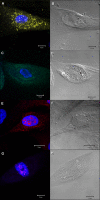
FIG. 1.
FIG. 1.
Primary human MEC express HIV-1 receptors, coreceptors, and an alternate receptor. Confocal microscopy images of MEC stained for HIV receptors (left) and the same cells imaged by DIC (right). Nuclei are stained blue with DAPI. An immunomagnetic bead can be observed in the DIC image shown in panel D. (A) GalCer expression (yellow). (C) CCR5 expression (green). (E) CXCR4 expression (red). (G) CD4 expression (pink). Z-stack imaging confirmed the presence of receptors within the cytoplasm and/or nucleus as shown. Similar results were obtained in two additional subjects, with the exception of CD4 expression. CD4 mRNA was confirmed in three subjects by use of real-time PCR.
FIG. 2.
FIG. 2.
Primary human MEC do not support HIV reverse transcription or integration of the CCR5-, CXCR4-, or dual-tropic strains tested. PCR results for HIV-1 reverse transcription and integration of CCR5-tropic HIV-1BaL, CXCR4-tropic HIV-1HC4, and dual-tropic HIV-1C7/86 in both primary human MEC and PBMC controls. The bottom row shows PCR results for human beta-actin genomic DNA. Lanes 1 to 4, MEC; lanes 5 to 8, PBMCs; lane 9, negative control (no DNA). Experiments were performed in triplicate under each condition. Similar results were obtained with MEC from three additional subjects.
FIG. 3.
FIG. 3.
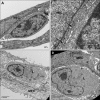
MEC endocytose HIV-1 virions from infected PBMCs. Transmission electron microscopy images showing HIV-1 virions inside MEC endocytic compartments, with panels from left to right exhibiting progressive enlargement of the boxed area. The image shows HIV-1HC4 virions. Experiments were performed in duplicate with both CCR5-tropic HIV-1BaL and CXCR4-tropic HIV-1HC4. Similar experiments were repeated with MEC from two additional subjects. L, lymphocyte.
FIG. 4.
FIG. 4.
MEC maintain close physical contact with CD4+ T lymphocytes. (A) Transmission electron microscopy image showing junctions and clefts between MEC and lymphocytes (L). (B) Enlarged image of boxed area, showing junctions between cells (black arrows). Junctions between MEC and lymphocytes were observed in all samples from two individual subjects, with conditions tested in duplicate, irrespective of HIV-1 infection of lymphocytes. (C and D) Images of separate observations of lymphocyte engulfment by MEC from two individual subjects. Lymphocyte endocytosis was not dependent on HIV-1 infection of lymphocytes.
FIG. 5.
FIG. 5.
Activated CD4+ T lymphocytes are productively infected with HIV-1 after coculture with HIV-exposed MEC. Mean p24 antigen concentrations in culture supernatants up to 14 days after coculture of activated versus resting CD4+ T lymphocytes with HIV-exposed and washed MEC. Each experiment was conducted in triplicate wells. Data represent the means ± standard deviations (SD) from three individual subjects.
FIG. 6.
FIG. 6.
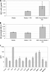
MEC secretions from the apical surface increase HIV-1 replication and cellular proliferation in infected PBMCs. (A) HIV-1-infected PBMCs were cultured in MEC conditioned medium and compared to controls cultured in medium alone. Mean HIV p24 antigen concentrations on day 7 of the culture (*, P < 0.02). Each experiment was conducted in triplicate wells. Data represent mean values ± SD from two subjects. (B) Mean proliferation of HIV-1-infected PBMCs, measured by a 3-(4,5 dimethylthiazol-2-yl)-5-(3-carboxymethoxyphenyl)-2-(4-sulfophenyl)-2H-tetrazolium, inner salt (MTS)-based assay (*, P < 0.0001). Data represent mean values ± SD from the same two subjects represented in panel A. (C) Cytokines, chemokines, and growth factors (>10 pg/ml) measured in MEC apical-surface conditioned medium. Data represent the mean values ± SD from four subjects. IL-1ra, IL-1 receptor antagonist; G-CSF, granulocyte colony-stimulating factor; IFN-g, gamma interferon; MCP-1, monocyte chemoattractant protein 1; PDGF, platelet-derived growth factor.

References

    1. Alfsen, A., P. Iniguez, E. Bouguyon, and M. Bomsel. 2001. Secretory IgA specific for a conserved epitope on gp41 enveloped glycoprotein inhibits epithelial transcytosis of HIV-1. J. Immunol. 166:6257-6265. - PubMed
    1. Aronica, S. M., P. Fanti, K. Kaminskaya, K. Gibbs, L. Raiber, M. Nazareth, R. Bucelli, M. Mineo, K. Grzybek, M. Kumin, K. Poppenberg, C. Schwach, and K. Janis. 2004. Estrogen disrupts chemokine-mediated chemokine release from mammary cells: implications for the interplay between estrogen and IP-10 in the regulation of mammary tumor formation. Breast Cancer Res. Treat. 84:235-245. - PubMed
    1. Asin, S. N., M. W. Fanger, D. Wildt-Perinic, P. L. Ware, C. R. Wira, and A. L. Howell. 2004. Transmission of HIV-1 by primary human uterine epithelial cells and stromal fibroblasts. J. Infect. Dis. 190:236-245. - V体育官网入口 - PubMed
    1. Becquart, P., N. Chomont, P. Roques, A. Ayouba, M. D. Kazatchkine, L. Belec, and H. Hocini. 2002. Compartmentalization of HIV-1 between breast milk and blood of HIV-infected mothers. Virology 300:109-117. - PubMed
    1. Bhaskaram, P., and V. Reddy. 1981. Bactericidal activity of human milk leukocytes. Acta Paediatr. Scand. 70:87-90. - PubMed

Publication types

MeSH terms