Skip to main page content
U.S. flag

An official website of the United States government

Dot gov

The . gov means it’s official. Federal government websites often end in VSports app下载. gov or . mil. Before sharing sensitive information, make sure you’re on a federal government site. .

Https

The site is secure. The https:// ensures that you are connecting to the official website and that any information you provide is encrypted and transmitted securely. V体育官网.

. 2010 Oct;59(10):1355-62.
doi: 10.1136/gut.2010.207456. Epub 2010 Jun 29.

Abnormally expressed ER stress response chaperone Gp96 in CD favours adherent-invasive Escherichia coli invasion

Affiliations

Abnormally expressed ER stress response chaperone Gp96 in CD favours adherent-invasive Escherichia coli invasion (V体育平台登录)

Nathalie Rolhion et al. Gut. 2010 Oct.

Abstract

Background and aims: Crohn's disease (CD) ileal lesions are colonised by pathogenic adherent-invasive Escherichia coli (AIEC) producing outer membrane vesicles (OMVs) that contribute to the bacterial invasion process VSports手机版. In addition, increased expression of endoplasmic reticulum (ER)-localised stress response proteins, due to ER stress, is observed in patients with CD. The expression of the ER-localised stress response protein Gp96 in patients with CD and its biological role with regards to the ability of AIEC to invade intestinal epithelial cells were analysed. .

Methods and results: Immunohistochemistry on tissue arrays showed that, together with CEACAM6 (carcinoembryonic antigen-related cell adhesion molecule 6) or the ER stress protein Grp78, Gp96 is also strongly expressed at the apical plasma membrane of the ileal epithelial cells of 50% of patients with CD. Invasion experiments in the presence of antibodies raised against Gp96, or after transfection of Intestine-407 cells with gp96 small interfering RNA (siRNA), indicated that Gp96 is essential to promote AIEC LF82 invasion, allowing, via the recognition of the outer membrane protein OmpA, OMVs to fuse with intestinal epithelial cells. V体育安卓版.

Conclusions: Gp96 is overexpressed on the apical surface of ileal epithelial cells in patients with CD and acts as a host cell receptor for OMVs, promoting AIEC invasion V体育ios版. From the results shown here, it is speculated that AIEC could take advantage of the abnormal expression of Gp96 in patients with CD to invade the ileal mucosa. .


Conflict of interest statement

Competing interests: None

VSports - Figures

Figure 1
Figure 1
Gp96 expression in the intestinal biopsies of patients with Crohn's disease (CD) and controls. (A) Gp96 immunohistochemical staining of tissues microarrays (TMAs) from ileum and colon biopsies of controls (a, b), patients in the acute inflamed phase (c, d) or patients in the quiescent phase (e, f) of CD. Each spot shows representative tissue immunostaining for Gp96 (a–f, immunoperoxidase ×40; c and e, inset, immunoperoxidase ×200). (B) Quantification of Gp96 immunostaining using the Spot Browser software, in TMAs from colon and ileum biopsies of controls, and patients in the acute or quiescent phase of CD, **p<0.01. (C) Carcinoembryonic antigen-related cell adhesion molecule 6 (CEACAM6), Grp78 and Gp96 immunostaining of TMAs from ileum biopsies of patients with CD, magnification ×400. (D) Western blot analysis of whole protein extracts from ileal biopsies taken in involved areas of six patients with CD and from six ileal biopsies from controls using anti-Gp96 and anti-β-actin antibodies.
Figure 2
Figure 2
Gp96 expression in Caco-2 cells. (A) Fold variation of gp96 mRNA levels in Caco-2 cells after 24 or 48 h of stimulation with tumour necrosis factor α (TNFα) or interferon γ (IFNγ) relative to that in non-treated cells (NT) using RT–PCR. gapdh (glyceraldehyde phosphate dehydrogenase) mRNA levels were measured as controls. Data are the mean±SEM of three separate experiments. (B and C). Western blot analysis showing expression levels of Gp96 and carcinoembryonic antigen-related cell adhesion molecule 6 (CEACAM6) by Caco-2 cells after 24 or 48 h of stimulation with 50 ng/ml TNFα or IFNγ (B) or after a 3 h infection period with adherent-invasive Escherichia coli (AIEC) strain LF82 (C) at a multiplicity of infection of 10 or 100. As loading control, labelling was performed using anti-β-actin polyclonal antibodies.
Figure 3
Figure 3
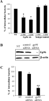
Gp96 expression supports LF82 invasion. (A) Effect of pretreatment of Intestine-407 epithelial cells with anti-Gp96 antibodies on the invasive level of LF82. Intestine-407 cells were pretreated with rabbit polyclonal antibodies raised against Gp96 (Gp96 Ab) or with rabbit polyclonal antibodies (isotype control) diluted 1:200 or 1:500 for 30 min and then infected by LF82 bacteria. Invasion was determined after a 3 h infection period and after gentamicin treatment for an additional hour. Results are expressed as intracellular bacteria relative to those obtained for strain LF82 on non-treated cells (NT), taken as 100%. Each value is the mean±SEM of at least four separate experiments. *p<0.05 compared with the wild-type strain on untreated cells. (B) Western blot analysis of whole protein extracts from Intestine-407 cells using anti-Gp96 and anti-β-actin antibodies. Intestine-407 cells were non-transfected (NT), or transfected with 10 ng of small interfering RNA blocking Gp96 (gp96 siRNA), or non-specific siRNA as control. (C) Effect of gp96 siRNA on the invasive level of the wild-type strain LF82. Invasive bacteria were quantified as described in A. **p<0.01 compared with the wild-type strain on untreated cells.
Figure 4
Figure 4
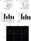
Gp96-dependent invasion involves adherent-invasive Escherichia coli (AIEC) outer membrane protein A (OmpA). Analysis of the interaction between OmpA and Gp96 by pull-down assay and ligand overlay. (A) For pull-down, total cell extracts from Caco-2 cells were incubated with Omp extracted from AIEC LF82 (1), LF82-ΔompA (2) and E. coli K-12 MG1655 (3), and immunoprecipitated with anti-Gp96 and Sepharose beads. In the control assay, the incubation step with anti-Gp96 was omitted. Captured proteins were separated on a 12% polyacrylamide gel, transferred onto a nitrocellulose membrane and probed with serum raised against OmpA. (B) For overlay, total extracts of Caco-2 cells were separated on a 12% polyacrylamide gel, transferred onto a nitrocellulose membrane and probed successively with Omp extracted from AIEC LF82 (1), LF82-ΔompA (2) and E. coli K-12 MG1655 (3), with anti-OmpA and detected with peroxidase-conjugated secondary antibody. (C) Invasion abilities with Intestine-407 epithelial cells of the LF82-ΔompA mutant, the LF82-ΔompA mutant transformed with the cloned LF82 ompA gene or the cloned K-12 MG1655 ompA gene, or the pBAD33 vector alone. Invasive bacteria were quantified as described in figure 3A. **p<0.01 compared with the wild-type strain. Expression of type 1 pili was determined visually by yeast aggregation and the titre was recorded as the last dilution giving a positive aggregation results. Whole-cell lysates of LF82, LF82-ΔompA bacteria transformed with the cloned LF82 ompA gene, with the cloned K-12 ompA gene, or with the pBAD33 vector alone and grown in medium with l-arabinose, were separated by sodium dodecylsulfate–polyacrylamide gel electrophoresis (SDS–PAGE) and stained with Coomassie blue. The positions of OmpC/F and OmpA are marked. (D) Effect of pretreatment of Intestine-407 epithelial cells with outer membrane vesicles (OMVs) and anti-Gp96 antibodies (Gp96 Ab) on the invasive level of LF82-ΔompA. Intestine-407 cells were pretreated with rabbit polyclonal antibodies raised against Gp96 diluted 1:500 for 30 min, then pretreated with OMVs from wild-type (WT) or LF82-ΔompA bacteria for 1 h and, after washing, cells were infected with bacteria. Invasive bacteria were quantified as described in figure 3A. (E) Analysis of the interaction of LF82 or ΔompA OMVs with the cytoplasmic membrane of Intestine-407 epithelial cells. The fusion of OMVs with the membrane Intestine-407 cells was analysed by confocal microscopy. Cells were incubated with OMVs at 37°C for 10 min. After washing and fixation, cells were labelled with rat antibodies raised against Gp96 and Cy3-conjugated goat anti-rat IgG secondary antibodies. OMVs fused with the cytoplasmic membrane of Intestine-407 cells were labelled with rabbit antibodies raised against E coli lipopolysaccharide O83 and fluorescein isothiocyanate (FITC)-conjugated donkey anti-rabbit immunoglobulin G (IgG). Each confocal microscopy image is representative of three independent experiments.

"V体育ios版" References

    1. Strober W, Fuss I, Mannon P. The fundamental basis of inflammatory bowel disease. J Clin Invest 2007;117:514–21 - VSports注册入口 - PMC - PubMed
    1. Xavier RJ, Podolsky DK. Unravelling the pathogenesis of inflammatory bowel disease. Nature 2007;448:427–34 - PubMed
    1. Sartor RB. Microbial influences in inflammatory bowel diseases. Gastroenterology 2008;134:577–94 - PubMed (V体育2025版)
    1. Baumgart M, Dogan B, Rishniw M, et al. Culture independent analysis of ileal mucosa reveals a selective increase in invasive Escherichia coli of novel phylogeny relative to depletion of Clostridiales in Crohn's disease involving the ileum. ISME J 2007;1:403–18 - PubMed
    1. Conte MP, Schippa S, Zamboni I, et al. Gut-associated bacterial microbiota in paediatric patients with inflammatory bowel disease. Gut 2006;55:1760–7 - PMC - PubMed

Publication types

MeSH terms

Substances