Skip to main page content
U.S. flag

An official website of the United States government

Dot gov

The . gov means it’s official. Federal government websites often end in VSports app下载. gov or . mil. Before sharing sensitive information, make sure you’re on a federal government site. .

Https

The site is secure V体育官网. The https:// ensures that you are connecting to the official website and that any information you provide is encrypted and transmitted securely. .

. 2010 May 7:11:288.
doi: 10.1186/1471-2164-11-288.

Analysis of microRNA transcriptome by deep sequencing of small RNA libraries of peripheral blood

Affiliations

Analysis of microRNA transcriptome by deep sequencing of small RNA libraries of peripheral blood

Candida Vaz (V体育官网) et al. BMC Genomics. .

Abstract (V体育平台登录)

Background: MicroRNAs are a class of small non-coding RNAs that regulate mRNA expression at the post - transcriptional level and thereby many fundamental biological processes. A number of methods, such as multiplex polymerase chain reaction, microarrays have been developed for profiling levels of known miRNAs VSports手机版. These methods lack the ability to identify novel miRNAs and accurately determine expression at a range of concentrations. Deep or massively parallel sequencing methods are providing suitable platforms for genome wide transcriptome analysis and have the ability to identify novel transcripts. .

Results: The results of analysis of small RNA sequences obtained by Solexa technology of normal peripheral blood mononuclear cells, tumor cell lines K562 and HL60 are presented. In general K562 cells displayed overall low level of miRNA population and also low levels of DICER. Some of the highly expressed miRNAs in the leukocytes include several members of the let-7 family, miR-21, 103, 185, 191 and 320a. Comparison of the miRNA profiles of normal versus K562 or HL60 cells revealed a specific set of differentially expressed molecules. Correlation of the miRNA with that of mRNA expression profiles, obtained by microarray, revealed a set of target genes showing inverse correlation with miRNA levels. Relative expression levels of individual miRNAs belonging to a cluster were found to be highly variable. Our computational pipeline also predicted a number of novel miRNAs. Some of the predictions were validated by Real-time RT-PCR and or RNase protection assay. Organization of some of the novel miRNAs in human genome suggests that these may also be part of existing clusters or form new clusters V体育安卓版. .

Conclusions: We conclude that about 904 miRNAs are expressed in human leukocytes. Out of these 370 are novel miRNAs. We have identified miRNAs that are differentially regulated in normal PBMC with respect to cancer cells, K562 and HL60 V体育ios版. Our results suggest that post - transcriptional processes may play a significant role in regulating levels of miRNAs in tumor cells. The study also provides a customized automated computation pipeline for miRNA profiling and identification of novel miRNAs; even those that are missed out by other existing pipelines. The Computational Pipeline is available at the website: http://mirna. jnu. ac. in/deep_sequencing/deep_sequencing. html. .


Figures
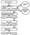
Figure 1
Figure 1
Flowchart describing the elimination pipeline used to filter out the indicated sequences from the library of sRNA sequences. The sequences were matched using an "in house" developed fast algorithm. Alignment with maximum of two mismatches was considered as hits. All the hits were removed before the next round of elimination. The databases used in this pipeline were either generated in house or downloaded from publicly available sites as described in "Methods".
Figure 2
Figure 2
Frequency of different classes of RNA species present in sRNA libraries. The sequences obtained from the sRNA libraries were subjected to a series of sequence similarity searches using specific databases (rRNAs, tRNA, sn/snoRNAs, miRNAs, other non-coding RNAs) and the pipeline described in Figure 1. The sequences that did not match with any known sequence were matched against databases of intergenic and intronic regions of the human genome. The pie-charts represent an overview of small RNA gene expression (shown in percentage) in normal PBMC and two cancer cell lines K562 and HL60. Small RNAs belonging to the miRNA family constitute the majority as in normal PBMC (61%) and HL60 (77%) samples. However, in K562 miRNAs constitute only 18% of the sRNA population.
Figure 3
Figure 3
Overall level of expression of known miRNAs. The distribution of known miRNA levels with respect to number of miRNAs is shown. Numbers of sequence reads are taken as miRNA levels and the values are represented in the form of range of values. The expression levels of the miRNAs span up to five orders of magnitude.
Figure 4
Figure 4
The abundance of selected miRNAs in human normal PBMC. The numbers of reads were used as expression level of respective miRNAs. [A]. Some of the highly expressing miRNAs (> 10,000 counts). [B]. Some of the low expressing miRNAs (< 10 counts).
Figure 5
Figure 5
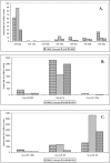
Differential expression of individual miRNAs present in the same cluster in different datasets. Here TPM (transcript per million) is used as a measure of expression. [A] miRNAs belonging to cluster miR-532, [B] cluster miR-99b and [C] cluster miR-106b in normal PBMC, K562 and HL60 cell lines. A large variation in expression levels of different miRNAs present within the same cluster is observed.
Figure 6
Figure 6
Differentially regulated known miRNAs. Up regulated/down regulated miRNAs are represented in the form of Venn diagrams. A subset of miRNAs that are differentially regulated but common in both cell lines as compared to normal PBMC is in the overlapped area and their expression levels can be seen in the heat map. Heat map of some of the differentially regulated known miRNAs with respect to datasets from normal PBMC and cancer cell lines K562 and HL60 is shown as an inset.
Figure 7
Figure 7
Expression levels of some of the known miRNAs determined by RNase protection assay. The relative expression levels of some of the differentially regulated miRNAs were determined using RPA. Briefly, total RNA from indicated cells was incubated with a labelled probe specific for a given miRNA and eventually treated with ribonuclease as described in the "Methods". The protected fragments, suggesting presence of specific transcripts, were first separated on 12% urea PAGE and then visualized by phosphorimager. Loading control was transcripts corresponding to RNU6B visualized using RPA.
Figure 8
Figure 8
A map of chromosome 9 showing locations of the differentially expressed HL60 miRNAs. The differentially expressed HL60 miRNAs were mapped to chromosomes based on the coordinates (GRCh37) available on miRBase version 14. The chromosome 9 is shown here as most of the miRNAs mapped to this chromosome.
Figure 9
Figure 9
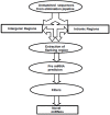
Flowchart describing the computational pipeline used for prediction of novel miRNAs. The sequencing reads that did not match with any of the databases of elimination pipeline, but matched with the human intergenic and intronic sequences, were extracted along with flanking sequences from human genome. These were then analysed by a number of miRNA precursor prediction algorithms and the hits were further analysed by a set of filters as described. The final output of the pipeline gives a list of novel miRNAs.
Figure 10
Figure 10
Detection of precursor novel miRNAs through Real-time PCR. Real-time PCR confirmation of the precursors of novel miRNAs predicted through CID, CSHMM, MiPred tools. A no-RT-PCR reaction is used as negative control.
Figure 11
Figure 11
Predicted novel miRNAs. A. A partial list of novel miRNAs predicted from deep sequencing data is displayed along with chromosomal location and the scores from different prediction tools. B. The precursor sequence and the secondary structure of the novel miRNAs. The highlighted regions in blue and yellow colour indicate the presence of 5p and 3p mature miRNA sequences, respectively. Note that the sequenced mature putative miRNAs map to the stem part of the structure. C. The expressions of these miRNAs were independently validated by RPA. RPA was carried out as described in the legend for Figure 7 using total RNA from normal PBMC and K562 cell lines. The phosphor imager images are shown. RNU6B transcripts were used as a control. Some of the miRNA star sequences were also detected. The brightness/contrast have been changed to normalize the signals across different probes.
Figure 12
Figure 12
Clustering of the novel miRNAs. A. Novel miRNAs occurring in the vicinity of the known miRNAs. B. Novel miRNAs forming a new cluster.

References

    1. Alvarez-Garcia I, Miska EA. MicroRNA functions in animal development and human disease. Development. 2005;132:4653–4662. doi: 10.1242/dev.02073. - DOI - PubMed
    1. Leung AK, Sharp PA. microRNAs: a safeguard against turmoil? Cell. 2007;130:581–585. doi: 10.1016/j.cell.2007.08.010. - DOI - PubMed
    1. Kulshreshtha R, Davuluri RV, Calin GA, Ivan M. A microRNA component of the hypoxic response. Cell Death Differ. 2008;15:667–671. doi: 10.1038/sj.cdd.4402310. - DOI (V体育平台登录) - PubMed
    1. Spizzo R, Nicoloso MS, Croce CM, Calin GA. SnapShot: MicroRNAs in Cancer. Cell. 2009;137:586–586. doi: 10.1016/j.cell.2009.04.040. e1. - "VSports" DOI - PubMed
    1. Ghildiyal M, Zamore PD. Small silencing RNAs: an expanding universe. Nat Rev Genet. 2009;10:94–108. doi: 10.1038/nrg2504. - DOI - PMC - PubMed

Publication types

Substances

"VSports在线直播" Associated data