Skip to main page content
U.S. flag

An official website of the United States government

Dot gov

The . gov means it’s official. Federal government websites often end in VSports app下载. gov or . mil. Before sharing sensitive information, make sure you’re on a federal government site. .

Https

The site is secure. The https:// ensures that you are connecting to the official website and that any information you provide is encrypted and transmitted securely V体育官网. .

. 2009 Dec 21;206(13):2897-906.
doi: 10.1084/jem.20090889. Epub 2009 Nov 23.

Diabetic retinopathy is associated with bone marrow neuropathy and a depressed peripheral clock

Affiliations

Diabetic retinopathy is associated with bone marrow neuropathy and a depressed peripheral clock

Julia V Busik et al. J Exp Med. .

V体育ios版 - Abstract

The present epidemic of diabetes is resulting in a worldwide increase in cardiovascular and microvascular complications including retinopathy. Current thinking has focused on local influences in the retina as being responsible for development of this diabetic complication VSports手机版. However, the contribution of circulating cells in maintenance, repair, and dysfunction of the vasculature is now becoming appreciated. Diabetic individuals have fewer endothelial progenitor cells (EPCs) in their circulation and these cells have diminished migratory potential, which contributes to their decreased reparative capacity. Using a rat model of type 2 diabetes, we show that the decrease in EPC release from diabetic bone marrow is caused by bone marrow neuropathy and that these changes precede the development of diabetic retinopathy. In rats that had diabetes for 4 mo, we observed a dramatic reduction in the number of nerve terminal endings in the bone marrow. Denervation was accompanied by increased numbers of EPCs within the bone marrow but decreased numbers in circulation. Furthermore, denervation was accompanied by a loss of circadian release of EPCs and a marked reduction in clock gene expression in the retina and in EPCs themselves. This reduction in the circadian peak of EPC release led to diminished reparative capacity, resulting in the development of the hallmark feature of diabetic retinopathy, acellular retinal capillaries. Thus, for the first time, diabetic retinopathy is related to neuropathy of the bone marrow. This novel finding shows that bone marrow denervation represents a new therapeutic target for treatment of diabetic vascular complications. .


Figures
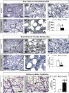
Figure 1.
Figure 1.
Dramatic decrease in humerus bone marrow innervation in diabetic rats is associated with an increase in retinal acellular capillaries. (A and B) NF200-positive nerve processes (arrowheads) are visible in bone marrow of control rats (A and A′) but significantly decreased in diabetic rats (B and B′). (C and D) TH-positive nerve processes running along blood vessels (arrows) were present in the bone marrow of controls (C and C′) but very rare in diabetic animals (D and D′). (E and G) No staining was found in negative controls treated only with secondary antibody. Bars: (A–D) 20 µM; (A′–D′, E, and G) 5 µM. (F and H) Quantification of the NF200 and TH data are shown in F and H, respectively. Diaphysis of the humerus bone from control (n = 4) and diabetic (n = 3) animals were assessed. At least 10 fields per humerus were analyzed on duplicate slides from each bone by three independent individuals. *, P < 0.05. The experiment was repeated on two independent sets of animals. (I and J) Retinal vasculature from control (I) and diabetic (J) animals was prepared using trypsin digestion and stained with hematoxylin and periodic acid–Schiff. Dramatically increased number of acellular capillaries (black arrows) was observed in retinal vasculature isolated from diabetic animals (I) compared with control (J). (K) Quantification of I and J. Bars, 10 µM. The data represent mean ± SD of three independent sets of animals, with nine control and nine diabetic animals total. At least eight fields per retina were counted in duplicates by two independent investigators. ***, P < 0.001.
Figure 2.
Figure 2.
Denervation of the bone marrow precedes acellular capillaries formation in diabetic rats after 2 mo of diabetes. (A and B) TH-positive nerve processes running along blood vessel (arrow) were present in diabetic bone marrow at 2 mo of diabetes (A and A′) but were significantly decreased compared with controls (B). At least 10 fields were analyzed on duplicate slides from each bone by three independent individuals. Bars: (A) 20 µm; (A′) 5 µm. *, P < 0.05. The experiment was repeated on two independent sets of animals. Data are presented as the mean ± SD of four control and three diabetic (A and B) rats. (C) On the contrary, the number of acellular capillaries was not changed. Data in C are presented as the mean of two independent sets of animals, with six control and six diabetic rats total. At least eight fields per retina were counted in duplicates by two independent investigators. *, P < 0.05.
Figure 3.
Figure 3.
Rat thy-1+ cell characterization. Thy-1+ cells isolated from rat bone marrow were analyzed by FACS for progenitor marker expression. (A–C) Isolated cells express CD133 (A), Thy-1 (B), and VEGFR-2 (C). The red line represents the samples incubated with respective antibody. The black line in the representative histogram plot corresponds to the samples incubated with the appropriate isotope control antibodies. (D) Thy-1+ progenitors express endothelial nitric oxide synthase (green) as detected by immunofluorescence with nuclei stained with DAPI (blue). Bar, 10 µM. (E and F) Cells in culture form colonies (E) and incorporate Dil-acLDL (red; F). Thy-1+ cells incorporate into capillary tubes with human retinal endothelial cells. (G) Fluorescently labeled Thy-1 cells (red) incorporating into HRECs. (H) Phase contrast image of same region as in G. (I) Merged phase contrast and fluorescence images of Thy-1+ cells (red) participating in tube formation. Bar, 100 µM. Flow cytometry data are represented as percentage of cells expressing respective marker for rats (n = 3). Images are representative of n = 4 of individual experiments.
Figure 4.
Figure 4.
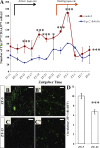
Diabetes decreases circadian release of EPCs. (A) Control (n = 4) and diabetic (n = 3) rats were maintained on a 12/12 h light/dark cycle (lights on at ZT-0, lights off at ZT-12). 100 µl of blood was taken every 2 h and analyzed for the number of EPCs by flow cytometry. EPCs were determined as number of thy-1+CD3CD4CD8 cells per µl of blood. There is a clear peak in the EPC number at ZT-3 in control animals (red line). However, the response is blunted in Type 2 (blue line) diabetic animals. The number of EPCs in blood was consistently lower in type 2 diabetic animals compared with control animals. The data represent the mean ± SD. The experiment was performed on two independent sets of animals, with 12 repetitions per animal. Statistical analysis was performed using two-way ANOVA for diabetes and time effect (***, P < 0.0001) and Bonferroni post-test to compare replicates by row (*, P < 0.05; ***, P < 0.001). (B) Circadian variation in the number of bone marrow–derived cells in the retinal circulation of mice. Retinal flat mounts from gfp+ chimeric control mice show increased numbers of bone marrow–derived circulating cells (green) in the retinal capillaries at ZT-5 (B and B′) as compared with ZT-13 (C and C′). 10 fields per retina were evaluated and both retinas per mouse were analyzed. Magnification is 4× in B and C and 20× in B′ and C′. Bars: (B and C) 100 µm; (B′ and C′) 10 µm. (D) Quantification of gfp+ cells shows an increase in green fluorescence in retinal capillaries at ZT-5 as compared with ZT-13 (n = 4; ***, P < 0.001). The data represents mean ± SE. The experiment was performed on three independent sets of animals, with the total number of mice per time point equal to nine.
Figure 5.
Figure 5.
Characterization of thy-1+ EPC number and function in type 2 diabetic rats. (A) Diabetic rats (n = 3; black) with decreased peripheral blood EPCs showed a marked increase in the number of bone marrow EPCs compared with controls (n = 6; white). (B) Diabetic bone marrow (black) showed a reduction in CFU compared with healthy controls (white). (C and D) Typical colony formation observed from explanted control (C) and diabetic EPCs (D). Bars, 100 µM. (E) Both blood and bone marrow EPCs of diabetic origin (black) demonstrate reduced migration to VEGF compared with control (white). (F) Diabetic bone marrow EPCs (black) show reduced proliferation compared with control cells (white). The data represent the mean ± SE of minimum of four separate experiments. *, P < 0.05.
Figure 6.
Figure 6.
Clock gene expression analysis. Rats used in the circadian rhythm study were sacrificed during the peak time of EPC release, ZT1–5, 24 h after blood collection. (A–D) RNA extracted from total retina (A), peripheral blood thy-1+ (B), bone marrow thy-1+ populations (C) and SCN (D) were analyzed for the clock genes Clock, Bmal1, Per1, Per2, CRY1, CRY2, ERB, and RORA. Clock genes demonstrate reduced expression in the diabetic retinas and thy-1+ cells. In the bone marrow fraction, Bmal1 and Per2 were significantly reduced in diabetic animals. Although changes in SCN were not significant, means for all clock genes were lower in diabetes. The mRNA expression levels were normalized to cyclophilin and expressed as fold change over control animals. (E) Western blotting from the retina demonstrated reduction of BMAL in diabetes. The quantitative PCR data are presented as the mean ± SE and the Western blotting represents a mean ± SD of four control and three diabetic animals. The experiment was performed on two independent sets of animals with triplicate measurements for quantitative PCR and duplicate Western blots. *, P < 0.05 compared with control.

V体育官网 - References

    1. Asahara T., Murohara T., Sullivan A., Silver M., van der Zee R., Li T., Witzenbichler B., Schatteman G., Isner J.M. 1997. Isolation of putative progenitor endothelial cells for angiogenesis. Science. 275:964–967 10.1126/science.275.5302.964 - DOI - PubMed
    1. Ellis E.A., Grant M.B., Murray F.T., Wachowski M.B., Guberski D.L., Kubilis P.S., Lutty G.A. 1998. Increased NADH oxidase activity in the retina of the BBZ/Wor diabetic rat. Free Radic. Biol. Med. 24:111–120 10.1016/S0891-5849(97)00202-5 - DOI - PubMed
    1. Ellis E.A., Guberski D.L., Somogyi-Mann M., Grant M.B. 2000. Increased H2O2, vascular endothelial growth factor and receptors in the retina of the BBZ/Wor diabetic rat. Free Radic. Biol. Med. 28:91–101 10.1016/S0891-5849(99)00216-6 - DOI - PubMed
    1. Grant M.B., May W.S., Caballero S., Brown G.A., Guthrie S.M., Mames R.N., Byrne B.J., Vaught T., Spoerri P.E., Peck A.B., Scott E.W. 2002. Adult hematopoietic stem cells provide functional hemangioblast activity during retinal neovascularization. Nat. Med. 8:607–612 10.1038/nm0602-607 - DOI - PubMed
    1. Ikeda H., Yong Q., Kurose T., Todo T., Mizunoya W., Fushiki T., Seino Y., Yamada Y. 2007. Clock gene defect disrupts light-dependency of autonomic nerve activity. Biochem. Biophys. Res. Commun. 364:457–463 10.1016/j.bbrc.2007.10.058 - DOI - PubMed