Skip to main page content (V体育安卓版)
U.S. flag

An official website of the United States government

Dot gov

The . gov means it’s official. Federal government websites often end in . gov or . mil. Before sharing sensitive information, make sure you’re on a federal government site VSports app下载. .

Https

The site is secure V体育官网. The https:// ensures that you are connecting to the official website and that any information you provide is encrypted and transmitted securely. .

. 2009 Nov 3;19(20):1741-6.
doi: 10.1016/j.cub.2009.08.042. Epub 2009 Oct 8.

Autophagy, not apoptosis, is essential for midgut cell death in Drosophila

Affiliations

"V体育官网" Autophagy, not apoptosis, is essential for midgut cell death in Drosophila

Donna Denton et al. Curr Biol. .

Abstract

Most developmentally programmed cell death in metazoans is mediated by caspases. During Drosophila metamorphosis, obsolete tissues, including the midgut and salivary glands, are removed by programmed cell death [1]. The initiator caspase Dronc and its activator Ark are required for the death of salivary glands, but not for midgut removal [2, 3]. In addition to caspases, complete removal of salivary glands requires autophagy [4]. However, the contribution of autophagy to midgut cell death has not been explored. Examination of combined mutants of the main initiator and effector caspases revealed that the canonical apoptotic pathway is not required for midgut cell death. Further analyses revealed that the caspase Decay is responsible for most of the caspase activity in dying midguts, yet inhibition of this activity has no effect on midgut removal. By contrast, midgut degradation was severely delayed by inhibition of autophagy, and this occurred without a decrease in caspase activity. Surprisingly, the combined inhibition of caspases and autophagy did not result in an additional delay in midgut removal. Together, our results indicate that autophagy, not caspases, is essential for midgut programmed cell death, providing the first in vivo evidence of caspase-independent programmed cell death that requires autophagy despite the presence of high caspase activity VSports手机版. .


Figures

Figure 1
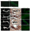
Figure 1. Midgut cell death occurs in the absence of the main effector and initiator caspases
Histolysis of (A) wild type (WT); (B) dcp-1Prev1; droncI24/I29 driceΔ1; and (C) strica4; droncI24/I29 driceΔ1 midguts occur similarly. Detection of TUNEL-positive nuclei (left) from -4 to -1 hr RPF larvae and Hoechst staining of DNA (middle). Morphology of midgut at +4 hr RPF, note the contraction of gastric caeca (arrows) as midguts undergo PCD.
Figure 2
Figure 2. Decay is required for caspase activity but not midgut degradation
(A) Expression of effector caspases drice, decay, dcp-1 and damm during midgut morphogenesis from third instar larvae, L3 (-8 hr RPF), late third instar larvae, late L3 (-4 hr to -1 hr RPF), and white prepupal, WPP (0 hr RPF) by qPCR. (B) Verification of decay knockdown in the midgut from late third instar larvae (-4 to -1 hr RPF) by qPCR. NP1-GAL4 UAS-decay-IR (NP1 decay) results in greatly reduced levels of decay compared to wild type (WT) and UAS-decay-IR alone (+/decay). In (A) and (B) the transcript levels were normalized against the internal control gene rp49 and represented as relative expression. (C) Compared to the high levels of caspase activity detected in wild type (WT) and droncI24/I29 driceΔ1 midgut, NP1-GAL4 UAS-decay-IR (decay), NP1-GAL4 UAS-decay-IR; dronc drice (decay dronc drice) and NP1-GAL4 UAS-p35 (p35) midgut have greatly reduced activity. The combined decay knockdown and p35 expression (NP1/+; UAS-decay-IR/UAS-p35) had no detectable activity. Caspase activity was measured from -4 to -1 hr RPF midgut lysates on DEVD-AMC and represented as pmol DEVD/min. In (A-C) the data are mean from three experiments, with error bars representing SEM. Histolysis of (D) wild type (WT); (E) NP1-GAL4/UAS-decay-IR (decay-IR); (F) NP1-GAL4/UAS-decay-IR; dronc drice (decay-IR dronc drice); (G) NP1-GAL4/+;UAS-p35/+ (p35); and (H) NP1-GAL4;UAS-decay-IR/UAS-p35 (decay-IR p35). Morphology of midgut at +4 hr RPF (left) showing (D) WT; (E) decay-IR; (F) decay-IR; dronc drice; (G) p35; and (H) decay-IR p35 undergo histolysis at similar rates, with contraction of gastric caeca (arrows). Histological analysis of paraffin sections at +12 hr RPF (right) shows that larval midgut contraction is similar in (D) WT; (E) decay-IR; (F) decay-IR; dronc drice; (G) p35; and (H) decay-IR p35.
Figure 3
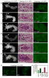
Figure 3. Atg gene mutants affect midgut histolysis
(A) pGFP-Atg8a expression as a marker for autophagy induction. pGFP-Atg8a is very low in early 3rd instar larval midgut and high levels of puncta are observed in +2 hr RPF midgut gastric caeca. Histolysis of (B) Atg2 control (Atg2EP3697/+); (C) Atg2 mutants (Atg2EP3697/Df{Exel}6091); (D) Atg18 control (Atg18KG03090/+); and (E) Atg18 mutants (Atg18KG03090/Df{Exel}6112). Morphology of midguts at +4 hr RPF (left) show a significant delay in midgut histolysis in (C) Atg2 and (E) Atg18 (E) compared to (B, D) controls as seen by the presence of less contracted gastric caeca (arrows). Histological analysis of paraffin sections at +12 hr RPF (middle) showing larval midgut contraction is dramatically delayed in (C) Atg2 and (E) Atg18 mutants compared to (B, D) controls, in particular the maintained presence of gastric caeca and proventriculus (arrows). GFP-Atg8a localisation in midgut +2 hr RPF (right) shows a dramatic reduction in puncta indicating that autophagy is inhibited in (C) Atg2 and (E) Atg18 mutants compared to (B, D) controls.
Figure 4
Figure 4. Knockdown of caspases fails to enhance Atg phenotypes
Histolysis of (A) NP1-GAL4/+; UAS-decay-IR/UAS-GFP-Atg5 (decay-IR); (B) NP1-GAL4/UAS-Atg1-IR; UAS-GFP-Atg5/+ (Atg1-IR); (C) NP1-GAL4/UAS-Atg1-IR; UAS-decay-IR/UAS-GFP-Atg5 (decay-IR, Atg1-IR); (D) NP1-GAL4/UAS-GFP-LC3; UAS-decay-IR/+ (decay-IR); (E) NP1-GAL4/UAS-GFP-LC3; UAS-Atg18-IR/+ (Atg18-IR); and (F) Atg18-IR (NP1-GAL4/UAS-GFP-LC3; UAS-decay-IR/UAS-Atg18-IR (decay-IR). Morphology of midguts at +4 hr RPF (left) show a significant delay in midgut histolysis in (B) Atg1-IR and (E) Atg18-IR compared to decay-IR (A and D respectively) as seen by the presence of less contracted gastric caeca (arrows). The combined knockdown of (C) decay-IR, Atg1-IR or (F) decay-IR, Atg18-IR show a delay in midgut histolysis, as seen by the presence of gastric caeca (arrows) that is similar to (B) Atg1-IR or (E) Atg18-IR alone. Histological analysis of paraffin sections at +12 hr RPF (middle). Incomplete degradation occurs in midguts of (B) Atg1-IR; (C) decay-IR Atg1-IR; (E) Atg18-IR; and (F) decay-IR Atg18-IR. The delay in midgut histolysis in (B) Atg1-IR and (E) Atg18-IR is not significantly affected by decay-IR (C and F). Autophagy is indicated by the presence of GFP-LC3 or GFP-Atg5 puncta in midgut at +2 hr RPF (right). A reduction in puncta indicating that autophagy is inhibited in (B) Atg1-IR and (E) Atg18-IR and not affected by (C, F) decay-IR. (G) Punctate localisation of GFP-Atg8a in midgut +2 hr RPF shows that autophagy is not affected by expression of p35 (NP1-GAL4 UAS-p35). (H) Caspase activity was measured from -4 to -1 hr RPF midgut lysates on DEVD-AMC, represented as pmol DEVD/min. There is an increase in caspase activity in Atg1-IR and Atg18-IR, which decreased significantly in decay-IR. Data are mean from three independent experiments, with error bars representing SEM.

References

    1. Baehrecke EH. Steroid regulation of programmed cell death during Drosophila development. Cell Death Differ. 2000;7:1057–1062. - PubMed
    1. Daish TJ, Mills K, Kumar S. Drosophila caspase DRONC is required for specific developmental cell death pathways and stress-induced apoptosis. Dev Cell. 2004;7:909–15. - PubMed
    1. Mills K, Daish T, Harvey KF, Pfleger CM, Hariharan IK, Kumar S. The Drosophila melanogaster Apaf-1 homologue ARK is required for most, but not all, programmed cell death. J Cell Biol. 2006;172:809–815. - "VSports在线直播" PMC - PubMed
    1. Berry DL, Baehrecke EH. Growth arrest and autophagy are required for salivary gland cell degradation in Drosophila. Cell. 2007;131:1137–48. - PMC - PubMed
    1. Chew SK, Akdemir F, Chen P, Lu WJ, Mills K, Daish T, Kumar S, Rodriguez A, Abrams JM. The apical caspase, dronc, governs programmed and unprogrammed cell death in Drosophila. Dev Cell. 2004;7:897–907. - PubMed

Publication types

MeSH terms