Skip to main page content
U.S. flag

An official website of the United States government

Dot gov

The . gov means it’s official. Federal government websites often end in . gov or . mil VSports app下载. Before sharing sensitive information, make sure you’re on a federal government site. .

Https

The site is secure. The https:// ensures that you are connecting to the official website and that any information you provide is encrypted and transmitted securely V体育官网. .

. 2008 May;47(5):1725-36.
doi: 10.1002/hep.22187.

"V体育平台登录" Impaired autophagy: A mechanism of mitochondrial dysfunction in anoxic rat hepatocytes

Affiliations

Impaired autophagy: A mechanism of mitochondrial dysfunction in anoxic rat hepatocytes

Jae-Sung Kim et al. Hepatology. 2008 May.

VSports - Abstract

Autophagy selectively removes abnormal or damaged organelles such as dysfunctional mitochondria. The mitochondrial permeability transition (MPT) is a marker of impaired mitochondrial function that is evident in hepatic ischemia/reperfusion (I/R) injury. However, the relationship between mitochondrial dysfunction and autophagy in I/R injury is unknown. Cultured rat hepatocytes and mouse livers were exposed to anoxia/reoxygenation (A/R) and I/R, respectively. Expression of autophagy-related protein 7 (Atg7), Beclin-1, and Atg12, autophagy regulatory proteins, was analyzed by western blots VSports手机版. Some hepatocytes were incubated with calpain 2 inhibitors or infected with adenoviruses encoding green fluorescent protein (control), Atg7, and Beclin-1 to augment autophagy. To induce nutrient depletion, a condition stimulating autophagy, hepatocytes were incubated in an amino acid-free and serum-free medium for 3 hours prior to onset of anoxia. For confocal imaging, hepatocytes were coloaded with calcein and tetramethylrhodamine methyl ester to visualize onset of the MPT and mitochondrial depolarization, respectively. To further examine autophagy, hepatocytes were infected with an adenovirus expressing green fluorescent protein-microtubule-associated protein light chain 3 (GFP-LC3) and subjected to A/R. Calpain activity was fluorometrically determined with succinyl-Leu-Leu-Val-Tyr-7-amino-4-methylcoumarin. A/R markedly decreased Atg7 and Beclin-1 concomitantly with a progressive increase in calpain activity. I/R of livers also decreased both proteins. However, inhibition of calpain isoform 2, adenoviral overexpression, and nutrient depletion all substantially suppressed A/R-induced loss of autophagy proteins, prevented onset of the MPT, and decreased cell death after reoxygenation. Confocal imaging of GFP-LC3 confirmed A/R-induced depletion of autophagosomes, which was reversed by nutrient depletion and adenoviral overexpression. .

Conclusion: Calpain 2-mediated degradation of Atg7 and Beclin-1 impairs mitochondrial autophagy, and this subsequently leads to MPT-dependent hepatocyte death after A/R V体育安卓版. .


Figures

Fig. 1
Fig. 1
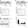
Suppression of Atg7 and Beclin-1 loss by nutrient depletion. Hepatocytes were incubated in anaerobic KRH buffer at pH 6.2 for up to 4 hours to simulate ischemia and then reoxygenated in aerobic KRH at pH 7.4 to simulate reperfusion. Nutrient depletion was simulated by incubation of cells in aerobic KRH at pH 7.4 for 3 hours prior to anoxia. (A) Atg7, Beclin-1, and Atg12 expression during A/R was analyzed by western blotting. (B) After 4 hours of anoxia in the presence and absence of nutrient depletion, hepatocytes were reoxygenated for 2 hours. Necrosis was measured by PI fluorometry. Values are means ± standard error. Cell death was significantly reduced by nutrient depletion (P < 0.0001 versus no treatment). Changes in (C) Atg7 and (D) Beclin-1 expression in the presence and absence of nutrient depletion were determined by western blotting. (E) Mouse livers were exposed to 45 minutes of total ischemia (I) and subsequently 30 minutes of reperfusion (R). Changes in Atg7 and Beclin-1 were analyzed by western blotting and compared with a sham control (S).
Fig. 2
Fig. 2
Cytoprotection by AdAtg7 and AdBeclin-1 against A/R. Hepatocytes were infected with AdAtg7 or AdBeclin-1 (10 plaque performing units/cell) and then subjected to A/R. AdGFP was used for viral control. Western blot analysis of (A) Atg7 and (B) Beclin-1 was performed during A/R. Infection of hepatocytes with (C) AdAtg7 or (D) AdBeclin-1 prevented reoxygenation-induced necrotic death (P < 0.0001 versus AdGFP). Values are means ± standard error. (E) Hepatocytes were subjected to 4 hours of anoxia and subsequently reoxygenated for 9 hours. Western blot analysis of procaspase 3 and caspase 3 was performed under the conditions of normoxia, A/R, zVAD (50 μM), nutrient depletion (N.D.), and adenoviral overexpression. (F). Hepatocellular ATP concentration was determined after 20 minutes of reoxygenation. *P < 0.05 versus A/R.
Fig. 3
Fig. 3
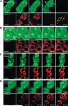
MPT inhibition by enhanced autophagy. After 4 hours of anoxia, hepatocytes were reoxygenated, and confocal images of calcein (green) and TMRM and PI (red) were simultaneously collected at 10, 20, 40, and 60 minutes. (A) Hepatocytes were reoxygenated with no other treatment. At the end of anoxia, calcein was excluded by mitochondria, and this was indicative of permeability pore closure. After 10 minutes of reoxygenation, mitochondria became repolarized without calcein redistribution into mitochondria. After 20 minutes, calcein redistributed into mitochondria, indicating onset of the MPT, and TMRM began to disappear, indicating mitochondrial depolarization. Within minutes, cytosolic calcein was lost, and PI labeled nuclei (arrows), indicating cell death. (B) Under the condition of nutrient depletion, mitochondrial voids in the calcein fluorescence were persistently present, and TMRM was continuously evident throughout reoxygenation. (C) AdAtg7 or (D) AdBeclin-1 infection prevented the MPT and cell death.
Fig. 4
Fig. 4
Autophagosome formation after A/R. Hepatocytes infected with AdGFP-LC3 were exposed to A/R, and changes in autophagosome formation were monitored by confocal microscopy. (A) After 8 minutes of anoxia, autophagosomes (green-fluorescing, punctate structures) were evident. After prolonged anoxia, most autophagosomes disappeared. Note a further decrease in autophagosomes after reoxygenation. (B) Nutrient depletion delayed loss of autophagosome. Autophagosomes were persistently present in hepatocytes coinfected with (C) AdAtg7 or (D) AdBeclin-1.
Fig. 5
Fig. 5
Sequestration of polarized mitochondria in autophagosomes. Confocal images of AdGFP-LC3, TMRM, and PI were collected after reoxygenation. (A) In the control group, green-fluorescing structures surrounding repolarized mitochondria were markedly smaller than the neighboring mitochondria, and this indicated mitophagic failure. Mitochondria released TMRM, and the cell lost viability thereafter (arrowheads). (B) After nutrient depletion, some mitochondria after reoxygenation were enveloped by autophagosomes but remained polarized until 40 to 60 minutes (arrows).
Fig. 6
Fig. 6
Lysosomes after A/R. Hepatocytes were labeled with rhoda-mine dextran and PI and subsequently subjected to A/R. The number and location of the lysosomes were assessed by confocal microscopy. At 35 minutes of reoxygenation, nuclei were labeled with PI (arrows).
Fig. 7
Fig. 7
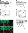
Suppression of Atg7 and Beclin-1 loss by calpain 2 inhibition. Hepatocytes were subjected to A/R in the presence and absence of 50 μM ALLM or zLLY-fmk, specific calpain 2 inhibitors. Changes in (A) Atg7 and (B) Beclin-1 were determined by western blotting. (C) Inhibition of calpain 2 prevented cell death after reoxygenation (P < 0.0001 versus no treatment). (D) Calpain activity was determined by SLLVY-AMC fluorometry. (E) Confocal images of calcein and TMRM in control (upper panels) and ALLM-treated cells (lower panels). (F) Changes in calpain 1 and 2 expression were analyzed by western blotting after A/R.
Fig. 8
Fig. 8
Scheme of A/R-induced impairment of mitophagy. Stimulation of calpain 2 after A/R hydrolyzes Atg 7 and Beclin-1, causing defective mitophagy. Because impaired mitophagy fails to remove dysfunctional mitochondria, the mitochondria laden with ROS and calcium undergo the MPT, which in turn leads to uncoupling of oxidative phosphorylation, energetic failure, ATP depletion, and ultimately cell death. Because the MPT causes cell death after A/R, blockade of the MPT onset by calpain 2 inhibition prevents cell death. Likewise, enhanced autophagy by nutrient depletion and Atg7 or Beclin-1 overexpression increases auto-phagy expression, which facilitates the formation of autophagosomes and mitophagy, leading to cell survival.

References

    1. Kim JS, He L, Qian T, Lemasters JJ. Role of the mitochondrial permeability transition in apoptotic and necrotic death after ischemia/reperfusion injury to hepatocytes. Curr Mol Med. 2003;3:527–535. - PubMed
    1. Jaeschke H, Lemasters JJ. Apoptosis versus oncotic necrosis in hepatic ischemia/reperfusion injury. Gastroenterology. 2003;125:1246–1257. - PubMed (VSports注册入口)
    1. Kim J-S, Qian T, Lemasters JJ. Mitochondrial permeability transition in the switch from necrotic to apoptotic cell death in ischemic rat hepatocytes. Gastroenterology. 2003;124:494–503. - PubMed
    1. Kim J-S, Ohshima S, Pediaditakis P, Lemasters JJ. Nitric oxide protects rat hepatocytes against reperfusion injury mediated by the mitochondrial permeability transition. HEPATOLOGY. 2004;39:1533–1543. - PubMed
    1. Kim J-S, Jin Y, Lemasters JJ. Reactive oxygen species, but not Ca2+ overloading, trigger pH- and mitochondrial permeability transition-dependent death of adult rat myocytes after ischemia/reperfusion. Am J Physiol Heart Circ Physiol. 2006;290:H2024–H2034. - PubMed

Publication types

MeSH terms (VSports)

LinkOut - more resources (V体育安卓版)