Skip to main page content
U.S. flag

An official website of the United States government

Dot gov

The . gov means it’s official. Federal government websites often end in . gov or . mil. Before sharing sensitive information, make sure you’re on a federal government site VSports app下载. .

Https

The site is secure. The https:// ensures that you are connecting to the official website and that any information you provide is encrypted and transmitted securely. V体育官网.

. 2008 Jul;23(7):1084-96.
doi: 10.1359/jbmr.080234.

HMGB1 regulates RANKL-induced osteoclastogenesis in a manner dependent on RAGE

Affiliations

HMGB1 regulates RANKL-induced osteoclastogenesis in a manner dependent on RAGE

Zheng Zhou et al. J Bone Miner Res. 2008 Jul.

Abstract

High-mobility group box 1 (HMGB1), a nonhistone nuclear protein, is released by macrophages into the extracellular milieu consequent to cellular activation. Extracellular HMGB1 has properties of a pro-inflammatory cytokine through its interaction with receptor for advanced glycation endproducts (RAGE) and/or toll-like receptors (TLR2 and TLR4) VSports手机版. Although HMGB1 is highly expressed in macrophages and differentiating osteoclasts, its role in osteoclastogenesis remains largely unknown. In this report, we present evidence for a function of HMGB1 in this event. HMGB1 is released from macrophages in response to RANKL stimulation and is required for RANKL-induced osteoclastogenesis in vitro and in vivo. In addition, HMGB1, like other osteoclastogenic cytokines (e. g. , TNFalpha), enhances RANKL-induced osteoclastogenesis in vivo and in vitro at subthreshold concentrations of RANKL, which alone would be insufficient. The role of HMGB1 in osteoclastogenesis is mediated, in large part, by its interaction with RAGE, an immunoglobin domain containing family receptor that plays an important role in osteoclast terminal differentiation and activation. HMGB1-RAGE signaling seems to be important in regulating actin cytoskeleton reorganization, thereby participating in RANKL-induced and integrin-dependent osteoclastogenesis. Taken together, these observations show a novel function of HMGB1 in osteoclastogenesis and provide a new link between inflammatory mechanisms and bone resorption. .


Figures

FIG. 1
FIG. 1
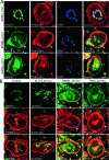
RANKL stimulation of HMGB1 translocation and release from macrophages in a time-dependent manner. Western blot analysis of HMGB1 in media and total cell lysates from cultures of RAW264.7 macrophages (A and B) and bone marrow macrophages (BMMs) (C and D) exposed to RANKL (A–C) or M-CSF (D) for the indicated times or concentrations. In C, BMMs were also harvested and isolated into nuclear and cytoplasmic fractions with the CelLytic NuCLEAR Extraction Kit (Sigma, St Louis, MO, USA). Lysates were subjected to Western blot analysis. (E) Immunostaining of HMGB1 in BMMs exposed to RANKL for the indicated times. BMMs were fixed and immunostained using antibodies against HMGB1 (rabbit polyclonal antibody, green), phalloidin to label actin filaments (red), and DAPI to label nuclei (blue). Marker bar, 10 μm.
FIG. 2
FIG. 2
RANKL, but not M-CSF, regulation of HMGB1 translocation in OCs. (A) Time course of HMGB1 translocation in OCs in response to RANKL. Polyclonal rabbit anti-HMGB1 antibody (green), phalloidin (red to label F-actin), and DAPI (blue) were used for immunostaining of OCs differentiated from BMMs. (B) Immunostaining analysis of HMGB1 in OCs exposed to M-CSF, RANKL, and TNFα for 30 min. Marker bars, 25 μm.
FIG. 3
FIG. 3
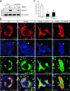
Requirement of extracellular HMGB1 for RANKL-induced actin ring formation in OCs in vitro. (A) RANKL-stimulated HMGB1 release in BMMs, as described in Fig. 1, was inhibited in the presence of nicotine (10 μM). (B) Immunostaining analysis of talin, phalloidin, and HMGB1 in OCs exposed to RANKL in the presence or absence of nicotine or anti-HMGB1 antibodies. Marker bars, 25 μm. (C) Quantitative analysis of data from B. Data were presented as percentage of OCs with actin ring formation (mean ± SE). **p < 0.01, significant difference from control (treatment with RANKL alone; t-test).
FIG. 4
FIG. 4
Requirement of HMGB1 for RANKL-induced osteoclastogenesis in vitro and in vivo. (A) HMGB1 is required for RANKL-induced osteoclastogenesis in vitro. Purified BMMs from WT mice were incubated with M-CSF (10%) for 2–3 days and cultured in the presence of RANKL (100 ng/ml) and M-CSF (1%), with or without anti-HMGB1 antibody (0.3 μg/ml) or nicotine (10 μM) for 4 days. The arrows denote a representative TRACP+ cell in each panel. (B) Quantitative analysis of TRACP+ multinuclei cells (MNCs) based on data from A. Data were presented as percentage of total number of BMMs (mean ± SE). **p < 0.01, significant difference from control (treatment with RANKL alone; t-test). (C) Representative resorption caused by RANKL-induced OCs in the absence or presence of anti-HMGB1 antibodies (0.3 μg/ml) was visualized by von Kossa staining. The resorption assay was performed by culturing OCs on coverslips coated with calcium phosphate matrix for 9 days and staining coverslips with von Kossa to display resorption pits (white). (D) Quantitative analysis of the average resorption area based on data from C. (E) HMGB1 is required for RANKL-induced osteoclastogenesis in vivo. C57/BL6 mice (3 mo old) were supracalvarially injected daily for 5 days with vehicle (PBS), RANKL (2 μg/mouse/d) + nonspecific (NS) IgG (3 μg/mouse/d), or RANKL (2 μg/mouse/d) + anti-HMGB1 IgG (3 μg/mouse/d). Animals were killed on day 6, and histological sections of calvariae were stained for TRACP and analyzed histomorphometrically for osteoclast erosion surface. Arrows denote an area displaying TRACP+ cells in each panel. (F) Histomorphometric analysis of TRACP+ OCs covered surface area based on data from E. **p < 0.01 significant difference from RANKL alone (t-test).
FIG. 5
FIG. 5
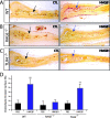
HMGB1 induction of osteoclastogenesis in vivo in a manner dependent on RAGE C57/BL6, RAGE−/−, and TLR4−/− mice (3 mo old) were intraperitoneally injected daily for 5 days with vehicle (PBS) and purified recombinant HMGB1 (10 μg/mouse/d). Animals were killed on day 6, and histologic sections of calvariae were stained for TRACP and analyzed histomorphometrically for osteoclast erosion surface. Representative images from WT (A), RAGE−/− (B), and TLR4−/− (C) mice. (D) Histomorphometric analysis of TRACP+ osteoclast erosion surface. (**p < 0.01 vs. PBS control, Student's t-test). Arrows in A–C denote an area of displaying TRACP+ cells.
FIG. 6
FIG. 6
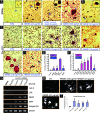
HMGB1 increases TRACP+ cells from BMMs but stimulates osteoclastogenesis from pre-OCs. BMMs of WT (A) and RAGE−/− (B) were cultured in the presence of vehicle (control) or the indicated concentrations of HMGB1 or RANKL. Where indicated, a permissive concentration of RANKL (10 ng/ml) was added. TRACP staining was performed. Arrow denotes a region with TRACP+ cells (insets show this area at higher power in the first three panels). Pre-OCs of wildtype (C) and RAGE−/− (D) were cultured in the presence of M-CSF (10 ng/ml) and HMGB1 at the indicated concentrations or vehicle alone. TRACP (C and D) staining was performed on day 5. Quantitative analysis of TRACP+ BMMs (E) and TRACP+ multinuclei cells (MNCs) (F) based on data from A–D. Data were presented as percentage of total number of BMMs (mean ± SE). **p < 0.01, significant difference from the control (t-test). #p < 0.01, significant difference from wildtype. (G) RT-PCR analysis to assess expression of transcripts for cathepsin K (Cath K), calcitonin R (CTR), MMP9, TRACP, and integrin β3 in BMMs, pre-OCs, and OCs formed in response to HMGB1 at indicated concentrations for 4 days. (H) Representative resorption caused by control (CTL; pre-OCs incubated with buffer for 9 days) OCs formed in response to indicated concentrations of HMGB1 or RANKL was visualized by von Kossa staining. The resorption assay was performed as described in the legend of Fig. 4C. (I) Quantitative analysis of the average resorption area based on data from H.
FIG. 7
FIG. 7
HMGB1 regulation of actin cytoskeleton reorganization in BMMs in a manner dependent on RAGE. (A) RAGE expression in BMMs and in differentiating OCs derived from wildtype and RAGE−/− mice was examined by Western blot analysis. (B) Co-immunostaining analysis of RAGE, HMGB1, and F-actin (by phalloidin) distribution in mature OCs. (C) Immunostaining analysis of F-actin structures in OCs derived from wildtype and RAGE−/− mice. DAPI was used to label nuclei, and GFP was a marker for RAGE−/− cells.(26) (D) Defective HMGB1 signaling in RAGE−/− BMMs cultured onto VN-coated dishes. Pre-OCs from WT and RAGE−/− mice were lifted and replated on VN-coated dishes in the absence of serum. Adherent cells were treated with HMGB1 at the indicated doses for the indicated times and lysed. Equal amounts of total proteins were immunoblotted with the indicated antibodies.
FIG. 8
FIG. 8
A working hypothesis for HMGB1 regulation of osteoclastogenesis. HMGB1 is released from BMMs in response to RANKL stimulation. The extracellular HMGB1, through RAGE in large, regulates osteoclastic actin cytoskeleton remodeling, differentiation, and function.

References

    1. Boyle WJ, Simonet WS, Lacey DL. Osteoclast differentiation and activation. Nature. 2003;423:337–342. - PubMed
    1. Tanaka S, Takahashi N, Udagawa N, Tamura T, Akatsu T, Stanley ER, Kurokawa T, Suda T. Macrophage colony-stimulating factor is indispensable for both proliferation and differentiation of osteoclast progenitors. J Clin Invest. 1993;91:257–263. - PMC - PubMed
    1. Yasuda H, Shima N, Nakagawa N, Yamaguchi K, Kinosaki M, Mochizuki S, Tomoyasu A, Yano K, Goto M, Murakami A, Tsuda E, Morinaga T, Higashio K, Udagawa N, Takahashi N, Suda T. Osteoclast differentiation factor is a ligand for osteoprotegerin/osteoclastogenesis-inhibitory factor and is identical to TRANCE/RANKL. Proc Natl Acad Sci USA. 1998;95:3597–3602. - PMC - PubMed
    1. Lacey DL, Timms E, Tan HL, Kelley MJ, Dunstan CR, Burgess T, Elliott R, Colombero A, Elliott G, Scully S, Hsu H, Sullivan J, Hawkins N, Davy E, Capparelli C, Eli A, Qian YX, Kaufman S, Sarosi I, Shalhoub V, Senaldi G, Guo J, Delaney J, Boyle WJ. Osteoprotegerin ligand is a cytokine that regulates osteoclast differentiation and activation. Cell. 1998;93:165–176. - PubMed
    1. Teitelbaum SL. Osteoclasts, integrins, and osteoporosis. J Bone Miner Metab. 2000;18:344–349. - "V体育平台登录" PubMed

Publication types