Skip to main page content
U.S. flag

An official website of the United States government

Dot gov

The . gov means it’s official. Federal government websites often end in . gov or . mil. Before sharing sensitive information, make sure you’re on a federal government site. VSports app下载.

Https

The site is secure. The https:// ensures that you are connecting to the official website and that any information you provide is encrypted and transmitted securely. V体育官网.

. 2008 Feb;172(2):454-69.
doi: 10.2353/ajpath.2008.070876. Epub 2008 Jan 10.

Inhibition of autophagy prevents hippocampal pyramidal neuron death after hypoxic-ischemic injury

Affiliations

Inhibition of autophagy prevents hippocampal pyramidal neuron death after hypoxic-ischemic injury

Masato Koike et al. Am J Pathol. 2008 Feb.

Abstract

Neonatal hypoxic/ischemic (H/I) brain injury causes neurological impairment, including cognitive and motor dysfunction as well as seizures. However, the molecular mechanisms regulating neuron death after H/I injury are poorly defined and remain controversial. Here we show that Atg7, a gene essential for autophagy induction, is a critical mediator of H/I-induced neuron death. Neonatal mice subjected to H/I injury show dramatically increased autophagosome formation and extensive hippocampal neuron death that is regulated by both caspase-3-dependent and -independent execution VSports手机版. Mice deficient in Atg7 show nearly complete protection from both H/I-induced caspase-3 activation and neuron death indicating that Atg7 is critically positioned upstream of multiple neuronal death executioner pathways. Adult H/I brain injury also produces a significant increase in autophagy, but unlike neonatal H/I, neuron death is almost exclusively caspase-3-independent. These data suggest that autophagy plays an essential role in triggering neuronal death execution after H/I injury and Atg7 represents an attractive therapeutic target for minimizing the neurological deficits associated with H/I brain injury. .


Figures

Figure 1
Figure 1
Activation of caspases and TUNEL staining in the hippocampus of wild-type neonatal mice after H/I injury. A–J: Histological sections of the left hippocampus of an untreated control (Cont) mouse at P7 (A, F) and ipsilateral hippocampus obtained 3 (B, G), 8 (C, H), 24 (D, I), and 72 hours (E, J) after H/I injury and stained with H&E (A–E) or for TUNEL and the active form of caspase-3 (F–J). An arrow in F shows a cleaved caspase-3-positive neuron undergoing programmed cell death. K and L: Double staining for TUNEL and the cleaved form of caspase-7 in the ipsilateral hippocampus at 8 (K) and 24 (L) hours after H/I injury. M: Number of neurons with pyknotic nuclei stained by H&E and the number of neurons positive for the active form of caspase-3 (Casp3), TUNEL (TU), or both in the pyramidal layers per hippocampal section at 3, 8, 24, and 72 hours after H/I injury. Counts were taken from three sections per animal, and the final value presented is the mean ± SD for three animals. P < 0.001. N: Proteolytic activity of the DEVDase for caspases-3 and -7 in the ipsilateral and contralateral hippocampi 3, 8, 24, and 72 hours after H/I injury. The activity in the nontreated hippocampus was used as a control. The final value presented is the mean ± SD for three animals. O: The cleavage of caspases-3 and -7 in the contralateral (CL) and ipsilateral (IL) hippocampi of wild-type neonatal mouse brains at 3, 8, 24, and 72 hours after H/I injury. Hippocampal tissues of the right (Rt) and left (Lt) sides from untreated neonatal mice were used as controls. Scale bars = 50 μm.
Figure 2
Figure 2
Pyramidal neuron death in the hippocampus of neonatal mouse brains after H/I injury. A–C: Coronal histological sections of wild-type (Wild) (A), caspase-3-deficient (Casp3−/−) (B), and CAD-deficient (CAD−/−) (C) brains including ipsilateral and contralateral hippocampi 24 hours after H/I injury. Staining with NeuN. D: Genomic DNA fragmentation detected by LMPCR. Samples were obtained from the hippocampus of an untreated wild-type brain (Cont) (lane 1), and the contralateral (CL) and ipsilateral (IL) hippocampi of wild-type (lanes 2 and 3), caspase-3-deficient (lanes 4 and 5), and CAD-deficient (lanes 6 and 7) brains 24 hours after H/I injury. E–G: A nucleus with small patches of chromatin clumping was observed in the pyramidal neurons of not only wild-type (E) but also caspase-3-deficient (F) and CAD-deficient (G) mouse hippocampi 8 hours after H/I injury. Scale bars: 500 μm (C); 2 μm (E–G).
Figure 3
Figure 3
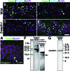
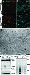
H/I injury-induced pyramidal neuron death in the hippocampi of the wild-type and caspase-3-deficient neonatal mouse brains. A: Double staining of cleaved caspase-7 (Casp7) and DAPI in the contralateral hippocampus of a caspase-3-deficient mouse brain 8 hours after H/I injury. An arrow shows a cleaved caspase-7-positive neuron. B–E: Triple staining of cleaved caspase-3 (B, D) or cleaved caspase-7 (C, E), TUNEL, and DAPI in the ipsilateral hippocampi of wild-type (Wild) (B, C) and caspase-3-deficient (D, E) neonatal mouse brains 8 hours after H/I injury. F and G: The cleavage of caspase-7 (F) and GAPDH (G) in the contralateral (CL) and ipsilateral (IL) hippocampi of caspase-3-deficient mouse brains 8 hours after H/I injury. In the ipsilateral hippocampus of a caspase-3-deficient mouse brain, a cleaved form appears intensely at ∼27 kDa but not at 18 kDa (F, middle), whereas an activated form of caspase-7 can be detected at ∼18 kDa under a longer exposure (F, right). These cleaved forms, along with the proform of caspase-7, correspond with those detected in a control lane (Cont) showing primary hepatocytes treated with tumor necrosis factor-α and actinomycin D for 6 hours (F, left). Scale bars: 10 μm (A); 30 μm (E).
Figure 4
Figure 4
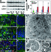
H/I injury induces autophagy in the hippocampi of wild-type neonatal mouse brains. A: Western blotting of LC3 in the untreated hippocampus (Cont) and the ipsilateral (IL) and contralateral (CL) hippocampi of neonatal mouse brains at 3, 8, and 24 hours after H/I injury. B: Quantification of A. The ratios of the amounts of LC3-II to LC3-I were calculated at each corresponding time. The final values represent the mean ± SD for three animals. *P < 0.05 and **P < 0.01 (Student’s t-test) for the comparison of the values between the ipsilateral and contralateral sides. C–H: Triple staining of LC3, TUNEL (TU), and DAPI in the ipsilateral (C, E, G) and contralateral (D, F, H) hippocampi of neonatal mouse brains 3 (C, D), 8 (E, F) and 24 (G, H) hours after H/I injury. Intense granular staining for LC3 is indicated by arrowheads, and granular staining of LC3 in boxed areas are shown in insets (C, E, G). I–K: Electron micrographs of a CA1 pyramidal neuron in the hippocampi of wild-type neonatal mouse brains 3 hours after H/I injury. Abundant vacuolar structures (I, arrows) and nascent autophagosomes (I, boxed areas) (J and K, arrowheads) were detected in the perikarion. Scale bars: 10 μm (A–H); 3 μm (I); 0.5 μm (J, K).
Figure 5
Figure 5
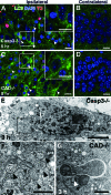
Induction of autophagy in the hippocampus of neonatal mutant mouse brains after H/I injury. A–D: Triple staining of LC3, TUNEL (TU), and DAPI in the CA1 pyramidal layer of ipsilateral (A, C) and contralateral (B, D) hippocampi deficient in caspase-3 (Casp3−/−) (A, B) and CAD (CAD−/−) (C, D), respectively, 8 hours after H/I injury. Intense granular staining for LC3 is indicated by arrowheads, and granular staining of LC3 in boxed areas are shown in insets (A, C). E–G: Electron micrographs of pyramidal neurons of the hippocampus in mice deficient in caspase-3 (E, F) and CAD (CAD−/−) (G) obtained 3 hours after H/I injury. E and F: Pyramidal neuron containing vacuolar structures in the perikaryal region and showing numerous patches of chromatin clumping in the nucleus (E). Nascent autophagosomes that contained part of the cytoplasm and wrapped by endoplasmic reticulum-like tubular structures were abundantly observed in the boxed area of E (arrowheads in F). G: Part of the perikaryal area of a pyramidal neuron of the hippocampus in a CAD-deficient mouse showing a nascent autophagosome wrapped by an endoplasmic reticulum-like tubule (arrowhead). Scale bars: 10 μm (A–D); 3 μm (E); 1 μm (F and G).
Figure 6
Figure 6
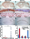
Atg7 deficiency prevents neuron death in the hippocampus of neonatal mouse brains 3 days after H/I injury. A–H: Saggital (A–D) and frontal (E–H) sections of the Atg7-deficient mouse brains (Atg7flox/flox; Nes) in the CNS (A, C, E, G) and its littermate (Atg7flox/flox) (B, D, F, H) 3 days after H/I injury. NeuN (A–F) and TUNEL (G, H) staining. Damaged areas in the hippocampus of A and B are shown in C and D and encircled by broken lines. I and J: Evaluation of damaged areas. I: Ratios (%) of damaged areas to the total areas in the hippocampal layers of Atg7-deficient (n = 17) and littermate control (n = 32) mouse brains 3 days after H/I injury. P < 0.001 (Mann-Whitney U-test for the comparison of the median values between the two groups). J: Incidence of hippocampal damage 3 days after H/I injury. The severity of the H/I injury was graded according to the number of TUNEL-positive neurons in the pyramidal layers of the hippocampus persection (less than 10 for grade 0, 11 to 100 for grade I, 101 to 200 for grade II, and more than 201 for grade III). The percentage of mice classified into each grade was determined from 17 Atg7 flox/flox; nestin-Cre (Atg7flox/flox; Nes), and 32 control littermate (Atg7flox/flox) mice. The number of mice belonging to each grade was described in the top of each bar. Scale bars: 500 μm (A, B, E, F); 50 μm (C, D); 100 μm (G, H, inset of G).
Figure 7
Figure 7
Prevention of neuronal loss in the pyramidal layers of the hippocampus is further sustained in Atg7-deficient mice until 7 days after H/I injury. A–H: Frontal sections of Atg7-deficient (Atg7flox/flox; Nes) (A, C, E, G) and its control littermate (Atg7flox/flox) (B, D, F, H) mouse brains 7 days after H/I injury. H&E (A–D) and NeuN (E–H) staining. I: Evaluation of the area loss in the ipsilateral hippocampi of Atg7-deficient (n = 11) and littermate control (n = 12) mouse brains 7 days after H/I injury. The mean value of the area loss in the ipsilateral hippocampus was significantly lower in Atg7-deficient mice (14.0) than in their littermate control mice (48.8). P < 0.0001 (Mann-Whitney U-test) for the comparison of mean values between the two groups. Scale bars: 1000 μm (A, B, E, F); 200 μm (C, D, G, H).
Figure 8
Figure 8
The apoptotic pathway is inhibited in the neonatal hippocampus after H/I injury by the Atg7 deficiency. A–D: Triple staining for cleaved caspase-3 (Casp3) (A, C), TUNEL (A, C), and DAPI (B, D) in the ipsilateral hippocampi of Atg7-deficient (Atg7flox/flox; Nes) (A, B) and control littermate (Atg7flox/flox) (C, D) mouse brains 24 hours after H/I injury. E: The neuronal cell architecture as well as morphological features of the neurons appeared intact in the hippocampal pyramidal layer of an Atg7-deficient mouse brain 24 hours after H/I injury. F: Western blotting of caspases-3 and -7 in the ipsilateral (IL) and contralateral hippocampi of littermate control (Atg7flox/flox) and Atg7-deficient (Atg7flox/flox; Nes) mouse brains 8 hours after H/I injury. Cleaved caspase-3, but not caspase-7, was detected only in the control IL hippocampus. GAPDH was used as a loading control. G: The DNA fragmentation in the ipsilateral (IL) and contralateral (CL) hippocampi from Atg7-deficient (Atg7flox/flox; Nes) and littermate (Atg7flox/flox) mouse brains 24 hours after H/I injury. Scale bars: 20 μm (A–D); 5 μm (E).
Figure 9
Figure 9
Pyramidal neuron death in the hippocampus of adult mouse brains after H/I injury. A–C: Frontal histological sections of wild-type (A), and caspase-3-deficient (Casp3−/−) (B) and CAD-deficient (CAD−/−) (C) brains including ipsilateral and contralateral hippocampi 24 hours after H/I injury. Staining with TUNEL. D: Genomic DNA fragmentation detected by LMPCR. Samples were obtained from the hippocampus of an untreated wild-type brain (Cont) (lane 1), and the contralateral (CL) and ipsilateral (IL) hippocampi of wild-type (lanes 2 and 3), caspase-3-deficient (lanes 4 and 5), and CAD-deficient (lanes 6 and 7) brains 24 hours after H/I injury. E–G: A nucleus with small patches of chromatin clumping was observed in the pyramidal neurons of not only wild-type (E) but also caspase-3-deficient (F) and CAD-deficient (G) mouse hippocampi 24 hours after H/I injury. H: The cleavage of caspases-3 and -7 in the contralateral (CL) and ipsilateral (IL) hippocampi of wild-type adult mouse brains at 3 hours, 8 hours, 24 hours, and 72 hours after H/I injury. Hippocampal tissues of the right (Rt) and left (Lt) sides from untreated adult mice were used as controls (Cont). Scale bars: 500 μm (C); 2 μm (E–G).
Figure 10
Figure 10
H/I injury induces autophagy in the hippocampi of wild-type and mutant adult mouse brains. A: Western blotting of LC3 in the untreated hippocampus (Cont) and the ipsilateral (IL) and contralateral (CL) hippocampi of adult mouse brains at 3, 8, and 24 hours after H/I injury. B: Quantification of A. The ratios of the amounts of LC3-II to LC3-I were calculated at each corresponding time. The final values represent the mean ± SD for three animals. *P < 0.05 and **P < 0.01 (Student’s t-test) for the comparison of the values between the ipsilateral and contralateral sides. C and D: Double staining of LC3 and DAPI in the ipsilateral (C) and contralateral (D) hippocampi of adult mouse brains 8 hours after H/I injury. E–G: Electron micrographs of a CA1 pyramidal neuron in the hippocampi of wild-type adult mouse brains 8 (E, F) and 24 (G) hours after H/I injury. Vacuolar structures with amorphous structures (arrows) are seen in E and F. F: A nascent autophagosome (arrowhead) (boxed areas in E). Scale bars: 10 μm (C, D); 3 μm (E, G); 0.5 μm (F).

Comment in

References

    1. du Plessis AJ, Volpe JJ. Perinatal brain injury in the preterm and term newborn. Curr Opin Neurol. 2002;15:151–157. - V体育安卓版 - PubMed
    1. Hamrick SE, Ferriero DM. The injury response in the term newborn brain: can we neuroprotect? Curr Opin Neurol. 2003;16:147–154. - PubMed
    1. Ashwal S, Pearce WJ. Animal models of neonatal stroke. Curr Opin Pediatr. 2001;13:506–516. - "VSports app下载" PubMed
    1. Ferriero DM. Neonatal brain injury. N Engl J Med. 2004;351:1985–1995. - PubMed
    1. Hagberg H, Bona E, Gilland E, Puka-Sundvall M. Hypoxia-ischaemia model in the 7-day-old rat: possibilities and shortcomings. Acta Paediatr Suppl. 1997;422:85–88. - PubMed

Publication types

MeSH terms