<font draggable="XJ2y7VI"></font><var lang="4YMYbGb"><style lang="4FeDOy"></style></var> Skip to main page content
U.S. flag

An official website of the United States government

Dot gov

The . gov means it’s official VSports app下载. Federal government websites often end in . gov or . mil. Before sharing sensitive information, make sure you’re on a federal government site. .

Https

The site is secure. The https:// ensures that you are connecting to the official website and that any information you provide is encrypted and transmitted securely. V体育官网.

. 2007 Sep 7;27(5):829-41.
doi: 10.1016/j.molcel.2007.06.029.

"V体育官网" HMGB1 is a cofactor in mammalian base excision repair

Affiliations

"VSports手机版" HMGB1 is a cofactor in mammalian base excision repair

"V体育2025版" Rajendra Prasad et al. Mol Cell. .

Abstract (VSports)

Deoxyribose phosphate (dRP) removal by DNA polymerase beta (Pol beta) is a pivotal step in base excision repair (BER). To identify BER cofactors, especially those with dRP lyase activity, we used a Pol beta null cell extract and BER intermediate as bait for sodium borohydride crosslinking. Mass spectrometry identified the high-mobility group box 1 protein (HMGB1) as specifically interacting with the BER intermediate. Purified HMGB1 was found to have weak dRP lyase activity and to stimulate AP endonuclease and FEN1 activities on BER substrates. Coimmunoprecipitation experiments revealed interactions of HMGB1 with known BER enzymes, and GFP-tagged HMGB1 was found to accumulate at sites of oxidative DNA damage in living cells. HMGB1(-/-) mouse cells were slightly more resistant to MMS than wild-type cells, probably due to the production of fewer strand-break BER intermediates. The results suggest HMGB1 is a BER cofactor capable of modulating BER capacity in cells. VSports手机版.


Figures

Figure 1
Figure 1. NaBH4 Cross-linking of Proteins in Cell Extracts
(A) Diagram of BER intermediate with 32P-labeled uracil at position 16 and a nick between positions 15 and 16, and the reaction scheme. (B) Phosphorimager scan of a NuPAGE Bis-Tris gel illustrating the NaBH4 cross-linking of proteins in various cell extracts; lanes 1-5, 32P-labeled DNA (200 nM) was mixed with either dilution buffer (lane 1), Pol β null expressing Flag epitope-tagged Pol β MEF extract (lane 2), Pol β null MEF extract (lane 3), BTNE (lane 4), or purified Pol β (lane 5), and 5 mM NaBH4. (C) Phosphoimager scan of a NuPAGE Bis-Tris gel illustrating the NaBH4 cross-linking in various cell extracts; reaction conditions and extracts are as in (B), except that the DNA substrate was not pre-treated with UDG. The positions of protein markers and the cross-linked protein-DNA products are indicated. (D) Photograph of an immunoblot illustrating the presence of HMGB1 in various extracts.
Figure 2
Figure 2. Identification of the Cross-linked Protein-DNA Complex by Mass Spectrometry
Photograph of a SYPRO Ruby stained gel (A) of the purified cross-linked protein-DNA complexes. The gel was scanned for imaging (“Supplemental Data”) and stained with SYPRO Ruby fluorescent dye before excising the protein bands designated as 32P-labeled proteins, “1-3.” (B) MALDI mass spectrum from the in-gel digestion of Band 1 in (A). The ions labeled as 842.51, 1045.56, and 2211.11 correspond in mass to autolysis products of trypsin. The ions labeled with an asterisk (*) correspond in mass to tryptic peptides of HMGB1 protein. (C) Observed masses in MALDI mass spectrum correspond in mass to theoretical tryptic peptides of HMGB1. “X” indicates the ion 1520.84 analyzed in (B). The inset shows the mass range of 900-1750. (D) MALDI/MS/MS data of the (M+H)+ ion of m/z 1520.84 shown in (B). Fragment ions observed confirm the sequence of this peptide as tryptic peptide T28-29 (aa113-127) of the HMGB1 protein.
Figure 3
Figure 3. Accumulation of GFP-tagged Proteins at Sites of DNA Damage in Living Cells
GFP-tagged human OGG1, NTH1, HMGB1, KU70, and RAD52 were transfected into HeLa cells as described under “Experimental Procedures.” Cells were irradiated with 405-nm scanning laser micro-irradiation system combined with a confocal microscope. Either 500 or 10 scans of 405 nm laser light were applied without or with photosensitizer (8-MOP, 100 μM), respectively. Two representative cells 3 min after irradiation (10 min for RAD52) are shown. These time periods are necessary to obtain the maximum accumulation of each protein. Scanned lines are indicated with arrows.
Figure 4
Figure 4. Characterization of HMGB1 from HeLa Cells
(A) Schematic representation of the dRP lyase substrate (19-mer with 5′-sugar phosphate) generated by pre-treatment of the 32P-labeled 34-bp oligonucleotide duplex with UDG and APE and the expected product formed as a result of dRP lyase (19-mer without sugar phosphate). (B) The dRP lyase reaction was performed with Pol β and HMGB1, or both, as indicated. The positions of the substrate and the product are indicated. (C) A portion of the purified HMGB1 (lanes 2 and 3, 0.45 and 0.9 μg, respectively) was renatured (lane 4, ~0.2 μg) and analyzed by NuPAGE Bis-Tris gel electrophoresis. The renatured protein was examined for (D) NaBH4 cross-linking and (E) dRP lyase activity. (D) The reaction mixtures in lanes 1-4 contained DNA alone (lane 1), 500 nM purified HMGB1 (lane 2), or 140 (lane 3) and 280 nM renatured HMGB1 (lane 4). (E) The dRP lyase reaction was performed as in (B) with ~420 nM renatured HMGB1, and samples were withdrawn at 20, 40, and 60 min (lanes 2, 3, and 4, respectively), as indicated. A control reaction in lane 1 was incubated with substrate alone for 60 min. (F) The dRP lyase activities of purified HMGB1 (B) and the renatured HMGB1 (E) were quantified. Data were plotted as product formed (%) against incubation time and fitted to a straight-line equation. The initial rates of purified HMGB1 and renatured HMGB1 were 0.008 and 0.0008/min, respectively. (G) Effect of HMGB1 was evaluated in a standard BER system that contained purified human proteins, UDG (10 nM), APE (1 nM), Pol β (10 nM), and DNA ligase I (200 nM). The reaction mixtures were supplemented with either no HMGB1 (lane 1) or 10 to 200 nM HMGB1 (lanes 2-6) and incubated for 6 min at 37 °C. The positions of HMGB1, BSA, cross-linked HMGB1, dRP lyase substrate and product, and the ligated and unligated BER products are indicated. All enzyme dilutions were made in a buffer containing 50 mM HEPES, pH 7.5, 100 mM NaCl, 1 mM DTT, 0.1 mM EDTA, 20% glycerol, and 1.5 μM BSA. Control reactions contained the same volume of dilution buffer as enzyme solutions.
Figure 5
Figure 5. Stimulation of APE and FEN1 Incision Activities by HMGB1
Reaction conditions and products analyses are described under “Experimental Procedures.” (A) Incision activity of APE was measured on 32P-labled UDG-pretreated DNA substrate with 0.1 nM APE in the absence (lane 2) or presence (lanes 3-5) of increasing concentrations of HMGB1 (10-50 nM). Lanes 1 and 6-8 represent DNA and HMGB1 alone controls, respectively. Incision products in controls were subtracted from the APE products for quantification. PhosphorImage of PAGE illustrating the APE incision products. (B) Quantification of APE activity (fold-increase) plotted as a function of HMGB1 concentration. (C) Effect of HMGB1 in a reconstituted BER system was evaluated under limiting APE concentration. The reaction mixture contained purified human proteins, UDG (10 nM), APE (0.05 nM), Pol β (10 nM), and DNA ligase I (200 nM). Reaction mixtures in lanes 1-6 also contained either no HMGB1 (lane 1) or 10 to 200 nM HMGB1 (lanes 2-6) and were incubated at 37 °C for 6 min. HMGB1 concentrations, and ligated and unligated BER products are indicated. FEN1 cleavage activity was measured on either 3 nt-nicked-THF flap (D and F) or nicked-THF flap (E and G) substrate in the absence (lanes 1-2) or in the presence (lanes 3-7) of increasing concentrations of HMGB1 (5-50 nM), respectively, and a constant concentration of FEN1 (5 nM) (lanes 3-7). Control reactions (without FEN1) in lanes 1 and 2 contained either DNA alone (lane 1) or 50 nM HMGB1(lane 2), respectively. All reactions were performed in a buffer containing 50 mM HEPES, pH 7.5, 50 mM KCl, 5 mM MgCl2, 0.1 mg/ml BSA, and 0.1 mM EDTA. Fold-increase in FEN1 activity in (D) and (E) was quantified and plotted as a function of HMGB1 concentration (F and G). The substrates are schematically depicted above the gel images. The positions of the substrate and FEN1 cleavage products are indicated.
Figure 6
Figure 6. Co-immunoprecipitation Demonstrating Interaction of HMGB1 with BER Proteins
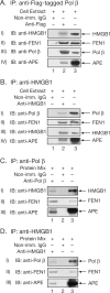
(A and B) extract from MEF cells overexpressing Flag epitope-tagged Pol β was immunoprecipitated (IP) with either non-immune IgG (Panels I-IV, lane 1), anti-Flag (A; Panels I-IV, lane 2) or anti-HMGB1 (B; Panels I-IV, lane 2) antibodies, respectively, and the immunoblots (IB) were developed with the antibodies, as indicated. Lanes 3 in all panels represent a positive control with 1/50th of the cell extract processed directly in a 4-12% NuPAGE Bis-Tris gel. (C and D) to the mixture of Pol β, HMGB1, APE, and FEN1 (1.5 μM each) in a final volume of 50 μl either anti-Pol β (C; Panels I-III, lane 1), anti-HMGB1 (D; Panels I-III, lane 1) or non-immune IgG (lane 2) were added, and the proteins were IP as described in “Supplemental Data.” Lane 3 represents a positive control where the proteins mixture was processed directly. The immunoblots were developed with respective antibodies, as indicated. Protein identity is shown on the right-hand side of the photographs.
Figure 7
Figure 7. Biological and Biochemical Analyses of HMGB1 +/+ and HMGB1 −/− MEFs after treatment with MMS
(A) HMGB1 +/+ (red filled circles) and HMGB1 −/− (blue open circles) MEFs were treated for 1 h with MMS and cell sensitivity was determined by growth inhibition experiments as described under “Experimental Procedures.” Data represent the mean ± S.E. of six independent experiments. (Panel A, inset) Immunoblot analysis of HMGB1 +/+, HMGB1 −/− and Pol β +/+ MEF extracts with specific antibodies to either (i) HMGB1 or (ii) Pol β. Purified HMGB1 and Pol β proteins were loaded as positive controls as indicated. (B) HMGB1 +/+ (red filled symbols) and HMGB1 −/− (blue open symbols) were treated for 1 h with MMS in the absence (circles) or presence of MX (30 mM) for 1 h (squares) or 4 h (diamonds). Data represent the mean ± S.E. of three independent experiments. (C) Comet assay was performed as described in “Supplemental Data.” HMGB1+/+ and HMGB1−/− cells were treated with MMS (0.5 mM for 30 min) or a combination of MMS and MX (30 mM), and harvested for analysis at 2 h. Medium (control) represents the results from untreated HMGB1+/+ and HMGB1−/− cells. Images were obtained with a fluorescence microscope and image analysis system using 20× magnification. (D) Quantification of Olive Tail Moment (OTM) from (C) in HMGB1 +/+ (red bars) and HMGB1 −/− cells (blue bars). Data represent the mean ± S.E. of three independent experiments; for each treatment, 150 cells were scored. (E) Measurement of incision activity of APE on MX-adducted DNA substrate. Reaction conditions and product analysis are described in “Supplemental Data.” 32P-labeled MX-adducted duplex oligonucleotide (50 nM) was incubated with 10 nM APE in the presence of increasing concentrations (25 to 200 nM) of HMGB1 (lanes 2-6) for 10 min at 37 °C. The positions of substrate and incised product and the HMGB1 concentration are indicated. (F) The incision activity of APE on MX-adducted substrate was quantified and plotted as arbitrary PhosphorImager units as a function of HMGB1 concentration.

References

    1. Beard WA, Wilson SH. Purification and domain-mapping of mammalian DNA polymerase β. Methods Enzymol. 1995;262:98–107. - V体育ios版 - PubMed
    1. Biade S, Sobol RW, Wilson SH, Matsumoto Y. Impairment of proliferating cell nuclear antigen-dependent apurinic/apyrimidinic site repair on linear DNA. J. Biol. Chem. 1998;273:898–902. - PubMed
    1. Bianchi ME. Significant (re)location: how to use chromatin and/or abundant proteins as messages of life and death. Trends Cell Biol. 2004;14:287–293. - PubMed (VSports最新版本)
    1. Bianchi ME, Beltrame M, Paonessa G. Specific recognition of cruciform DNA by nuclear protein HMG1. Science. 1989;243:1056–1059. - PubMed (V体育ios版)
    1. Biemann K. Contributions of mass spectrometry to peptide and protein structure. Biomed. Environ. Mass Spectrom. 1988;16:99–111. - PubMed (V体育安卓版)

Publication types (V体育平台登录)

MeSH terms (VSports最新版本)