Skip to main page content
U.S. flag

An official website of the United States government

Dot gov

The . gov means it’s official. Federal government websites often end in . gov or . mil. Before sharing sensitive information, make sure you’re on a federal government site. VSports app下载.

Https

The site is secure. The https:// ensures that you are connecting to the official website and that any information you provide is encrypted and transmitted securely. V体育官网.

. 2007 Sep;117(9):2477-85.
doi: 10.1172/JCI32054.

A Foxo/Notch pathway controls myogenic differentiation and fiber type specification

Affiliations

A Foxo/Notch pathway controls myogenic differentiation and fiber type specification

Tadahiro Kitamura (VSports app下载) et al. J Clin Invest. 2007 Sep.

Abstract

Forkhead box O (Foxo) transcription factors govern metabolism and cellular differentiation. Unlike Foxo-dependent metabolic pathways and target genes, the mechanisms by which these proteins regulate differentiation have not been explored. Activation of Notch signaling mimics the effects of Foxo gain of function on cellular differentiation. Using muscle differentiation as a model system, we show that Foxo physically and functionally interacts with Notch by promoting corepressor clearance from the Notch effector Csl, leading to activation of Notch target genes. Inhibition of myoblast differentiation by constitutively active Foxo1 is partly rescued by inhibition of Notch signaling while Foxo1 loss of function precludes Notch inhibition of myogenesis and increases myogenic determination gene (MyoD) expression. Accordingly, conditional Foxo1 ablation in skeletal muscle results in increased formation of MyoD-containing (fast-twitch) muscle fibers and altered fiber type distribution at the expense of myogenin-containing (slow-twitch) fibers VSports手机版. Notch/Foxo1 cooperation may integrate environmental cues through Notch with metabolic cues through Foxo1 to regulate progenitor cell maintenance and differentiation. .


V体育官网入口 - Figures
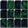
Figure 1
Figure 1. Regulation of myoblast differentiation by Foxo and Notch.
C2C12 cells were immunostained with anti-myosin antibody (green) and DAPI (blue). See text for panel description. Each experiment was repeated at least 6 times. Original magnification, ×10.
Figure 2
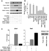
Figure 2. Quantitative analysis of C2C12 differentiation.
(A) Western blotting analysis of myosin expression in C2C12 cells. (B) Morphometric analysis of myosin-positive cells. Results from differentiation experiments were analyzed by scoring the number of myosin-immunostained cells as a percentage of all DAPI-positive cells. (C) DBD-Foxo1-ADA reporter gene assays. We carried out reporter gene assays using the canonical Foxo1-responsive Igfbp1 promoter (left panel) and the Hes1 promoter (right panel) in cells cotransfected with Foxo1-ADA or DBD-Foxo1-ADA. Western blot (inset) demonstrates that expression levels of the 2 proteins are similar. An asterisk indicates P < 0.01 by ANOVA.
Figure 3
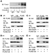
Figure 3. Foxo1 coimmunoprecipitates with Csl.
(A) Coimmunoprecipitation of endogenous Foxo1 and Csl in C2C12 cells cocultured with LacZ-expressing (denoted by the minus sign) or Jagged1-expressing HEK293 cells (denoted by the plus sign). (B and C) Coimmunoprecipitation experiments in C2C12 cells cotransfected with FLAG-Csl and HA-Foxo1. (D and E) Coimmunoprecipitation experiments in C2C12 cells cotransfected with FLAG-Csl and the truncated mutant Myc- or HA-tagged Δ256 Foxo1. TCL, total cellular lysate.
Figure 4
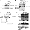
Figure 4. Foxo1 binds directly to Csl.
(A) GST pull-down assays of GST-Foxo1 fusion protein with Csl immunoprecipitated from HEK293 cells. (B and C) Binding of GST-Foxo1 and GST-FLAG-Csl in a cell-free system and mapping of the Csl interaction domain. Full-length and truncated fragments of GST-Foxo1 and GST-FLAG-Csl were purified from bacteria and coincubated. Thereafter, Csl was isolated using anti-FLAG antibody, and the immunoprecipitate was analyzed by immunoblotting with anti-Foxo1 or anti-FLAG antibodies. (D) Hes1 promoter ChIP assay spanning the Csl-binding site in C2C12 cells to detect endogenous Foxo1, Csl, and Notch1 (Endog) or following transduction with Foxo1-ADA during myoblast differentiation. Input represents DNA extracted from chromatin prior to immunoprecipitation. Hes1 (semiquantitative RT-PCR) and myosin (Western blot) expression corresponding to each time point are shown. Day 0 is defined as the time when cells were serum deprived to induce myoblast fusion.
Figure 5
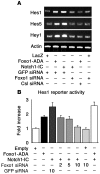
Figure 5. Foxo1 regulates Notch-induced Hes1, Hes5, and Hey1 expression.
(A) Hes1, Hes5, and Hey1 expression measured by semiquantitative RT-PCR in C2C12 cells transduced with Foxo1-ADA or Notch1-IC following transfection of GFP, Foxo1, or Csl siRNA as indicated. (B) Hes1 reporter gene assays in HEK293 cells transduced with Foxo1-ADA, Notch1-IC, Foxo1 siRNA, GFP siRNA, or control plasmid (empty). We measured luciferase activity and normalized it by β-galactosidase activity. The data represent arbitrary units relative to control empty vector.
Figure 6
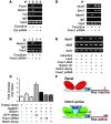
Figure 6. Foxo1 is required for Notch binding to the Hes1 promoter and activation of Hes1 target genes.
(A) ChIP assays of endogenous Foxo1 and Notch1 in C2C12 cells cocultured with LacZ-expressing (denoted by a minus sign) or Jagged1-expressing HEK293 cells (denoted by a plus sign) in the absence (lanes 1 and 2) and presence (lanes 3 and 4) of Csl siRNA. (B) ChIP assays of endogenous Notch1 in coculture system in the absence (lanes 1 and 2) and presence (lanes 3 and 4) of Foxo1 siRNA. (C) Hes1 promoter assays following coculture in the absence and presence of Foxo1 or GFP siRNA. (D) ChIP assays of NcoR and Smrt and Maml1 binding to Hes1 in the coculture system in the absence (lanes 1 and 2) and presence (lanes 3 and 4) of Foxo1 siRNA. (E) Expression of MyoD, Myf5, and β-actin in C2C12 cells by semiquantitative RT-PCR. (F) Model of Foxo1 and Notch regulation of Hes1 promoter.
Figure 7
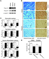
Figure 7. Conditional ablation of Foxo1 in skeletal muscle.
(A) Western blot analysis of Foxo1 and Foxo4 expression levels in various muscle types. Gastroc, gastrocnemius muscle; Vastus, vastus lateralis muscle. (B) Metachromatic and immunohistochemical analysis of soleus and plantaris muscle from Myog-Foxo1 mice and control (lox/lox) littermates. Original magnification, ×10. (C) Gene expression analysis of Myog-Foxo1 (black bars) and control mice (white bars); TropC, troponin-C; TropT, troponin-T; Mlc, myosin light chain; Myog, myogenin; Mck, muscle-type creatine kinase. Data are means ± SEM of 3 independent measurements (n = 6 for each genotype). An asterisk indicates P < 0.05 by ANOVA. (D) Treadmill performance test in 8-week-old Myog-Foxo1 mice and lox/lox littermates (n = 6 for each genotype). An asterisk indicates P < 0.05 by ANOVA.

"V体育官网入口" References

    1. Singec I., Jandial R., Crain A., Nikkhah G., Snyder E.Y. The leading edge of stem cell therapeutics. Annu. Rev. Med. 2007;58:313–328. doi: 10.1146/annurev.med.58.070605.115252. - DOI - PubMed
    1. Accili D., Arden K.C. FoxOs at the crossroads of cellular metabolism, differentiation, and transformation. Cell. 2004;117:421–426. doi: 10.1016/S0092-8674(04)00452-0. - "V体育平台登录" DOI - PubMed
    1. Hribal M.L., Nakae J., Kitamura T., Shutter J.R., Accili D. Regulation of insulin-like growth factor-dependent myoblast differentiation by Foxo forkhead transcription factors. J. Cell Biol. 2003;162:535–541. doi: 10.1083/jcb.200212107. - DOI - PMC - PubMed
    1. Nakae J., et al. The forkhead transcription factor foxo1 regulates adipocyte differentiation. Dev. Cell. 2003;4:119–129. doi: 10.1016/S1534-5807(02)00401-X. - DOI - PubMed
    1. Potente M., et al. Involvement of Foxo transcription factors in angiogenesis and postnatal neovascularization. J. Clin. Invest. 2005;115:2382–2392. doi: 10.1172/JCI23126. - DOI - PMC - PubMed

Publication types

MeSH terms