Skip to main page content (V体育官网)
U.S. flag

An official website of the United States government

Dot gov

The . gov means it’s official. Federal government websites often end in VSports app下载. gov or . mil. Before sharing sensitive information, make sure you’re on a federal government site. .

Https

The site is secure V体育官网. The https:// ensures that you are connecting to the official website and that any information you provide is encrypted and transmitted securely. .

. 2004 Apr;24(8):3430-44.
doi: 10.1128/MCB.24.8.3430-3444.2004.

Activated liver X receptors stimulate adipocyte differentiation through induction of peroxisome proliferator-activated receptor gamma expression

Affiliations

V体育平台登录 - Activated liver X receptors stimulate adipocyte differentiation through induction of peroxisome proliferator-activated receptor gamma expression

Jong Bae Seo et al. Mol Cell Biol. 2004 Apr.

Abstract

Liver X receptors (LXRs) are nuclear hormone receptors that regulate cholesterol and fatty acid metabolism in liver tissue and in macrophages. Although LXR activation enhances lipogenesis, it is not well understood whether LXRs are involved in adipocyte differentiation. Here, we show that LXR activation stimulated the execution of adipogenesis, as determined by lipid droplet accumulation and adipocyte-specific gene expression in vivo and in vitro. In adipocytes, LXR activation with T0901317 primarily enhanced the expression of lipogenic genes such as the ADD1/SREBP1c and FAS genes and substantially increased the expression of the adipocyte-specific genes encoding PPARgamma (peroxisome proliferator-activated receptor gamma) and aP2. Administration of the LXR agonist T0901317 to lean mice promoted the expression of most lipogenic and adipogenic genes in fat and liver tissues. It is of interest that the PPARgamma gene is a novel target gene of LXR, since the PPARgamma promoter contains the conserved binding site of LXR and was transactivated by the expression of LXRalpha. Moreover, activated LXRalpha exhibited an increase of DNA binding to its target gene promoters, such as ADD1/SREBP1c and PPARgamma, which appeared to be closely associated with hyperacetylation of histone H3 in the promoter regions of those genes. Furthermore, the suppression of LXRalpha by small interfering RNA attenuated adipocyte differentiation. Taken together, these results suggest that LXR plays a role in the execution of adipocyte differentiation by regulation of lipogenesis and adipocyte-specific gene expression. VSports手机版.


Figures
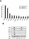
FIG. 1.
FIG. 1.
Expression of LXRα mRNA in adipocytes and mouse tissues. (A) Relative amounts of mRNA expression of adipogenic genes as determined by DNA microarray analysis. Total RNA was isolated from confluent preadipocytes and fully differentiated 3T3-F442A adipocytes and used for DNA microarray analysis. Relative amounts of mRNA expression of 11 genes are shown. Apo CI, apolipoprotein 1; Apo C2, apolipoprotein 2. (B) LXRα mRNA expression in C57BL/6 mouse tissues. Northern blot analysis was performed by using 20 μg of total RNA and cDNA probes for LXRα, ADD1/SREBP1c, PPARγ, aP2, and 36B4. B, brain; F, white adipose tissue (epididymal fat); K, kidney; M, muscle; Lu, lung; Li, liver; S, spleen; H, heart.
FIG. 2.
FIG. 2.
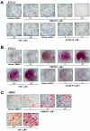
Adipogenic effect of LXR activation in preadipocytes. 3T3-L1 cells (A and B) and HSVCs (C) were differentiated into adipocytes in the presence or absence of the LXR agonist T0901317, 22(R)-hydroxycholesterol [22-(R) HC], or the PPARγ agonist TZD. (A and C) Microscopic pictures were taken 10 days after differentiation. (B and C) Differentiated adipocytes were stained with Oil Red O and photographed. EtOH, ethyl alcohol.
FIG. 3.
FIG. 3.
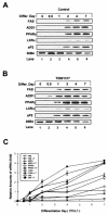
Stimulation of adipogenic marker gene expression following LXR activation during adipocyte differentiation (differ.). (A and B) 3T3-L1 cells were differentiated into adipocytes in the absence (A) or presence (B) of T0901317 (3 μM), and cells were harvested at the indicated time points. Northern blots (20 μg of total RNA) were hybridized with FAS, ADD1/SREBP1c, PPARγ, LXRα, aP2, and 36B4 cDNA probes. (C) Data from panels A and B were quantified and normalized relative to the loading control to show relative mRNA expression. Experiments were independently repeated three times.
FIG. 4.
FIG. 4.
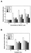
Induction of lipogenic and adipogenic genes following acute LXR activation in differentiated adipocytes. (A) Differentiated 3T3-L1 adipocytes were treated with 0, 1, 3, or 10 μM T0901317 for 24 h. Northern blot analysis results were quantified and normalized relative to 28S rRNA levels. (B) 3T3-L1 adipocytes were treated for 0, 24, and 48 h with 3 μM T0901317, after which cells were harvested for Northern blot analysis. The results obtained were quantified and normalized relative to 28S rRNA levels. Northern blots (20 μg of total RNA) were hybridized with FAS, ADD1/SREBP1c, PPARγ, LXRα, and aP2 cDNA probes. Each experiment was independently repeated two times.
FIG. 5.
FIG. 5.
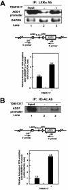
ChIP assay of mouse ADD1/SREBP1c promoter. Differentiated 3T3-L1 adipocytes were incubated in the absence (−) or presence (+) of 10 μM T0901317 for 24 h. Cells were cross-linked and immunoprecipitated with rabbit polyclonal antibodies against LXRα (LXRα Ab) (A) or polyclonal antibodies against acetylated histone H3 (H3-Ac Ab) (B). Immunoprecipitated DNA fragments were amplified by PCR (see Materials and Methods). Lane 1 shows the amplified mouse ADD1/SREBP1c promoter and GAPDH from 1% of the input DNA. GAPDH fragments were also amplified for the normalization of input DNA. PCR-amplified products of the mouse ADD1/SREBP1c promoter normalized with the amplified GAPDH fragments were quantitated. The putative sterol regulatory element (SRE; ellipse), LXRE (white square), and E-box (black triangle) are indicated. Data from a representative of three independent experiments are shown. IP, immunoprecipitant.
FIG. 6.
FIG. 6.
In vivo effects of LXR activation on adipogenic gene expression in white adipose tissue and liver. C57BL/6 mice were treated with T0901317 (50 mg/kg) or vehicle for 0, 1, 3, or 5 days. Epididymal fat and liver tissues were collected, and total RNA was isolated for Northern blotting (A and C) or RT-PCR analysis (E) to examine the mRNA expression of several genes, including the FAS, LXRα, ADD1/SREBP1c, PPARγ, and aP2 genes. (A) Expression in epididymal fat. (B) Data in panel A were quantified and normalized relative to 28S rRNA levels. (C and E) Expression in liver. (D) Data in panel C were quantified and normalized relative to 28S rRNA levels. Data from a representative of two independent experiments are shown.
FIG. 7.
FIG. 7.
Direct binding of LXRα to the mouse PPARγ promoter. (A) Sequence comparison of putative LXREs (DR4) in mouse and human PPARγ promoters. (B) In vitro-translated LXRα and RXRα proteins were used for EMSA with 32P-labeled PPARγ LXRE oligonucleotide. Sequence-specific competition assays were performed with the addition of a 100-fold molar excess of unlabeled sterol regulatory element (SRE) (lane 4), Cyp7A1 LXRE (lane 5), and PPARγ LXRE (lane 6) oligonucleotides. (C) ChIP assays of the mouse PPARγ promoter. Differentiated 3T3-L1 adipocytes were incubated with (+) or without (−) LXR agonist T0901317 (10 μM) for 0, 2, or 12 h. Cells were cross-linked and immunoprecipitated with rabbit polyclonal antibodies against LXRα or polyclonal antibodies against acetylated histone H3. Immunoprecipitated DNA fragments were amplified by PCR (see Materials and Methods). Lane 1 shows the amplified mouse PPARγ promoter and GAPDH from 1% of the input DNA. GAPDH fragments were also amplified for the normalization of input DNA. IP, immunoprecipitant. (D) h293 cells were cotransfected with the pADD1/SREBP1c −600-Luc reporter DNA (100 ng/well) and expression vectors for LXRα and RXRα (lanes 3 and 4). pADD1/SREBP1c −600-Luc is a luciferase reporter containing the region comprising bp −600 to +89 of the mouse ADD1/SREBP1c promoter. In parallel, h293 cells were cotransfected with the mouse pPPARγ −1,022-LXRE-Luc reporter (wild-type) DNA (100 ng/well) or the mutant pPPARγ −1,022-mLXRE-Luc reporter DNA (100 ng/well) and expression vectors for LXRα and RXRα (lanes 2 and 3). The pPPARγ −1,022-LXRE-Luc reporter is a luciferase reporter containing bp −1022 to +26 of the mouse PPARγ promoter. The pPPARγ −1,022-mLXRE-Luc reporter is a luciferase reporter containing the mutation in the LXRE motif of the mouse PPARγ promoter. After transfection, cells were treated with (+) or without (−) LXR agonist T0901317 (10 μM) for 24 h.
FIG. 8.
FIG. 8.
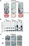
Effect of LXRα knockdown by siRNA during adipogenesis. 3T3-L1 cells were infected and selected with pSUPER retroviruses including mock, siLXRαSR933, and siLXRαSR1246 (see Materials and Methods). Those infected cells were differentiated into adipocytes and were harvested for total RNA preparation at the indicated time point. (A) The cells were stained with Oil Red O and photographed. (B) Northern blots (20 μg of total RNA) were hybridized with FAS, ADD1/SREBP1c, PPARγ, LXRα, LXRβ, and aP2 cDNA probes. Mock and siLXRαSR1246 were used as negative controls. (C) Effects of PPARγ ligand on 3T3-L1-LXRα siRNA stable cells. Mock and 3T3-L1-LXRα siRNA stable cell lines were differentiated into adipocytes under normal differentiation conditions (see Materials and Methods) in the absence (DMSO) or presence of T0901317 (1 μM) or rosiglitazone (0.1 μM).
FIG. 9.
FIG. 9.
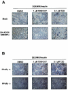
Effects of LXR or PPARγ ligand on DN-ADD1/SREBP1c-expressing cells or PPARγ-deficient MEF cells. (A) 3T3-L1 cells were infected and selected with pBabe retroviruses including mock and DN-ADD1/SREBP1c. (B) MEF cells deficient in the PPARγ or the PPARγ heterozygote mutant or both of them were differentiated into adipocytes in the absence (DMSO) or presence of T0901317 (1 μM) or rosiglitazone (0.1 μM).
FIG. 10.
FIG. 10.
Functional roles of LXR in adipocyte differentiation. Crosstalks between LXRs and other adipogenic transcription factors such as ADD1/SREBP1c and PPARγ would play key roles in the mediation of lipogenesis and adipocyte-specific gene expression during adipogenesis.

References

    1. Akiyama, T. E., S. Sakai, G. Lambert, C. J. Nicol, K. Matsusue, S. Pimprale, Y. H. Lee, M. Ricote, C. K. Glass, H. B. Brewer, Jr., and F. J. Gonzalez. 2002. Conditional disruption of the peroxisome proliferator-activated receptor gamma gene in mice results in lowered expression of ABCA1, ABCG1, and apoE in macrophages and reduced cholesterol efflux. Mol. Cell. Biol. 22:2607-2619. - PMC - PubMed
    1. Amemiya-Kudo, M., H. Shimano, T. Yoshikawa, N. Yahagi, A. H. Hasty, H. Okazaki, Y. Tamura, F. Shionoiri, Y. Iizuka, K. Ohashi, J. Osuga, K. Harada, T. Gotoda, R. Sato, S. Kimura, S. Ishibashi, and N. Yamada. 2000. Promoter analysis of the mouse sterol regulatory element-binding protein-1c gene. J. Biol. Chem. 275:31078-31085. - PubMed
    1. Barak, Y., M. C. Nelson, E. S. Ong, Y. Z. Jones, P. Ruiz-Lozano, K. R. Chien, A. Koder, and R. M. Evans. 1999. PPAR gamma is required for placental, cardiac, and adipose tissue development. Mol. Cell 4:585-595. - PubMed
    1. Cao, G., Y. Liang, C. L. Broderick, B. A. Oldham, T. P. Beyer, R. J. Schmidt, Y. Zhang, K. R. Stayrook, C. Suen, K. A. Otto, A. R. Miller, J. Dai, P. Foxworthy, H. Gao, T. P. Ryan, X. C. Jiang, T. P. Burris, P. I. Eacho, and G. J. Etgen. 2003. Antidiabetic action of a liver X receptor agonist mediated by inhibition of hepatic gluconeogenesis. J. Biol. Chem. 278:1131-1136. - PubMed (V体育平台登录)
    1. Chawla, A., W. A. Boisvert, C. H. Lee, B. A. Laffitte, Y. Barak, S. B. Joseph, D. Liao, L. Nagy, P. A. Edwards, L. K. Curtiss, R. M. Evans, and P. Tontonoz. 2001. A PPAR gamma-LXR-ABCA1 pathway in macrophages is involved in cholesterol efflux and atherogenesis. Mol. Cell 7:161-171. - V体育平台登录 - PubMed

"V体育平台登录" Publication types

VSports app下载 - MeSH terms

"VSports在线直播" LinkOut - more resources