Skip to main page content
U.S. flag

An official website of the United States government

Dot gov

The VSports app下载. gov means it’s official. Federal government websites often end in . gov or . mil. Before sharing sensitive information, make sure you’re on a federal government site. .

Https

The site is secure. The https:// ensures that you are connecting to the official website and that any information you provide is encrypted and transmitted securely. V体育官网.

. 2003 Nov;77(21):11603-15.
doi: 10.1128/jvi.77.21.11603-11615.2003.

The 3' end of Norwalk virus mRNA contains determinants that regulate the expression and stability of the viral capsid protein VP1: a novel function for the VP2 protein

Affiliations

The 3' end of Norwalk virus mRNA contains determinants that regulate the expression and stability of the viral capsid protein VP1: a novel function for the VP2 protein

Andrea Bertolotti-Ciarlet et al. J Virol. 2003 Nov.

Abstract

Norwalk virus (NV) is the prototype strain of a group of noncultivable human caliciviruses responsible for epidemic outbreaks of acute gastroenteritis. The capsid protein VP1 is synthesized from a subgenomic RNA that contains two open reading frames (ORFs), ORF2 and ORF3, and the 3' untranslated region (UTR). ORF2 and ORF3 code for the capsid protein (VP1) and a small structural basic protein (VP2), respectively. We discovered that the yields of virus-like particles (VLPs) composed of VP1 are significantly reduced when this protein is expressed from ORF2 alone VSports手机版. To determine how the 3' terminus of the NV subgenomic RNA regulates VP1 expression, we compared VP1 expression levels by using recombinant baculovirus constructs containing different 3' elements. High VP1 levels were detected by using a recombinant baculovirus that contained ORF2, ORF3, and the 3'UTR (ORF2+3+3'UTR). In contrast, expression of VP1 from constructs that lacked the 3'UTR (ORF2+3), ORF3 (ORF2+3'UTR), or both (ORF2 alone) was highly reduced. Elimination of VP2 synthesis from the subgenomic RNA by mutation resulted in VP1 levels similar to those obtained with the ORF2 construct alone, suggesting a cis role for VP2 in upregulation of VP1 expression levels. Comparisons of the kinetics of RNA and capsid protein expression levels by using constructs with or without ORF3 or the 3'UTR revealed that the 3'UTR increased the levels of VP1 RNA, whereas the presence of VP2 resulted in increased levels of VP1. Furthermore, VP2 increased VP1 stability and protected VP1 from disassembly and protease degradation. The increase in VP1 expression levels caused by the presence of VP2 in cis was also observed in mammalian cells. .


Figures
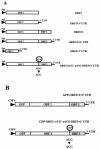
FIG. 1.
FIG. 1.
Schematic representation of recombinant baculoviruses and GFP contructs generated in the present study to monitor the expression levels of NV VP1 in insect cells and mammalian cells. (A) Baculovirus recombinants to analyze the expression of VP1 in Sf9 insect cells were generated by cloning into the pFastBac1 expression vector (Gibco-BRL). Since the same restriction enzyme site (BamHI) was used to generate all baculovirus recombinants, all seven constructs possess an identical 5′-terminal sequence. In addition, all of these constructs encode the poly(A) signal from SV40. These recombinant baculoviruses were used to study the effect of the different 3′-terminal elements of the NV subgenomic RNA on NV VP1 capsid protein expression. The stop signal indicates the site targeted by directed mutagenesis to block VP2 expression. (B) Constructs generated to analyze the effect of ORF3 or VP2 expression on VP1 expression in mammalian cells. The entire NV subgenomic RNA was placed, in frame, downstream of the GFP gene in the pEGFP-C1 vector (Clontech), and VP1 was expressed as a fusion protein with the GFP protein. The stop signal indicates a point mutation generated to block the expression of VP2. Abbreviations: Pph, polyhedron promoter; UTR, untranslated region; ORF, open reading frame.
FIG. 2.
FIG. 2.
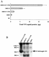
VP1 expression levels obtained with the baculovirus recombinants containing NV ORF2 only, NV ORF2+3, NV ORF2+3′UTR, or the entire NV subgenomic RNA (ORF2+3+3′UTR). (A) VP1 yield from 3 × 10⁶ Sf9 insect cells infected with each recombinant baculovirus measured at 36 hpi by ELISA from the cell lysate. Purified rNV VLPs were used as the positive antigen control at concentrations ranging from 0.5 to 62 ng. The values shown are the arithmetic mean of at least four independent experiments. Error bars represent one standard error of the mean. (B) Expression of VP1 analyzed by Western blotting with a rabbit hyperimmune serum specific to VP1. Infected cell lysates (3 × 10⁶ cells) were harvested at 36 hpi in SDS-PAGE sample buffer and analyzed by SDS-12% PAGE and Western blotting. To compare the relative levels of VP1 expression of all recombinants, the same volume of lysate was loaded for each recombinant baculovirus. The arrow indicates the bands that correspond to the uncleaved VP1. Molecular weight markers are indicated on the left. Two approximately 30K cleavage products were present in all of the samples but in larger amounts in the lysates from cells infected with the ORF2+3 and ORF2+3′UTR recombinant baculoviruses. The Western blotting and the ELISA analysis were performed with the same antibody; therefore, these 30K VP1-related degradation products were detected in the total protein quantification done by ELISA. BV, wild-type baculovirus.
FIG. 3.
FIG. 3.
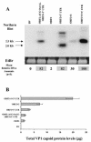
Comparison of steady-state levels of ORF2 mRNA and VP1 expressed from different constructs. (A) The steady-state levels of NV ORF2 mRNA were assessed at 36 hpi in 3 × 10⁶ cells infected with the different recombinant baculoviruses by Northern blotting with a 32P-labeled probe specific for the NV ORF2 gene. Ethidium bromide (EtBr) staining of the total amount of RNA run was used as a loading control. The ethidium bromide-stained bands shown correspond to the rRNA. The bands that corresponded to the expected molecular sizes, 2.0 kb for ORF2 alone and 2.5 kb for the entire subgenomic RNA, were quantitated by phosphorimaging. The values shown at the bottom of each lane of the gel are the average of three independent experiments and represent the relative amounts of RNA with respect to the amount obtained with the entire subgenomic RNA construct (ORF2+3+3′UTR). The highest values are shaded. The band corresponding to the RNA for the ORF2-alone construct was not high enough to be seen on this exposure of the gel, but it was detected by the phosphorimager. (B) The level (μg) of VP1 from the same samples was determined by analysis of duplicate samples by ELISA. Purified rNV VLPs were used as the positive antigen control at concentrations ranging from 0.5 to 62 ng. Each bar shows the arithmetic means of three independent experiments. Error bars represent one standard error of the mean.
FIG. 4.
FIG. 4.
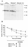
Analysis of VP1 stability. (A) For pulse-chase experiments performed in insect cells, cells were infected with the recombinant baculoviruses containing the entire NV subgenomic RNA (ORF2+3+3′UTR) or lacking the 3′UTR (ORF2+3) or the ORF3 gene (ORF2+3′UTR). After a 30-min pulse at 36 hpi, chase periods of 6, 12, and 24 h were analyzed. The samples from each of the indicated time points were immunoprecipitated with a rabbit hyperimmune serum against the NV VP1 capsid protein in RIPA buffer, and the samples were then analyzed by autoradiography on an SDS-12% polyacrylamide gel. The arrows indicate the bands that correspond to the uncleaved VP1. The asterisk indicates a VP1-related band that is not present in the NV VLPs and has not been detected previously. This band was not included in the VP1 quantitation and was not present in the wild-type baculovirus (BV WT). The band was not detected when a monoclonal antibody was used to immunoprecipitate the protein; however, the epitope that the monoclonal antibody used may not be present in this VP1-related protein. (B) Phosphorimaging was performed to quantitate each band, and the amount of VP1 present at the zero time point for each recombinant baculovirus was set to 100%. The amount of VP1 at additional time points was represented relative to the amount of protein at 0 h after pulse-labeling. The values shown are the arithmetic means of three independent experiments. The error bars represent one standard error of the mean. The half-life of VP1 expressed from each of the recombinant baculoviruses tested was calculated from the curves obtained.
FIG. 5.
FIG. 5.
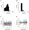
DLS analysis of the homogeneity of intact particles in preparations of VLPs composed of only VP1 or of both VP1and VP2 proteins (VP1/VP2). (A and B) Graphs show the percentage of intact particles from either VP1 VLP or VP1/VP2 VLP preparations, respectively. The particles were analyzed in conditions known to keep the particles assembled (Tris-HCl [pH 6.7]). Histograms of the percentage of scatter for VP1 (A) or VP1/VP2 particles (B) particles measured at a 90° scattering angle are shown. Because the total protein concentration was similar for all of the preparations, the percentage of scatter represents the molecular mass and effective protein concentration for each particle size. (C and D) In addition, the particle size distributions for VP1 (C) and VP1/VP2 (D) particles are shown. The vertical lines in panels C and D represent the distributions of different particle sizes in the sample for each reading. The top square represents the largest particle found in the sample (nanometers of radius), the middle square represents the average particle size, and the bottom square the smallest particle size found in the sample. A monodispersed, homogeneous sample is expected to have only one size of particle, depicted as one square and no line. Twenty-five readings were recorded for each sample. Three different samples for each type of particles (VP1 or VP1/VP2) were analyzed. The data from one representative reading are shown.
FIG. 6.
FIG. 6.
Analysis of the sensitivity of intact or partially denatured rNV VLPs to protease degradation. VLPs composed of only the VP1 capsid protein (VP1 VLPs) or composed of both VP1 and VP2 proteins (VP1/VP2 VLPs) were kept assembled (Tris-HCl [pH 6.7]) or disassembled by high-pH treatment (Tris-HCl [pH 8.9]) prior to protease digestion. (A) After protease digestion, the proteins were separated in SDS-12% polyacrylamide gels, and the bands were analyzed by Western blotting with a rabbit serum specific to VP1 (30). The arrows indicate the bands that correspond to the uncleaved 58K full-length VP1 and the 32K soluble protein formed by the protruding domain (26). V, VLPs; D, disassembled form of the NV VP1 capsid protein. (B) The arithmetic means of the densitometric values corresponding to the VP1 bands are shown for three experiments.
FIG. 7.
FIG. 7.
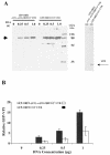
Regulatory effect of VP2 on expression of VP1 in mammalian cells. (A) Western blot analysis of a GFP-VP1 fusion protein expressed in HEK 293T cells after transfection with constructs containing the entire NV subgenomic RNA (GFP-ORF2+3+3′UTR) or lacking the ability to express VP2 (GFP-ORF2-AUG→ACG-ORF3+3′UTR). After transfection of cells with the indicated amount (in micrograms above each lane) of DNA into 293T cells, the same amount of cell lysate was used to analyze the expressed proteins. The NV VP1 capsid protein was detected by Western blotting with a rabbit serum specific to VP1. The sizes are shown in kilodaltons, and the arrow indicates the band that corresponds to the uncleaved GFP-VP1 fusion protein. The Western blot shown in the right panel was performed with rabbit sera specific to VP2 (15, 16). The band corresponding to the VP2 protein is indicated with an arrow and was only observed when the GFP-ORF2+3+3′UTR recombinant was expressed. (B) The bands corresponding to the expected molecular weight for the GFP-VP1 fusion protein in panel A were analyzed by densitometry. The densitometric values were normalized by using luciferase activity values and the relative amounts of GFP-VP1 are shown. Each bar shows the arithmetic means of three independent experiments performed with different DNA preparations. The error bars represent one standard error of the mean.
FIG. 8.
FIG. 8.
Proposed model for the mechanism of the VP2 stabilizing effect on NV VP1 capsid protein. Particles composed of both VP1 and VP2 proteins are more stable than those composed only of the VP1 capsid protein. Therefore, particles composed only of VP1 (A) could be more sensitive to various treatments (such as pressure, heat, pH, or changes in ionic concentration; represented by the shaded arrow) that cause disassembly of the particles compared to particles that also contain VP2 (B). Once the particles disassemble, the soluble VP1 capsid protein may be more sensitive to protease degradation (solid arrows) because of the exposure of more cleavage sites.

"V体育安卓版" References

    1. Agrawal, S., D. Gupta, and S. K. Panda. 2001. The 3′ end of hepatitis E virus (HEV) genome binds specifically to the viral RNA-dependent RNA polymerase (RdRp). Virology 282:87-101. - PubMed
    1. Arora, R., C. Priano, A. B. Jacobson, and D. R. Mills. 1996. cis-Acting elements within an RNA coliphage genome: fold as you please, but fold you must!! J. Mol. Biol. 258:433-446. - PubMed
    1. Bachurski, C. J., N. G. Theodorakis, R. M. Coulson, and D. W. Cleveland. 1994. An amino terminal tetrapeptide specifies cotranslational degradation of beta-tubulin but not alpha-tubulin mRNAs. Mol. Cell. Biol. 14:4076-4086. - PMC - PubMed
    1. Belliot, G., J. S. Noel, J.-F. Li, S. Yoshiyuki, Humphrey, T. M., Ando, T., Glass, R. I., and Monroe, S. S. 2001. Characterization of capsid genes, expressed in the baculovirus system, of three new genetically distinct strains of “Norwalk-like viruses.” J. Clin. Microbiol. 39:4288-4294. - V体育2025版 - PMC - PubMed
    1. Bertolotti-Ciarlet, A., L. J. White, R. Chen, B. V. Prasad, and M. K. Estes. 2002. Structural requirements for the assembly of Norwalk virus-like particles. J. Virol. 76:4044-4055. - PMC - PubMed

Publication types

MeSH terms (VSports在线直播)

LinkOut - more resources