Skip to main page content
U.S. flag

An official website of the United States government

Dot gov

The . gov means it’s official. Federal government websites often end in . gov or VSports app下载. mil. Before sharing sensitive information, make sure you’re on a federal government site. .

Https

The site is secure. The https:// ensures that you are connecting to the official website and that any information you provide is encrypted and transmitted securely. V体育官网.

. 2003 Jun;111(11):1723-32.
doi: 10.1172/JCI17220.

The secretory proprotein convertases furin, PC5, and PC7 activate VEGF-C to induce tumorigenesis

Affiliations

The secretory proprotein convertases furin, PC5, and PC7 activate VEGF-C to induce tumorigenesis

Geraldine Siegfried et al. J Clin Invest. 2003 Jun.

Abstract

The secretory factor VEGF-C has been directly implicated in various physiological processes during embryogenesis and human cancers. However, the importance of the conversion of its precursor proVEGF-C to mature VEGF-C in tumorigenesis, and vessel formation and the identity of the protease(s) that regulate these processes is/are not known. The intracellular processing of proVEGF-C that occurs within the dibasic motif HSIIRR(227)SL suggests the involvement of the proprotein convertases (PCs) in this process. In addition, furin and VEGF-C were found to be coordinately expressed in adult mouse tissues. Cotransfection of the furin-deficient colon carcinoma cell line LoVo with proVEGF-C and different PC members revealed that furin, PC5, and PC7 are candidate VEGF-C convertases. This finding is consistent with the in vitro digestions of an internally quenched synthetic fluorogenic peptide mimicking the cleavage site of proVEGF-C ((220)Q-VHSIIRR downward arrow SLP(230)). The processing of proVEGF-C is blocked by the inhibitory prosegments of furin, PC5, and PACE4, as well as by furin-motif variants of alpha2-macroglobulin and alpha1-antitrypsin. Subcutaneous injection of CHO cells stably expressing VEGF-C into nude mice enhanced angiogenesis and lymphangiogenesis, but not tumor growth. In contrast, expression of proVEGF-C obtained following mutation of the cleavage site (HSIIRR(227)SL to HSIISS(227)SL) inhibits angiogenesis and lymphangiogenesis as well as tumor growth VSports手机版. Our findings demonstrate the processing of proVEGF-C by PCs and highlight the potential use of PC inhibitors as agents for inhibiting malignancies induced by VEGF-C. .


Figures
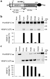
Figure 1
Figure 1
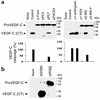
Processing of proVEGF-C by furin, PC5, and PC7. (a) Schematic representation of the primary structure of the 419-AA human proVEGF-C. Shown are the signal peptide (SP), PC-processing site (HSIIRR227SL), an unknown protease site (indicated by question mark) that generates the 21-kDa VEGF-C, and the Flag attached to the C-terminus. ProVEGF-C processing was analyzed by biosynthesis (b) and Western blotting (c) of LoVo-C5–conditioned media obtained from cells transiently transfected with either the empty vectors (None), pIRES2-EGFP vector and pcDNA3-zeo-Flag.cm5 vector containing proVEGF-C (Control), or with the pcDNA3-zeo-Flag.cm5 vector containing proVEGF-C and pIRES2-EGFP vector that expresses full-length human furin, PACE4, or SKI-1; mouse PC5A or PC5B; or rat PC7. The corresponding percentages of proVEGF-C cleavage calculated from the ratio of band intensities of VEGF-C/(proVEGF-C + VEGF-C) are indicated.
Figure 2
Figure 2
In vitro digestions of Q-h-VEGF-C with recombinant furin, PC5, and PC7. (a) RP-HPLC chromatogram of the crude digest following 4 hours of incubation at 37°C of 20 μg of QVEGF-C with furin, PC5, or PC7 in 25 mM Tris, 25 mM Mes, and 2.5 mM CaCl2, pH 7.4. The elution of the peaks was monitored on-line by UV absorbance at 214 nm as well as by fluorescence detectors (λex, 320 nm; λem, 420 nm). (b) MALDI-ToF mass spectra of the crude digests following 24 hours of incubation at 37°C of 20 μg of QVEGF-C with furin, PC5, and PC7 in 25 mM Tris, 25 mM Mes, and 2.5 mM CaCl2, pH 7.4. Note the absence of the peak at m/z 1,703 suggesting complete cleavage of QVEGF-C. The peaks at m/z 1,129 and 595 were attributed to the highly fluorescent N-terminal (NT) (Abz-Q-VHSIIRR-OH) and the nonfluorescent C-terminal (CT) [SLP(NO2)-A-CONH2] fragments, respectively. The peaks at m/z 972 and 663 are not PC-dependent since they are also present in the crude digest of QVEGF-C by wild-type medium (data not shown). NT-R, the N-terminal sequence without Arginine residue.
Figure 3
Figure 3
(a) Kinetic parameters Vmax (apparent) and Km (apparent) for the cleavage of QVEGF-C by furin, PC5, and PC7. Various concentrations of QVEGF-C (0.5–200 μM) were incubated in the presence of furin, PC5, or PC7, and the fluorescence released at various times was measured at λex 320 nm and λem 420. (b) The data collected after either 1 hour or 4 hours of incubation were used to calculate Vmax (apparent) and Km (apparent) using GraFit software as described in Methods. The amounts of furin, PC5, and PC7 used in this study were adjusted to show similar levels of enzymatic activity when measured in the presence of the universal PC substrate, the fluorogenic peptide pERTKR-MCA (100 μM). RFU, raw fluorescence unit; app, apparent.
Figure 4
Figure 4
Blockade of proVEGF-C processing. (a) Processing of proVEGF-C was analyzed by Western blotting in HEK 293 cells transiently cotransfected with the empty pIRES2-EGFP vector (None) and pcDNA3-zeo-Flag.cm5 vector containing proVEGF-C cDNA (Control), or with the pcDNA3-zeo-Flag.cm5 vector containing proVEGF-C and pIRES2-EGFP vector that expresses profurin, proPACE4, proPC5, and proPC7, wild-type or mutated α2-macroglobulin (α2-MG and α2-MG-F, respectively), and α1-antitrypsin. The corresponding percentages of proVEGF-C cleavage calculated from the ratio of band intensities of VEGF-C/(proVEGF-C + VEGF-C) is indicated. (b) CHO tumor cells were stably transfected with empty pcDNA3-zeo-Flag.cm5 vector (None), pcDNA3-zeo-Flag.cm5 vector containing wild-type proVEGF-C cDNA (HSIIRR227), or pcDNA3-zeo-Flag.cm5 vector containing mutated proVEGF-C cDNA (HSIISS227). Populations of stably transfected cells were selected using Zeocin resistance and analyzed by Western blot using the VEGF-C antibody.
Figure 5
Figure 5
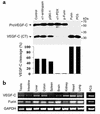
Endogenous VEGF-C processing by PC-like activity and coexpression of furin and VEGF-C in mouse tissues. (a) Endogenous proVEGF-C processing was analyzed by Western blotting of PC3 cell–conditioned media obtained from cells transiently transfected with either the pIRES2-EGFP empty vectors (Control) or vector expressing α1-antitrypsin, pSKI-1, α1-PDX, p-furin, furin, or PC5. The corresponding percentages of proVEGF-C cleavage calculated from the ratio of band intensities of VEGF-C/(proVEGF-C + VEGF-C) are indicated. (b) Total RNA was extracted from PC3 cells and the indicated tissues and organs, and RT-PCR analysis was performed using primers specific for VEGF-C, furin, and GAPDH (control) under the conditions described in Methods. Results shown are representative of three experiments.
Figure 6
Figure 6
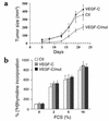
Blockade of proVEGF-C processing inhibits in vivo tumor cell growth. (a) Populations of control CHO cells (Ctl) or CHO cells expressing wild-type (VEGF-C) or mutant VEGF-C (VEGF-C/mut) were injected subcutaneously into 4-week-old male nude mice. Tumor size was measured every 3 days. (b) Starved populations of control CHO cells or CHO cells expressing wild-type (VEGF-C) or mutant VEGF-C (VEGF-C/mut) were incubated for 24 hours in medium containing increasing concentrations of serum (0–10% FCS). [3H]thymidine was added for the final 6 hours of incubation. [3H]thymidine incorporation was measured as described previously (9). Data are presented as mean ± SE of four experiments.
Figure 7
Figure 7
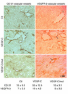
Immunohistochemical analyses. Tumors that developed 20 days after subcutaneous injection into nude mice of CHO cells stably transfected with empty vector (Ctl), vector containing wild-type or mutant proVEGF-C cDNA were analyzed for angiogenesis and lymphangiogenesis using an anti-mouse CD-31 monoclonal antibody and an anti–VEG-FR-C polyclonal antibody, respectively. The sections were observed at a magnification of ×200. Arrows indicate vessels. The number of intratumoral vascular and lymphatic vessels was counted in ten different fields for each tumor. Data are presented as mean ± SD.

References

    1. Joukov V, et al. A novel vascular endothelial growth factor, VEGF-C, is a ligand for the Flt4 (VEGFR-3) and KDR (VEGFR-2) receptor tyrosine kinases. [erratum 1996, 15:1751] EMBO J. 1996;15:290–298. - PMC - PubMed
    1. Huang HY, Ho CC, Huang PH, Hsu SM. Co-expression of VEGF-C and its receptors, VEGFR-2 and VEGFR-3, in endothelial cells of lymphangioma. Implication in autocrine or paracrine regulation of lymphangioma. Lab. Invest. 2001;81:1729–1734. - "V体育安卓版" PubMed
    1. Karkkainen MJ, Makinen T, Alitalo K. Lymphatic endothelium: a new frontier of metastasis research. Nat. Cell Biol. 2002;4:E2–E5. - PubMed
    1. Joukov V, et al. Proteolytic processing regulates receptor specificity and activity of VEGF-C. EMBO J. 1997;16:3898–3911. - PMC - PubMed
    1. Seidah NG, Chretien M. Proprotein and prohormone convertases: a family of subtilases generating diverse bioactive polypeptides. Brain Res. 1999;848:45–62. - PubMed

V体育ios版 - Publication types

MeSH terms