"VSports app下载" Skip to main page content
U.S. flag

An official website of the United States government

Dot gov

The . gov means it’s official. Federal government websites often end in . gov or . mil. Before sharing sensitive information, make sure you’re on a federal government site VSports app下载. .

Https

The site is secure V体育官网. The https:// ensures that you are connecting to the official website and that any information you provide is encrypted and transmitted securely. .

Comparative Study
. 2001 Sep 25;98(20):11598-603.
doi: 10.1073/pnas.181181198. Epub 2001 Aug 14.

"V体育官网入口" A phosphatidylinositol 3-kinase/Akt pathway promotes translocation of Mdm2 from the cytoplasm to the nucleus

Affiliations
Comparative Study

A phosphatidylinositol 3-kinase/Akt pathway promotes translocation of Mdm2 from the cytoplasm to the nucleus

"V体育安卓版" L D Mayo et al. Proc Natl Acad Sci U S A. .

Abstract

The Mdm2 oncoprotein promotes cell survival and cell cycle progression by inhibiting the p53 tumor suppressor protein. To regulate p53, Mdm2 must gain nuclear entry, and the mechanism that induces this is now identified. Mitogen-induced activation of phosphatidylinositol 3-kinase (PI3-kinase) and its downstream target, the Akt/PKB serine-threonine kinase, results in phosphorylation of Mdm2 on serine 166 and serine 186. Phosphorylation on these sites is necessary for translocation of Mdm2 from the cytoplasm into the nucleus. Pharmacological blockade of PI3-kinase/Akt signaling or expression of dominant-negative PI3-kinase or Akt inhibits nuclear entry of Mdm2, increases cellular levels of p53, and augments p53 transcriptional activity VSports手机版. Expression of constitutively active Akt promotes nuclear entry of Mdm2, diminishes cellular levels of p53, and decreases p53 transcriptional activity. Mutation of the Akt phosphorylation sites in Mdm2 produces a mutant protein that is unable to enter the nucleus and increases p53 activity. The demonstration that PI3-kinase/Akt signaling affects Mdm2 localization provides insight into how this pathway, which is inappropriately activated in many malignancies, affects the function of p53. .


Figures
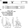
Figure 1
Figure 1
Akt phosphorylation sites in Mdm2. (a) Schematic of the Akt phosphorylation sites (bold) containing serine 166 and 186 in human Mdm2 and their relationship to the domains in other species. (b) In vitro phosphorylation of human Mdm2 and 2xSA by recombinant activated Akt (raAkt). One microgram of each recombinant protein was incubated with 150 ng of raAkt and 12 μCi (1 Ci = 37 GBq) γ 32P ATP for 30 min at 30°C and a Western blot was prepared. Autoradiography detected phosphorylation of Mdm2 (Top). The blot was probed with anti-Mdm2 (IF-2; Middle) and then anti-Akt (Bottom) to show the presence of equal amounts of each protein. (c) Calculated target ions for serine 166 (mass 1,001) and serine 186 (mass 1,018) from His-Mdm2 incubated in vitro with activated Akt were isolated by mass spectrometry using a quadrapole ion trap with a width of 3 m/z. The high-resolution scan is shown.
Figure 2
Figure 2
Phosphorylation of Mdm2. (a) Western blot analysis of insulin receptor (control, 2 μg), Akt, and Mdm2 (2 μg) immunoprecipitated from serum-starved MCF-7 cells probed with anti-Mdm2 (IF-2) or anti-Akt. (b) Serum-starved MCF-7 cells were stimulated with 50 ng/ml IGF-1 and a Western blot was probed with anti-phospho (activated)-Akt (Upper). The blot was stripped and reprobed with anti-Akt (Lower). (c) Serum-starved MCF-7 cells were treated with medium or LY294002 for 30 min and then IGF-1 for 30 min. Western blots were probed with anti-phospho Akt (Upper) or anti-Akt (Lower). (d) MCF-7 cells were stimulated with insulin before immunoprecipitation of Mdm2. A Western blot was probed with anti-Akt and anti-Mdm2. (e) Recombinant Mdm2 or 2xSA was incubated with GST or GST-Akt-agarose for 30 min in vitro, centrifuged, and washed three times. Vehicle or raAkt was incubated with the agarose complexes for 30 min at 37°C and these were centrifuged and washed three times. A Western blot probed with anti-Mdm2 or anti-Akt quantitated the amount of Mdm2 or 2xSA that had associated with GST-Akt agarose. (f) Mdm2 immunoprecipitated from MCF-7 cells incubated with vehicle or IGF-1 was analyzed by mass spectrometry. Predicted parent ions for unphosphorylated serine 166 and serine 186 were recovered from the control incubates (Left), whereas parent ions predicted for phosphorylated serine 166 and serine 186 were recovered from cells treated with IGF-1 (Right).
Figure 3
Figure 3
Localization of Mdm2 characterized by confocal microscopy. (a) Endogenous Mdm2 in serum-starved MCF-7 cells transfected with constitutively active Akt (CAAkt) or incubated in the absence or presence of LY294002 and then treated with IGF-1 (5 min). (b) HEK 293 cells transiently transfected with human Flag-Mdm2 and Flag-Mdm2, then treated with wortmannin (1 h), Flag-Mdm2 and KD-Akt, Flag-Mdm2 and Δp85 PI3-kinase, and Flag-Mdm2 and KD-Akt, and then treated with IGF-1. (c) Primary, serum-starved human keratinocytes were treated with growth factor-free medium (control) or medium containing IGF-1. Keratinocytes were also transfected with Flag-Mdm2, Flag-Mdm2, and KD-Akt, or treated with LY294002 (1 h). (d) HEK 293 cells transfected with Flag-Mdm2, Flag-S166A, Flag-186A, and Flag-2xSA. Additionally, cells were cotransfected with Flag-2xSA and CA-Akt or were transfected with Flag-2xSA and then treated with IGF-1.
Figure 4
Figure 4
Regulation of p53 by Akt. (a) MCF-7 cells were cultured in the absence or presence of serum for 24 h and then incubated in the absence or presence of LY294002 for 1 h before treatment with medium or IGF-1 (30 min). A Western blot was then probed for p53 expression. (b) Saos-2 cells were transiently transfected with pHook, p53 in the absence or presence of Mdm2, or Mdm2 with CA-Akt. After 24 h, p53 expression relative to pHook expression was determined by Western blotting. (c) Saos-2 cells were transiently transfected with pHook and p53 alone, or in combination with Mdm2 or mutants of Mdm2 (S166A, S186A, 2xSA). p53 was detected by probing a Western blot with anti-p53. (d) Serum-starved keratinocytes were incubated with IGF-1 for various times. Western blot analysis assayed the effect of such treatment on p53 and p21 protein levels. (e) MCF-7 cells (Left) were transiently transfected with 1 μg β-gal, 2 μg of the p53 luc reporter plasmid, and 7 μg of CA-Akt or KD-Akt. HEK 293 cells were transfected with 0.3 μg β-gal, 1 μg of the mutant or functional mdm2luc reporter plasmid, 1 μg p53, and 1 μg p53 and 5 μg Mdm2 in the absence or presence of 2.4 μg CA-Akt or KD-Akt. After 24 h, β-gal and luciferase activities were measured. Transfections were conducted in triplicate and two aliquots from each cell population were used for β-gal and luciferase assays. Relative luciferase activity was calculated from three independent experiments, normalized to β-gal expression, and standard deviation is derived from the mean. (f) MCF-7 (Left) or HEK 293 (Right) cells were transiently transfected with the mdm2luc reporter, the β-gal expression plasmid, and empty vector (control) or 1 μg of p53 alone, with 5 μg of Mdm2, or with 2xSA Mdm2. Relative luciferase activity was calculated and fold induction is reported relative to control.

Comment in

V体育2025版 - References

    1. Agarwal M L, Taylor W R, Chernov M V, Chernova O B, Stark G R. J Biol Chem. 1997;273:1–4. - PubMed (V体育官网)
    1. Bates S, Vousden K H. Cell Mol Life Sci. 1999;55:28–37. - PMC - PubMed
    1. Levine A J. Cell. 1997;88:323–331. - PubMed
    1. Donehower L A, Harvey M, Slagle B L, McArthur M J, Montgomery C A, Jr, Butel J S, Bradley A. Nature (London) 1992;356:215–221. - PubMed
    1. Harvey M, McArthur M J, Montgomery C A, Jr, Butel J S, Bradley A, Donehower L A. Nat Genet. 1993;5:225–229. - PubMed

Publication types

MeSH terms

Substances