Skip to main page content
U.S. flag

An official website of the United States government

Dot gov

The . gov means it’s official. Federal government websites often end in . gov or . mil. Before sharing sensitive information, make sure you’re on a federal government site. VSports app下载.

Https

The site is secure V体育官网. The https:// ensures that you are connecting to the official website and that any information you provide is encrypted and transmitted securely. .

. 1998 Jul 20;188(2):287-96.
doi: 10.1084/jem.188.2.287.

CD4+CD25+ immunoregulatory T cells suppress polyclonal T cell activation in vitro by inhibiting interleukin 2 production

Affiliations

CD4+CD25+ immunoregulatory T cells suppress polyclonal T cell activation in vitro by inhibiting interleukin 2 production

A M Thornton et al. J Exp Med. .

Abstract (V体育ios版)

Peripheral tolerance may be maintained by a population of regulatory/suppressor T cells that prevent the activation of autoreactive T cells recognizing tissue-specific antigens. We have previously shown that CD4+CD25+ T cells represent a unique population of suppressor T cells that can prevent both the initiation of organ-specific autoimmune disease after day 3 thymectomy and the effector function of cloned autoantigen-specific CD4+ T cells. To analyze the mechanism of action of these cells, we established an in vitro model system that mimics the function of these cells in vivo. Purified CD4+CD25+ cells failed to proliferate after stimulation with interleukin (IL)-2 alone or stimulation through the T cell receptor (TCR). When cocultured with CD4+CD25- cells, the CD4+CD25+ cells markedly suppressed proliferation by specifically inhibiting the production of IL-2 VSports手机版. The inhibition was not cytokine mediated, was dependent on cell contact between the regulatory cells and the responders, and required activation of the suppressors via the TCR. Inhibition could be overcome by the addition to the cultures of IL-2 or anti-CD28, suggesting that the CD4+CD25+ cells may function by blocking the delivery of a costimulatory signal. Induction of CD25 expression on CD25- T cells in vitro or in vivo did not result in the generation of suppressor activity. Collectively, these data support the concept that the CD4+CD25+ T cells in normal mice may represent a distinct lineage of "professional" suppressor cells. .


V体育官网入口 - Figures
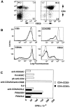
Figure 1
Figure 1
Characterization of CD4+CD25+ cells. (A) Total lymph node cells (left) or purified CD4+CD25+ cells (right) from BALB/c mice were stained with PE-conjugated anti-CD4 and biotin-conjugated anti-CD25 followed by FITC-conjugated streptavidin. (B) Purified CD4+ CD25 cells or purified CD4+CD25+ cells were double stained for CD25 and the indicated surface molecules. (C) Proliferative responses of CD4+ CD25+ cells. Cells (5 × 10⁴) were incubated with the indicated agents (10 ng/ml PMA, 1 mM ionomycin, 3 ng/ml IL-2, 3.0 μg/ml soluble anti-CD3, 3.0 μg/ml Con A, 10 μg/ml anti-CD28, or wells coated with 10 μg/ml anti-CD3) in the presence of AC (5 × 10⁴). Results are expressed as the mean of triplicate cultures.
Figure 2
Figure 2
CD4+CD25+ cells suppress the proliferation of CD4+ cells. (A) CD4+CD25 cells (5 × 10⁴) were incubated with plate bound anti-CD3 (triangles) or with 1.0 μg/ml soluble anti-CD3 (squares) in the presence of AC (5 × 10⁴) and the indicated number of CD4+CD25+ cells. (B) CD4+CD25+ cells do not suppress antigen-specific CD4+ cells from TCR transgenic mice. CD4+CD25 cells (5 × 10⁴) were purified from DO.11.10 SCID mice on a BALB/c background and stimulated with 0.5 μM ovalbumin peptide (amino acid 323–339; triangles) or 0.5 μg/ml anti-CD3 (squares) in the presence of AC (5 × 10⁴) and the indicated number of CD4+CD25+ cells. Results are expressed as the mean of triplicate cultures.
Figure 3
Figure 3
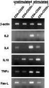
PCR analysis of CD4+CD25+ cells. RNA was purified from 6–8 × 10⁶ CD4+CD25 or CD4+CD25+ cells either unstimulated or stimulated with an equivalent number of AC and 0.5 μg/ml anti-CD3 for 15 h. RNA was reverse transcribed and primers for the indicated genes were used to amplify the cDNA. The number of cycles for each primer set are as follows: β-actin, 20 cycles; IL-2, 25 cycles; IL-4, IL-10, TNF-α, and FasL, 30 cycles.
Figure 4
Figure 4
A soluble factor does not mediate CD4+CD25+ induced suppression. (A) CD4+CD25 cells (5 × 10⁵) were cultured in 24-well plates in the presence of 3.0 μg/ml soluble anti-CD3 and AC (5 × 10⁵). The indicated number of CD4+CD25+ cells was added directly to the culture (squares) or to the transwell in the absence (triangles) or presence (circles) of AC (5 × 10⁵). (B) CD4+CD25 cells (5 × 10⁴) were cultured with AC (5 × 10⁴), 0.5 μg/ ml anti-CD3, and 10 μg/ml of the indicated antibodies in the absence (white bars) or presence (black bars) of CD4+ CD25+ cells (2.5 × 10⁴). (C) Supernatants were collected after a 48-h stimulation with soluble anti-CD3 and AC (5 × 10⁴) from CD4+CD25 cells alone (5 × 10⁴), CD4+CD25+ cells alone (5 × 10⁴), or CD4+CD25 (5 × 10⁴) cells coincubated with CD4+CD25+ cells (2.5 × 10⁴). Supernatants (0.1 ml) were then added to CD4+ cells (5 × 10⁴) stimulated with anti-CD3 and AC (5 × 10⁴). Results are expressed as the mean of triplicate cultures.
Figure 4
Figure 4
A soluble factor does not mediate CD4+CD25+ induced suppression. (A) CD4+CD25 cells (5 × 10⁵) were cultured in 24-well plates in the presence of 3.0 μg/ml soluble anti-CD3 and AC (5 × 10⁵). The indicated number of CD4+CD25+ cells was added directly to the culture (squares) or to the transwell in the absence (triangles) or presence (circles) of AC (5 × 10⁵). (B) CD4+CD25 cells (5 × 10⁴) were cultured with AC (5 × 10⁴), 0.5 μg/ ml anti-CD3, and 10 μg/ml of the indicated antibodies in the absence (white bars) or presence (black bars) of CD4+ CD25+ cells (2.5 × 10⁴). (C) Supernatants were collected after a 48-h stimulation with soluble anti-CD3 and AC (5 × 10⁴) from CD4+CD25 cells alone (5 × 10⁴), CD4+CD25+ cells alone (5 × 10⁴), or CD4+CD25 (5 × 10⁴) cells coincubated with CD4+CD25+ cells (2.5 × 10⁴). Supernatants (0.1 ml) were then added to CD4+ cells (5 × 10⁴) stimulated with anti-CD3 and AC (5 × 10⁴). Results are expressed as the mean of triplicate cultures.
Figure 5
Figure 5
CD4+CD25+ cells from IL-10−/− and IL-4−/− mice suppress proliferation. CD4+CD25+ cells were purified from (A) IL-4−/− and BALB/c control mice or (B) IL-10−/− and C57BL/10 mice, and the indicated numbers were coincubated with CD4+CD25 cells (5 × 10⁴) from control mice stimulated with 0.5 μg/ml anti-CD3 and AC (5 × 10⁴). Results are expressed as the mean of triplicate cultures.
Figure 6
Figure 6
Abrogation of suppression by IL-2 or anti-CD28. CD4+ CD25 cells (5 × 10⁴) were cultured in the presence or absence of CD4+CD25+ cells (2.5 × 10⁴) and 10 μg/ml of the indicated antibodies or 3 ng/ml IL-2. Results are expressed as the mean of triplicate cultures.
Figure 7
Figure 7
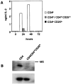
CD4+CD25+ cells suppress IL-2 production. (A) CD4+ CD25 cells (5 × 10⁴), AC (5 × 10⁴), and 0.5 μg/ml anti-CD3 were cultured in the presence or absence of CD4+CD25+ cells (2.5 × 10⁴), supernatants were taken at the indicated times and IL-2 was quantified. Each sample was tested in triplicate. (B) RNA was purified from CD4+ cells stimulated in the presence or absence of CD4+CD25+ cells after 16 h using RNAzol B (Tel-test). RNA was separated by gel electrophoresis, transferred to nitrocellulose and probed for IL-2 message (top) or β-actin message (bottom) by using an IL-2 PCR fragment or a β-actin PCR product as a probe.
Figure 8
Figure 8
(A) CD4+CD25+ cells were purified from BALB/c mice while CD4+CD25 cells were obtained by cell sorting and were cultured with AC and Con A for 24 h and were then used as induced CD4+CD25+ cells. Induced or control CD4+CD25+ cells were then coincubated with 0.5 μg/ml anti-CD3 and AC. (B) CD4+CD25+ cells were purified from 3dTx mice or BALB/c control mice and coincubated with CD4+CD25 cells from control mice stimulated with 0.5 μg/ml anti-CD3 and AC. Results are expressed as the mean of triplicate cultures.
Figure 8
Figure 8
(A) CD4+CD25+ cells were purified from BALB/c mice while CD4+CD25 cells were obtained by cell sorting and were cultured with AC and Con A for 24 h and were then used as induced CD4+CD25+ cells. Induced or control CD4+CD25+ cells were then coincubated with 0.5 μg/ml anti-CD3 and AC. (B) CD4+CD25+ cells were purified from 3dTx mice or BALB/c control mice and coincubated with CD4+CD25 cells from control mice stimulated with 0.5 μg/ml anti-CD3 and AC. Results are expressed as the mean of triplicate cultures.

References

    1. Schwartz RH. A cell culture model for T lymphocyte clonal anergy. Science. 1990;248:1349–1356. - PubMed
    1. Miller JFAP, Heath WR. Self-ignorance in the peripheral T-cell pool. Immunol Rev. 1993;133:131–150. - PubMed
    1. Wucherpfennig KW, Strominger JL. Molecular mimicry in T cell–mediated autoimmunity: viral peptides activate human T cell clones specific for myelin basic protein. Cell. 1995;80:695–705. - PMC - PubMed
    1. Möller, G., editor. 1996. Dominant immunological tolerance. Immunol. Rev. 149:1–243.
    1. Fowell D, Mason D. Evidence that the T cell repertoire of normal rats contains cells with the potential to cause diabetes. Characterization of the CD4+T cell subset that inhibits this autoimmune potential. J Exp Med. 1993;177:627–636. - PMC (V体育官网) - PubMed