Skip to main page content (V体育官网)
U.S. flag

An official website of the United States government

Dot gov

The . gov means it’s official. Federal government websites often end in . gov or . mil VSports app下载. Before sharing sensitive information, make sure you’re on a federal government site. .

Https

The site is secure. The https:// ensures that you are connecting to the official website and that any information you provide is encrypted and transmitted securely. V体育官网.

. 1998 Jun 29;141(7):1539-50.
doi: 10.1083/jcb.141.7.1539.

Claudin-1 and -2: novel integral membrane proteins localizing at tight junctions with no sequence similarity to occludin

Affiliations

Claudin-1 and -2: novel integral membrane proteins localizing at tight junctions with no sequence similarity to occludin

M Furuse et al. J Cell Biol. .

Abstract

Occludin is the only known integral membrane protein localizing at tight junctions (TJ), but recent targeted disruption analysis of the occludin gene indicated the existence of as yet unidentified integral membrane proteins in TJ. We therefore re-examined the isolated junction fraction from chicken liver, from which occludin was first identified. Among numerous components of this fraction, only a broad silver-stained band approximately 22 kD was detected with the occludin band through 4 M guanidine-HCl extraction as well as sonication followed by stepwise sucrose density gradient centrifugation. Two distinct peptide sequences were obtained from the lower and upper halves of the broad band, and similarity searches of databases allowed us to isolate two full-length cDNAs encoding related mouse 22-kD proteins consisting of 211 and 230 amino acids, respectively. Hydrophilicity analysis suggested that both bore four transmembrane domains, although they did not show any sequence similarity to occludin. Immunofluorescence and immunoelectron microscopy revealed that both proteins tagged with FLAG or GFP were targeted to and incorporated into the TJ strand itself. We designated them as "claudin-1" and "claudin-2", respectively. Although the precise structure/function relationship of the claudins to TJ still remains elusive, these findings indicated that multiple integral membrane proteins with four putative transmembrane domains, occludin and claudins, constitute TJ strands VSports手机版. .


Figures

Figure 1
Figure 1
Identification of the occludin band and nine guanidine-insoluble bands in the isolated junction fraction from chick liver. (a) Immunoabsorption of occludin from the isolated junction fraction. The isolated junction fraction was solubilized with 0.1% SDS. After dilution, immunoprecipitation was carried out with rabbit anti–chicken occludin pAb (F44) or control rabbit serum, and then the supernatant was processed for SDS-PAGE. In silver-stained gels, a 60-kD band (arrow) that was detected in the non-treated (Junction Fr.) as well as control serum-treated (+control IgG) fraction, disappeared in the F44-treated fraction (+anti-Oc pAb). Thus, occludin was identified as a 60-kD band in the junction fraction by silver staining. Accompanying immunoblots with anti-occludin pAb (F44) confirmed this notion. (b) Guanidine extraction of isolated junction fraction. To thoroughly extract peripheral membrane proteins from isolated junctions, the fraction was treated with 4 M guanidine-HCl. The silver-stained banding pattern was compared between non-treated (Junction Fr.) and guanidine-treated (4 M guanidine) samples. Occludin in the non-treated fraction (arrow) was concentrated by guanidine treatment (occludin). In addition to the occludin band, nine guanidine-insoluble bands were identified (bands 1–9), the amounts of which were similar to or greater than that of occludin. Bars indicate molecular masses of 200, 116, 97, 66, 45, 31, and 21 kD, respectively, from the top.
Figure 3
Figure 3
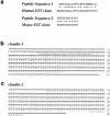
Cloning and sequencing of cDNAs encoding claudin-1 and -2. (a) Peptide sequencing. The lower and upper halves of band 9 (see Fig. 1 b) were separately subjected to the direct peptide sequencing, and yielded peptide sequence-1 and -2, respectively. Database searching identified human and mouse EST clones (these sequence data are available from GenBank/ EMBL/DDBJ under accession numbers AA305424 and AA116709, respectively), which encoded sequences showing significant similarity to peptide sequence-1 and -2, respectively. Identity and homology are indicated by asterisks and dots, respectively. (b and c) Nucleotide and deduced amino acid sequences of mouse claudin-1 and -2. Based on the above peptide sequences, two distinct cDNAs were isolated from a mouse liver cDNA library (for details, see Materials and Methods). Products encoded by these cDNAs were designated as claudin-1 and -2. Sequences homologous to peptide sequence-1 and -2 (a) are underlined. Since peptide sequence-1 was determined as the NH2-terminal sequence (for details, see Materials and Methods), the first ATG codon was conclusively determined in claudin-1 cDNA. Sequence similarity between claudin-1 and -2 (see Fig. 4 a) then enabled us to estimate the first ATG codon in claudin-2 cDNA. Claudin-1 and -2 cDNAs encoded 211– and 230– amino acid polypeptides with predicted molecular masses of 22.9 kD and 24.5 kD, respectively.
Figure 4
Figure 4
Structures of mouse claudin-1 and -2. (a) Comparison of amino acid sequences of mouse claudin-1 and -2 by the GENETYX program. Identity and homology are indicated by asterisks and dots, respectively. They showed 38% identity at the amino acid sequence level. (b) Hydrophilicity plots for claudin-1 and -2 prepared using the Kyte and Doolittle algorithm. The plot records the average hydrophilicity along the sequence over a window of eight residues. Hydrophilic and hydrophobic residues are in the lower and upper parts of the frames, respectively. The axes are numbered in amino acid residues. Both claudin-1 and -2 appeared to contain four major hydrophobic, potentially membrane-spanning regions (arrows), although the possibility was not excluded that five transmembrane domains were included in these molecules only from these plots as discussed in the text.
Figure 4
Figure 4
Structures of mouse claudin-1 and -2. (a) Comparison of amino acid sequences of mouse claudin-1 and -2 by the GENETYX program. Identity and homology are indicated by asterisks and dots, respectively. They showed 38% identity at the amino acid sequence level. (b) Hydrophilicity plots for claudin-1 and -2 prepared using the Kyte and Doolittle algorithm. The plot records the average hydrophilicity along the sequence over a window of eight residues. Hydrophilic and hydrophobic residues are in the lower and upper parts of the frames, respectively. The axes are numbered in amino acid residues. Both claudin-1 and -2 appeared to contain four major hydrophobic, potentially membrane-spanning regions (arrows), although the possibility was not excluded that five transmembrane domains were included in these molecules only from these plots as discussed in the text.
Figure 2
Figure 2
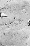
Behavior of the occludin and nine guanidine- insoluble bands on sonication followed by sucrose density gradient centrifugation. (a and b) Ultrathin sectional electron microscopic images of non-treated (a) and sonicated (b) junction fraction fixed in the presence of 0.1% tannic acid. The non-treated fraction was characterized by numerous actin filaments (*) and isolated junctions (arrow), which contained typical tight junctions (inset). After sonication followed by centrifugation, no actin filaments were detected, and isolated junctions were fragmented into small vesicular structures (arrow), some of which still contained typical tight junctions (inset). (c) Fractionation of sonicated junction fraction. After sonication, the isolated junctions were fractionated by stepwise sucrose density gradient centrifugation. 0:25%, 25:30%, 30:34%, 34:38%, 38:42%, and 42:50% interfaces were collected, and subjected to SDS-PAGE followed by silver staining. The distribution of the occludin band (occludin) was compared with those of nine guanidine-insoluble bands (band 1–9), and only band 9 was copartitioned with occludin. Silver staining and accompanying immunoblots with anti-occludin mAb (Oc-1) revealed that occludin was mainly recovered at 25:30%, 30:34%, and 34:38% interfaces, where band 9 was also characteristically accumulated. Bars in c indicate molecular masses of 200, 116, 97, 66, 45, 31, and 21 kD, respectively, from the top. Bar, 0.2 μm.
Figure 2
Figure 2
Behavior of the occludin and nine guanidine- insoluble bands on sonication followed by sucrose density gradient centrifugation. (a and b) Ultrathin sectional electron microscopic images of non-treated (a) and sonicated (b) junction fraction fixed in the presence of 0.1% tannic acid. The non-treated fraction was characterized by numerous actin filaments (*) and isolated junctions (arrow), which contained typical tight junctions (inset). After sonication followed by centrifugation, no actin filaments were detected, and isolated junctions were fragmented into small vesicular structures (arrow), some of which still contained typical tight junctions (inset). (c) Fractionation of sonicated junction fraction. After sonication, the isolated junctions were fractionated by stepwise sucrose density gradient centrifugation. 0:25%, 25:30%, 30:34%, 34:38%, 38:42%, and 42:50% interfaces were collected, and subjected to SDS-PAGE followed by silver staining. The distribution of the occludin band (occludin) was compared with those of nine guanidine-insoluble bands (band 1–9), and only band 9 was copartitioned with occludin. Silver staining and accompanying immunoblots with anti-occludin mAb (Oc-1) revealed that occludin was mainly recovered at 25:30%, 30:34%, and 34:38% interfaces, where band 9 was also characteristically accumulated. Bars in c indicate molecular masses of 200, 116, 97, 66, 45, 31, and 21 kD, respectively, from the top. Bar, 0.2 μm.
Figure 5
Figure 5
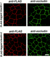
Colocalization of FLAG-tagged claudin-1 and -2 with occludin in MDCK transfectants. Confluent cultures of MDCK transfectants expressing FLAG–claudin-1 or FLAG–claudin-2 (FLAG-tagged claudin-1 or FLAG-tagged claudin-2) were doubly stained with mouse anti-FLAG mAb (anti-FLAG) and rat anti-occludin mAb MOC37 (anti-occludin). Images were obtained at the focal plane of the most apical region of lateral membranes by confocal microscopy. Both FLAG–claudin-1 and FLAG– claudin-2 were precisely colocalized with occludin at tight junction regions. Bar, 10 μm.
Figure 6
Figure 6
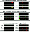
Comparison of the subcellular distributions of claudin-1 and -2 with those of occludin and E-cadherin in MDCK transfectants. Confluent sheets of MDCK transfectants expressing FLAG-tagged claudin-1 or FLAG-tagged claudin-2 were doubly stained with mouse anti-FLAG mAb (M2)/rat anti-occludin mAb (MOC37) or mouse anti-FLAG mAb (M2)/rat anti– E-cadherin mAb (ECCD2). For each sample, 22 optical sections were recorded with 0.5-μm intervals by confocal microscopy, and cross-sectional views were generated. The thickness of each cellular sheet is indicated by arrows. In computer-generated cross-sectional views, FLAG–claudin-1, FLAG– claudin-2 (anti-FLAG), and occludin (anti-occludin) were highly concentrated at the most apical portion of lateral membranes of MDCK cells, and overlaid images showed that FLAG–claudin-1 and FLAG–claudin-2 were precisely colocalized with occludin (composite). In contrast, E-cadherin was distributed along lateral membranes (anti-E-cadherin), and its distribution was more basally than those of FLAG–claudin-1 or FLAG–claudin-2 (composite). The same spatial relationships of claudin-1/occludin and claudin-1/E-cadherin were observed in MDCK transfectants expressing GFP-tagged claudin-1, indicating that tag-peptides did not affect the subcellular distributions of claudins.
Figure 7
Figure 7
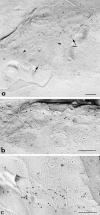
Immunolabeling of freeze-fracture replicas of MDCK transfectants expressing FLAG- (a and b) or GFP-tagged claudin-1 (c) with anti-FLAG mAb or anti-GFP pAb. TJ regions (arrows), but not other membrane domains such as desmosomes (arrowheads in a; asterisks in b and c), were specifically labeled with immunogold particles. Taking the intrinsic resolution of immunogold labeling (20–25 nm) into consideration, most of the immunogold particles appeared to directly label the TJ strands themselves, indicating that claudin-1 is incorporated into TJ strands. Bars, 0.1 μm.
Figure 8
Figure 8
Immunolabeling of freeze-fracture replicas of MDCK transfectants expressing FLAG-tagged claudin-2 with anti-FLAG mAb. TJ regions, but not other membrane domains such as desmosomes (asterisks in b), were specifically labeled with immunogold particles. Taking the intrinsic resolution of immunogold labeling (20–25 nm) into consideration, most of the immunogold particles appeared to directly label the TJ strands themselves, indicating that claudin-2 is incorporated into TJ strands. Bars, 0.1 μm.
Figure 9
Figure 9
Northern blots of mouse claudin-1 and -2 expression. Mouse Multiple Tissue Northern Blot (Clontech) was probed with claudin-1 or claudin-2 cDNA fragments. Claudin-1 mRNAs were detected as major 4.0 kb and minor 1.3 kb bands in all the tissues examined, and were seen in especially large amounts in the liver and kidney. Claudin-2 expression (major 4.0 kb and minor 2.0 kb mRNAs) was restricted to the liver and kidney (with small amounts in the brain).

References

    1. Anderson JM, Fanning AS, Lapierre L, Van Itallie CM. Zonula occludens (ZO)-1 and ZO-2: membrane-associated guanylate kinase homologues (MAGuKs) of the tight junction. Biochem Soc Trans. 1995;23:470–475. - PubMed
    1. Anderson JM, Van Itallie CM. Tight junctions and the molecular basis for regulation of paracellular permeability. Am J Physiol. 1995;269:467–475. - PubMed
    1. Ando-Akatsuka Y, Saitou M, Hirase T, Kishi M, Sakakibara A, Itoh M, Yonemura S, Furuse M, Tsukita Sh. Interspecies diversity of the occludin sequence: cDNA cloning of human, mouse, dog, and rat-kangaroo homologues. J Cell Biol. 1996;133:43–47. - PMC - PubMed
    1. Balda MS, Whitney JA, Flores C, González S, Cereijido M, Matter K. Functional dissociation of paracellular permeability and transepithelial electrical resistance and disruption of the apical-basolateral intramembrane diffusion barrier by expression of a mutant tight junction membrane protein. J Cell Biol. 1996;134:1031–1049. - PMC - PubMed
    1. Blashcuk OW, Manteuffel RL, Steinberg MS. Purification of desmoglein II: a method for the preparation and fractionation of desmosomal components. Biochim Biophys Acta. 1986;833:426–431. - PubMed

Publication types

MeSH terms

Substances

Associated data