Skip to main page content
U.S. flag

An official website of the United States government

Dot gov

The VSports app下载. gov means it’s official. Federal government websites often end in . gov or . mil. Before sharing sensitive information, make sure you’re on a federal government site. .

Https

The site is secure. The https:// ensures that you are connecting to the official website and that any information you provide is encrypted and transmitted securely. V体育官网.

. 1997 Sep 30;94(20):10705-10.
doi: 10.1073/pnas.94.20.10705.

Cell-cell interaction in prostate gene regulation and cytodifferentiation

Affiliations

V体育安卓版 - Cell-cell interaction in prostate gene regulation and cytodifferentiation

A Y Liu et al. Proc Natl Acad Sci U S A. .

"V体育ios版" Abstract

To examine the role of intercellular interaction on cell differentiation and gene expression in human prostate, we separated the two major epithelial cell populations and studied them in isolation and in combination with stromal cells. The epithelial cells were separated by flow cytometry using antibodies against differentially expressed cell-surface markers CD44 and CD57. Basal epithelial cells express CD44, and luminal epithelial cells express CD57. The CD57+ luminal cells are the terminally differentiated secretory cells of the gland that synthesize prostate-specific antigen (PSA). Expression of PSA is regulated by androgen, and PSA mRNA is one of the abundant messages in these cells VSports手机版. We show that PSA expression by the CD57+ cells is abolished after prostate tissue is dispersed by collagenase into single cells. Expression is restored when CD57+ cells are reconstituted with stromal cells. The CD44+ basal cells possess characteristics of stem cells and are the candidate progenitors of luminal cells. Differentiation, as reflected by PSA production, can be detected when CD44+ cells are cocultured with stromal cells. Our studies show that cell-cell interaction plays an important role in prostatic cytodifferentiation and the maintenance of the differentiated state. .


Figures

Figure 1
Figure 1
Flow analysis of cultured cells. Mixture of LNCaP and PC3 cells sorted by αCD44-PE. The left diagram of each top three panel shows scatter plots of fluorescence vs. forward light scatter (FLS) intensity. The right diagram displays the histogram along the fluorescence axis. Fluorescence is expressed in arbitrary units that are proportional to the logarithm of the fluorescence intensity. The cells used are identified for each panel. The bottom panel shows a sort of 103 PC3 cells from 106 LNCaP cells. The positive cells are circled and are represented by 45 events among the 52,216 total events (0.08%) scored in this dot plot. PLS is right-angle light scatter.
Figure 2
Figure 2
Tissue specificity of antibodies and flow analysis of prostate cells. Immunohistochemistry of αCD44 (A) and αCD57 (B). The former stains basal epithelial cells and the latter luminal epithelial cells. (C) Flow cytometry of prostate cells prepared from specimens HM and KM. KM cells were cultured overnight prior to flow analysis. Autofluorescence, along the diagonal, was observed when no antibody was used. It overlaps to some extent the FITC population. The PE-positive population is well separated. The circled populations were sorted.
Figure 3
Figure 3
Gene-expression analysis of sorted CD44+ epithelial cells. The single cells were stained with αCD44-PE and αCD57-FITC, followed by a 30-min incubation on ice with 7-aminoactinomycin D. A total of 300,000 CD44+ and 160,000 CD57+ events were sorted. About 25% of the unstained population was 7-aminoactinomycin D positive. The CD44+ cells were homogenized and RNA prepared for gene-expression analysis. The selected genes are identified above the ethidium bromide-stained gel containing PCR products resolved by agarose gel electrophoresis. DNA size markers in kb to the left are from λHindIII. Expected PCR product sizes are as follows: CD44, 600 bp, PSA, 460 bp, EGP, 890 bp, ETS2, 850 bp, Gsα, 980 bp, PSP94, 430 bp, PAP, 670 bp, Znα2GP, 590 bp, FNRβ, 1210 bp, and COL, 750 bp. The stained material in the PSA and PAP lanes are nonspecific products <200 bp.
Figure 4
Figure 4
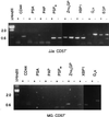
Gene-expression analysis of sorted CD57+ epithelial cells. Sorted cells from specimens JJa and MG are shown. The epithelial fraction after collagenase digestion was cultured overnight and sorted with αCD57-PE the next day into CD57+ and CD57 populations. A total of 740,000 CD57+ and 680,000 CD57 cells from JJa; and 160,000 CD57+ and 1,050,000 CD57 cells from MG respectively were obtained. Gene markers are identified above the gel lanes; + represents CD57+ cells and − represents CD57 cells. Correct product sizes in bp are as indicated in the legend to Fig. 3; XBP1 is 570 bp. Stained material near the bottom of the gel represent small nonspecific products.
Figure 5
Figure 5
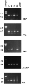
Prostate RNA stability. RT-PCR products of cDNA prepared from tissue pieces kept at room temperature for 24, 48, 72, and 96 h (indicated above the lanes) are resolved in lanes 2–5. Lane 1 contains DNA size markers. Lane 6 marked dH2O is a blank control. The expected PCR product sizes are as indicated in the legend to Fig. 3.

References

    1. Cunha G R, Donjacour A A, Cooke P S, Mee S, Bigsby R M, Higgins S J, Sugimura Y. Endocr Rev. 1987;8:338–362. - "V体育官网入口" PubMed
    1. Aumüller G. Bull Assoc Anatom. 1991;75:39–42. - "VSports" PubMed
    1. Lilja H, Abrahamsson P A. Prostate. 1988;12:29–38. - "V体育2025版" PubMed
    1. Bonkhoff H, Remberger K. Prostate. 1996;28:98–106. - PubMed
    1. Walensky L D, Coffey D S, Chen T, Wu T, Pasternack G R. Cancer Res. 1993;53:4720–4726. - "V体育安卓版" PubMed

V体育官网入口 - Publication types

MeSH terms

Substances

VSports手机版 - LinkOut - more resources