Skip to main page content
U.S. flag

An official website of the United States government

Dot gov

The . gov means it’s official VSports app下载. Federal government websites often end in . gov or . mil. Before sharing sensitive information, make sure you’re on a federal government site. .

Https

The site is secure V体育官网. The https:// ensures that you are connecting to the official website and that any information you provide is encrypted and transmitted securely. .

. 1997 Jul 15;17(14):5509-27.
doi: 10.1523/JNEUROSCI.17-14-05509.1997.

GABAergic neurons in barrel cortex show strong, whisker-dependent metabolic activation during normal behavior

Affiliations

GABAergic neurons in barrel cortex show strong, whisker-dependent metabolic activation during normal behavior (VSports最新版本)

J S McCasland et al. J Neurosci. .

Abstract

Electrophysiological data from the rodent whisker/barrel cortex indicate that GABAergic, presumed inhibitory, neurons respond more vigorously to stimulation than glutamatergic, presumed excitatory, cells. However, these data represent very small neuronal samples in restrained, anesthetized, or narcotized animals or in cortical slices. Histochemical data from primate visual cortex, stained for the mitochondrial enzyme cytochrome oxidase (CO) and for GABA, show that GABAergic neurons are more highly reactive for CO than glutamatergic cells, indicating that inhibitory neurons are chronically more active than excitatory neurons but leaving doubt about the short-term stimulus dependence of this activation. Taken together, these results suggest that highly active inhibitory neurons powerfully influence relatively inactive excitatory cells but do not demonstrate directly the relative activities of excitatory and inhibitory neurons in the cortex during normal behavior. We used a novel double-labeling technique to approach the issue of excitatory and inhibitory neuronal activation during behavior. Our technique combines high-resolution 2-deoxyglucose (2DG), immunohistochemical staining for neurotransmitter-specific antibodies, and automated image analysis to collect the data VSports手机版. We find that putative inhibitory neurons in barrel cortex of behaving animals are, on average, much more heavily 2DG-labeled than presumed excitatory cells, a pattern not seen in animals anesthetized at the time of 2DG injection. This metabolic activation is dependent specifically on sensory inputs from the whiskers, because acute trimming of most whiskers greatly reduces 2DG labeling in both cell classes in columns corresponding to trimmed whiskers. Our results provide confirmation of the active GABAergic cell hypothesis suggested by CO and single-unit data. We conclude that strong activation of inhibitory cortical neurons must confer selective advantages that compensate for its inherent energy inefficiency. .


Figures
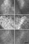
Fig. 2.
Fig. 2.
Whisker-dependent 2DG labeling of the barrel field. Computer-generated montages depict the laminar distribution of overall 2DG labeling in sections of barrel cortex stained for GAD. Montages represent 2 × 2 mm regions of sections from supragranular (top), granular (middle), and infragranular layers (bottom) of behaving animals with all whiskers present (left) or all but row C whiskers acutely trimmed (right). White or lightly shaded areas represent heavy label, whereas darker areasrepresent relatively sparse label. Note the heavier labeling and the clear barrel representation in layer IV of both cases. Barrel rows are indicated with letter in these panels. Scale bar, 500 μm.
Fig. 10.
Fig. 10.
Measures of cell populations detected by automated methods. A, Numbers of GAD+ and GAD cells detected for each neural network score. GAD+ cell numbers decline monotonically, whereas the GAD counts appear as a positively skewed normal distribution. Cells with scores of 3 and above were subject to additional analysis. B, Total GAD+ and GAD cells detected, a summary of the information in A. A total of ∼100,000 GAD+ cells and 1,000,000 GAD cells received scores of 3 or above. C, Cell densities in each lamina, by cell type. Eachbar represents the mean for 10 hemispheres (4 normal, 6 acute, row C spared), as shown in Table 1. Error bars represent SD for the 10 hemispheres. These counts show slight but not statistically significant trends toward greater densities in granular layers.
Fig. 8.
Fig. 8.
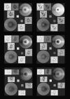
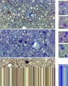
Gallery of six randomly selected GAD+ heavily 2DG-labeled neurons from the layer IV–V boundary. The cells were detected by automated template-matching algorithm and scored >3 by neural network. For each cell the five color-filtered raw image portions are reproduced (upper leftand lower right), along with small squaresindicating the strengths of two types of template correlation (center) and disks representing statistical/spatial models of the distributions of GAD stain (lower left) and 2DG silver grains (upper right). To generate the disks, we used the RS/1 function “PROBNORM-INV,” which computes the value of a normal distribution corresponding to a given probability. For each pixel in the disk, the distance from disk center was computed and normalized to a range of 1–32 to determine which concentric ring density values to apply. The angle of the pixel relative to disk center in radians was divided by π to normalize to a range of 0–1. This number served as the probability input to the PROBNORM-INV function, which returned values from approximately −3 to +3, representing the number of SD from the mean (for the computed data ring) that the pixel should represent. Then the pixel value was computed as the algebraic sum of ring mean and the product of ring SD and the value returned by the PROBNORM-INV function. In effect, the disks created by this algorithm represent the range of values expected from the computed mean and SD for all 32 data rings. Our convention in generating the 2DG disks was that pixel values representing dense 2DG grains (white) or GAD stain (dark) were plotted to the right, both above and below the axis of bilateral symmetry used for making the plot.
Fig. 9.
Fig. 9.
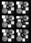
Gallery of six randomly selected GAD lightly 2DG-labeled neurons. These images, computed by the same algorithm as those in Figure 8, illustrate the generally very low 2DG labeling over GAD neuronal somata.
Fig. 11.
Fig. 11.
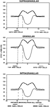
Cross-sectional profiles of 2DG labeling in GAD+ and GAD neurons. This analysis was restricted to cells from barrel rows B, C, andD in four hemispheres from normal behaving hamsters and depicts cells from supragranular (top), granular (middle), and infragranular layers (bottom). Thecurves were generated from the concentric ring density measurements and drawn as a mirror image from the detected cell center. They represent averaged 2DG densities (in arbitrary units representing the gray scale dynamic range of the raw images) for thenumbers of cells indicated in the key. These curves and those in Figure 12 reveal several features of detected cells, as discussed in the text. Vertical dashed linesindicate the apparent mean cell perimeter for GAD+and GAD cells. The contrast between curves for GAD+ and GAD neurons is most pronounced in layer IV (middle), less so in infragranular layers (bottom), and relatively subtle in supragranular layers (top).
Fig. 12.
Fig. 12.
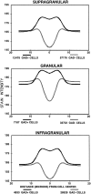
Cross-sectional profiles of GAD stain intensity in GAD+ and GAD neurons in supragranular (top), granular (middle), and infragranular layers (bottom) of barrel cortex from a normal behaving hamster. These curves represent the same cell groups as those portrayed in Figure 11 and were generated in the same manner. See legend to Figure 11 and text for discussion.
Fig. 1.
Fig. 1.
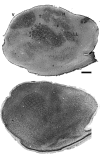
Somatotopically appropriate 2DG labeling in normal hamster barrel field. Top, Low-magnification photomicrograph of a 2DG-labeled/GAD-stained section from hamster, including barrel cortex, cut tangential to the barrel representation in layer IV. The image was contrast-enhanced to show the pattern of 2DG label, which reveals a full body map of 2DG label (a “hamsterunculus”), including the whisker-related barrel field, in somatosensory cortex. Note the clear demarcation of individual barrels near the center of the section, marked A–E to denote barrel rows corresponding to rows of whiskers on the contralateral face. V, Visual cortex; T, trunk representation; F, forelimb;H, hindlimb; LL, lower lip. Scale bar, 500 μm.Bottom, A similar low-magnification photomicrograph of a Nissl-stained section from another hamster, also cut tangential to the barrels. Individual barrels are defined clearly with this stain, which is denser in barrel walls than in the septum between barrels. This pattern is similar to that shown with 2DG.
Fig. 3.
Fig. 3.
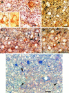
Strong metabolic activation of GABAergic neurons during normal behavior. High-magnification photomicrographs of double-labeled GAD+ and GADsomata in supragranular (top), granular (middle), and infragranular layers (bottom) of barrel cortex from a normal behaving hamster show a stark contrast between heavily 2DG-labeled mostly GAD+ (black dots) neurons and lightly labeled GAD “headlights” (smaller blank patches in the silver grain emulsion; absence of brown GAD stain). These phenomena are recognizable but somewhat more subtle in supragranular and infragranular layers. Several examples of heavy 2DG labeling in ridges surrounding GAD somata can be seen in the bottom panel, representing infragranular layers (see also Fig. 4). Smaller photos at right, taken from the same laminae but different specimens with less heavily exposed autoradiograms, better indicate the GAD stain underlying 2DG silver grains. Scale bar, 25 μm.
Fig. 4.
Fig. 4.
Top. Additional examples of ridges of 2DG grains closely apposed to perisomatic GAD+ puncta. These photomicrographs, taken from infragranular layers of barrel cortex stained for GAD, show many examples of GADneurons with curvilinear arrays of 2DG grains and GAD+ puncta at their somal perimeters (some of these are indicated by filled arrows). We refer to these grain arrays as “inhibitory ridges” (see Results and Discussion).Inset to A shows one example of such a ridge at two different focal depths, one at the plane of the silver grains (left) and one just below the grains, showing the GAD+ puncta (right). B, Two examples of GAD cells (open arrows), which exhibit inhibitory ridges but are nevertheless moderately 2DG-labeled (the labeling is comparable to that of the surrounding neuropil). In our interpretation these cases show that GAD cells can be activated metabolically during normal behavior although subject to strong proximal inhibition. Scale bars: 10 μm for inset to A; 25 μm for all other panels.
Fig. 6.
Fig. 6.
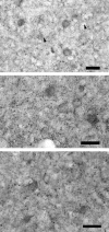
Absence of 2DG labeling bias in GAD+ and GAD cells from animals anesthetized simultaneously with 2DG injection. Each of the three panels shows a different subfield of barrel cortex (layer IV), in which very sparse silver grains (some indicated by arrows) show no obvious spatial relationship with GAD+ or GAD cells. A similar figure has been published previously (McCasland, 1996). Scale bars, 25 μm.
Fig. 7.
Fig. 7.
Detected cell map (montage) from a layer IV section of barrel cortex in a normal behaving hamster. Blue shading represents 2DG density from the silver grain montage (as in Fig. 2; see Materials and Methods); green stars represent candidate GAD+ cells; red dots represent candidate GAD cells. For each cell our automated routines quantify grain and stain densities in an annular array about the detected cell center (Figs. 8, 9, 11).
Fig. 13.
Fig. 13.
Laminar distribution of 2DG labeling in somata of GAD+ and GAD neurons.Curves depict relative 2DG grain densities (in arbitrary units as in Figs. 11, 12) for detected GAD+ cells (filled squares), GAD cells (filled triangles), and overall mean 2DG labeling (filled circles) in barrel cortex from a single normal hemisphere. Numbers along the abscissacorrespond to tangential section numbers from the specimen, starting just deep to the pia. GAD+ neurons are more heavily 2DG-labeled than GAD neurons in every layer. For each tissue section this difference was highly significant (t test, p < 0.001); error bars are omitted for clarity of presentation. The difference in 2DG labeling of GAD+ and GAD cells is most pronounced in layer IV (sections 9–13), less so in infragranular layers, and relatively subtle in supragranular layers.

References

    1. Agmon A, Connors BW. Correlation between intrinsic firing patterns and thalamocortical synaptic responses of neurons in mouse barrel cortex. J Neurosci. 1992;12:319–329. - PMC (V体育官网入口) - PubMed
    1. Armstrong-James M. The nature and plasticity of sensory processing within adult rat barrel cortex. In: Jones EG, Diamond IT, editors. Cerebral cortex. Plenum; New York: 1995. pp. 333–373.
    1. Beitz AJ, Larson AA, Monaghan P, Altschuler RA, Mullett MM, Madl JE. Immunohistochemical localization of glutamate, glutaminase, and aspartate aminotransferase in neurons of the pontine nuclei of the rat. Neuroscience. 1986;17:741–753. - VSports在线直播 - PubMed
    1. Bracewell RN. The Fourier transform and its applications, 2nd Ed. McGraw-Hill; New York: 1986.
    1. Bush PC, Sejnowski TJ. Effects of inhibition and dendritic saturation in simulated neocortical pyramidal cells. J Neurophysiol. 1994;71:2183–2193. - PubMed

Publication types

Substances