Skip to main page content
U.S. flag

An official website of the United States government

Dot gov

The . gov means it’s official VSports app下载. Federal government websites often end in . gov or . mil. Before sharing sensitive information, make sure you’re on a federal government site. .

Https

The site is secure V体育官网. The https:// ensures that you are connecting to the official website and that any information you provide is encrypted and transmitted securely. .

. 1996 Dec 10;93(25):14559-63.
doi: 10.1073/pnas.93.25.14559.

BAX-induced cell death may not require interleukin 1 beta-converting enzyme-like proteases

Affiliations

BAX-induced cell death may not require interleukin 1 beta-converting enzyme-like proteases

J Xiang et al. Proc Natl Acad Sci U S A. .

Abstract

Expression of BAX, without another death stimulus, proved sufficient to induce a common pathway of apoptosis. This included the activation of interleukin 1 beta-converting enzyme (ICE)-like proteases with cleavage of the endogenous substrates poly(ADP ribose) polymerase and D4-GDI (GDP dissociation inhibitor for the rho family), as well as the fluorogenic peptide acetyl-Asp-Glu-Val-Asp-aminotrifluoromethylcoumarin (DEVD-AFC). The inhibitor benzyloxycarbonyl-Val-Ala-Asp-fluoromethyl ketone (zVAD-fmk) successfully blocked this protease activity and prevented FAS-induced death but not BAX-induced death. Blocking ICE-like protease activity prevented the cleavage of nuclear and cytosolic substrates and the DNA degradation that followed BAX induction. However, the fall in mitochondrial membrane potential, production of reactive oxygen species, cytoplasmic vacuolation, and plasma membrane permeability that are downstream of BAX still occurred VSports手机版. Thus, BAX-induced alterations in mitochondrial function and subsequent cell death do not apparently require the known ICE-like proteases. .


Figures
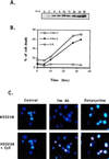
Figure 1
Figure 1
BAX-induced apoptosis in Jt-Bax cells. (A) Jt-Bax-21 cells were incubated with doxycycline (Sigma) at 1 μg/ml for various times. BAX expression was examined by immunoblot analysis with anti-BAX antibody 651. BAX levels were not substantially greater than that obtained in stable clones. (B) Cell viability was assayed by propidium iodide exclusion. Jt-Bax-13 and -21 were two independent Jt-Bax clones, while Jt-H was an empty vector containing control clone. (C) Jt-Bax-21 cells were treated with anti-FAS antibody (100 ng/ml, Upstate Biotechnology) for 4 hr or doxycycline (1 μg/ml) for 20 hr or left untreated (control). Apoptotic nuclei were visualized by H33258 or H33258 plus dUTP-cyanine-3-TdT-TUNEL (Cy3). The cells with blue or blue plus pink fragmented and condensed nuclei represent apoptotic cells.
Figure 2
Figure 2
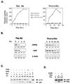
Activation of ICE-like proteases in Jt-Bax cells. (A) Jt-Bax-21 or Jt-H cells were treated with doxycycline or anti-FAS antibody in the presence (solid circle) or absence (open square and diamond) of zVAD-fmk (50 μM, Enzyme Systems Products). ICE and CPP32-like activities were measured using fluorogenic substrates YVAD-AFC and DEVD-AFC, respectively. (B) The cleavage of ICE-like proteases was examined by immunoblot analysis with the antibodies indicated and quantitated by densitometer scanning (Ultroscan XL) using β-actin as a control. Apoptotic degradation of PARP (C) or D4-GDI (D) was analyzed by immunoblot. p85, the apoptotic fragment of PARP; p20, the apoptotic fragment of D4-GDI; Dox, doxycycline; F, anti-FAS antibody.
Figure 3
Figure 3
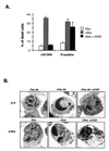
zVAD-fmk blocks DNA fragmentation but not cell death after BAX induction. (A) Jt-Bax-21 cells were incubated with doxycycline (1 μg/ml) for 24 hr in the presence or absence of zVAD-fmk (50 μM). DNA fragmentation (<2N DNA) and cell viability (propidium iodide exclusion) were measured as described (1). (B) Jt-H and Jt-Bax-21 cells were treated with either anti-FAS antibody (100 ng/ml) for 4 hr or doxycycline (1 μg/ml) for 20 hr, respectively, in the presence or absence of zVAD-fmk. The ultrastructure of the cells was analyzed by electron microscopy.
Figure 4
Figure 4
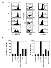
Reduction of mitochondrial membrane potential (ΔΨm) and production of ROS after BAX induction. (A) Jt-Bax-21 cells were treated with doxycycline for indicated periods of time. Aliquots of 1 × 10⁶ cells were incubated with 50 nM 3,3′-dihexyloxacarbocynine iodide [DiOC6(3)], 2 μM hydroethidine, or 5 μM DCFH-DA and analyzed by cytofluorometry. The percentages reflect the reduction of ΔΨm [DiOC6 (3)] (Left), ROS production (hydroethidine converted to ethidum) (Middle), and production of peroxides (DCFH) (Right). Data are representative of three experiments. The open peak in the DCFH panel of 0 hour was a positive control after 10 mM H202 treatment. (B) Jt-Bax cells were induced with Dox (1 μg/ml) or anti-FAS antibody (100 ng/ml) for 24 hr in the presence or absence of 50 μM zVAD-fmk. The reduction of ΔΨm (Left) and ROS production (Right) were measured as described above.
Figure 5
Figure 5
Schematic model of BAX-induced apoptosis. The ICE-like protease-dependent and -independent pathways may be parallel or sequential.

References

    1. Oltvai Z N, Milliman C L, Korsmeyer S J. Cell. 1993;74:609–619. - PubMed
    1. Sedlak T W, Oltvai Z N, Yang E, Wang K, Boise L H, Thompson C B, Korsmeyer S J. Proc Natl Acad Sci USA. 1995;92:7834–7838. - PMC (VSports app下载) - PubMed
    1. Oltvai Z N, Korsmeyer S J. Cell. 1994;79:189–192. - "V体育2025版" PubMed
    1. Knudson C M, Tung K S, Tourtellotte W G, Brown G A, Korsmeyer S J. Science. 1995;270:96–99. - PubMed
    1. Deckwerth T L, Ellitott J L, Knudson C M, Johnson E M, Snider W D, Korsmeyer S J. Neuron. 1996;17:401–411. - PubMed