Skip to main page content
U.S. flag

An official website of the United States government

Dot gov

The . gov means it’s official. Federal government websites often end in . gov or VSports app下载. mil. Before sharing sensitive information, make sure you’re on a federal government site. .

Https

The site is secure. The https:// ensures that you are connecting to the official website and that any information you provide is encrypted and transmitted securely V体育官网. .

. 1996 Jan 17;88(2):100-8.
doi: 10.1093/jnci/88.2.100.

Loss of functional beta 2-microglobulin in metastatic melanomas from five patients receiving immunotherapy

Affiliations

"VSports注册入口" Loss of functional beta 2-microglobulin in metastatic melanomas from five patients receiving immunotherapy

N P Restifo et al. J Natl Cancer Inst. .

Abstract

Background: In a subset of patients with metastatic melanoma, T lymphocytes bearing the cell-surface marker CD8 (CD8+ T cells) can cause the regression of even large tumors VSports手机版. These antitumor CD8+ T cells recognize peptide antigens presented on the surface of tumor cells by major histocompatibility complex (MHC) class I molecules. The MHC class I molecule is a heterodimer composed of an integral membrane glycoprotein designated the alpha chain and a noncovalently associated, soluble protein called beta2-microglobulin (beta 2m). Loss of beta 2m generally eliminates antigen recognition by antitumor CD8+ T cells. .

Purpose: We studied the loss of beta 2m as a potential means of tumor escape from immune recognition in a cohort of patients receiving immunotherapy V体育安卓版. .

Methods: We successfully grew 13 independent tumor cell cultures from tumor specimens obtained from 13 patients in a cohort of 40 consecutive patients undergoing immunotherapy for metastatic melanoma and for whom tumor specimens were available V体育ios版. These cell lines, as well as another melanoma cell line (called 1074mel) that had been derived from tumor obtained from a patient in a cytokine-gene therapy study, were characterized in vitro cytofluorometrically for MHC class I expression and by northern and western blot analyses for messenger RNA (mRNA) and protein expression, respectively, and ex vivo by immunohistochemistry. .

Results: After one melanoma cell line (1074mel) was found not to express functional beta 2m by cytofluorometric analysis, four (31%) of the 13 newly established melanoma cell lines were found to have an absolute lack of functional MHC class I expression. Northern blot analysis of RNA extracted from the five cell lines exhibiting no functional MHC class I expression showed that these cells contained normal levels of alpha-chain mRNA but variable levels of beta 2m mRNA. In addition, no immunoreactive beta 2m protein was detected by western blot analysis. When human beta 2m was transiently expressed with the use of a recombinant vaccinia virus, cell-surface MHC class I expression was reconstituted and the ability of these five cell lines to present endogenous antigens was restored VSports最新版本. Immunohistochemical staining of tumor sections revealed a lack of immunoreactive MHC class I in vivo, supporting the notion that the in vitro observations were not artifactual. Furthermore, archival tumor sections obtained from patients prior to immunotherapy were available from three patients and were found to be beta 2m positive. This result was consistent with the hypothesis that loss of beta 2m resulted from immunotherapy. .

Conclusions: These data suggest that the loss of beta 2m may be a mechanism whereby tumor cells can acquire immunoresistance. This study represents the first characterization of a molecular route of escape of tumors from immune recognition in a cohort of patients being treated with immunotherapy V体育平台登录. .


Figures
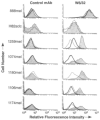
Fig. 1
Fig. 1
Absence of major histocompatibility complex class I on the surfaces of metastatic human melanoma cell lines and restoration by transient expression of beta2-microglobulin (β2m). Fluorescence-activated cell sorter analysis was performed on tumor cell lines alone (heavy solid line), infected with wild-type vaccinia virus (dashed line), and infected with the recombinant vaccinia virus expressing β2m (light solid line, shaded gray under the curve); cell lines were sequentially incubated with either the control immunoglobulin G2a antibody (left panels) or the W6/32 monoclonal antibody (right panels) and then with goat anti-mouse fluorescein isothiocyanate-conjugated Fab fragments. The cell lines analyzed were the positive control cell line 888mel (that is normal in its antigen-presenting capacity), the negative-control H82sclc (a small-cell lung cancer cell line that does not process antigens), and the five melanoma cell lines (1259mel, 1074mel, 1180mel, 1106mel, and 1174mel) that have lost functional β2m. For two cell lines (H82sclc and 1180mel), only two curves are shown in this experiment because the tumor-cell-alone control was not performed because of inadequate numbers of cells.
Fig. 2
Fig. 2
Northern blot analysis of total RNA extracted from the indicated cells (624[mel], 397[mel], FO-1, 1106[mel], 1074[mel], 1l74[mel], 1180[mel], 1259[mel]) and probed for the expression of beta2-microglobulin (β2m) messenger RNA. The northern blot probed for β2m messenger RNA (top panel) was stripped and probed for β-actin to ensure sufficient loading of all lanes (bottom panel).
Fig. 3
Fig. 3
Western blot analysis of total protein extracts for expression of immunoreactive beta2-microglobulin (β2m). Lysates of melanoma cell lines 1074mel, 1106mel, 1180mel, 1174mel, and 1259mel do not express any measurable immunoreactive β2m. Line l074mel is shown after infection with recombinant vaccinia virus expressing β2m (left-most panel) or after infection with wild-type control vaccinia virus (second from left).
Fig. 4
Fig. 4
Microcytotoxicity assay evaluating the antigen-presenting capacities of beta2-microglobulin (β2m)-deficient cell lines. Cell lines were screened for their capacities to present vaccinia virus antigens in the context of a transiently expressed H-2 Kd molecule. Effector cells (E) were vaccinia virus-specific, H-2 Kd-restricted murine cytotoxic T lymphocytes. Target cells (T) were either uninfectcd (▲), infected with vaccinia virus expressing H-2 Kd alone (●), or in combination with recombinant vaccinia virus expressing β2m (△). The E:T ratios for the six points in each curve, reading from left to right, were 100:1, 50:1, 25:1, 12.5:1, 6.25:1, and 3.125:1. The melanoma cell line 526 (the positive control) is an antigen-processing intact cell line that processes vaccinia antigens efficiently in the absence of β2m. The small-cell lung cancer cell line H82sclc fails to process antigens even in the presence of β2m, since the antigen-processing lesions in this cell line are elsewhere (14). Note that 1259mel, 1180mel, 1106mel, 1174mel, and 1074mel all fail to present vaccinia virus antigens but can do so efficiently when functional β2m is expressed endogenously.
Fig. 5
Fig. 5
Microcytotoxicity assay evaluating the capacity of a beta2-microblobulin (β2m)-deficient cell line to present tumor-associated antigen after restoration of β2m. Melanoma cell lines were tested for recognition by HLA-A2.1-restricted antimelanoma effector (E) cells. Target cells (T) were the HLA-A2.1-positive, β2m-positive 526 cell line (◆); the HLA-A2.1-positive but β2m-negative 1074 cell line infected with control vaccinia virus (▲) or infected with recombinant vaccinia virus-expressing β2m (△); the HLA-A2.1-negative but β2m-positive 888 melanoma cell line with control (□) or β2m vaccinia virus (■); and the HLA-A2.1-negative and β2m- negative 1259 cell line with control (●) or β2m vaccinia virus (○).
Fig. 6
Fig. 6
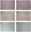
Immunohistochemical staining of sections of melanoma in situ. Sections from tumor specimens described in Table 1 from patient Nos. 1106 (panels A-C) and 1180 (panels D-F) are shown. Immunohistochemical staining in panels B, C, E, and F was done at the same time, under exactly the same conditions with anti-human beta2-microglobulin (β2m) monoclonal antibody followed by immunoperoxidase staining (see “Materials and Methods” section for details). Strongly positive and weakly positive staining for β2m is shown in panels B and E, respectively. Panels C and F are examples of specimens that stain negative for β2m. Hematoxylin–eosin stains are shown in panels A and D, where melanin deposits that appear dark brown can be seen.

References

    1. Topalian SL. Cell transfer therapy: preclinical studies. In: DeVita VT Jr, Hellman S, Rosenberg SA, editors. Biologic therapy of cancer. 2nd ed. Lippincott; Philadelphia: 1995. pp. 467–86.
    1. Peoples GE, Goedegebuure PS, Smith R, Linehan DC, Yoshino I, Everlein TJ. Breast and ovarian cancer-specific cytotoxic T lymphocytes recognize the same HER2/neu-derived peptide. Proc Natl Acad Sci U S A. 1995;92:432–6. - PMC - PubMed
    1. Hoppe RT, Medeiros LJ, Warnke RA, Wood GS. CD8-positive tumor-infiltrating lymphocytes influence the long-term survival of patients with mycosis fungoides. J Am Acad Dermatol. 1995;32:448–53. - PubMed
    1. Rosenberg SA, Yannelli JR, Yang JC, Topalian SL, Schwartzentruber DF, Weber JS, et al. Treatment of patients with metastatic melanoma with autologous tumor-infiltrating lymphocytes and interleukin 2 [see comment citation in Medline] J Natl Cancer Inst. 1994;86:1159–66. - PubMed
    1. Ikarashi H, Fujita K, Takakuwa K, Kodama S, Tokunaga A, Takahashi T, et al. Immunomodulation in patients with epithelial ovarian cancer after adoptive transfer of tumor-infiltrating lymphocytes. Cancer Res. 1994;54:190–6. - PubMed

MeSH terms