<tt lang="haNjmHY"></tt> "VSports最新版本" Skip to main page content
U.S. flag

An official website of the United States government

Dot gov

The . gov means it’s official. Federal government websites often end in . gov or . mil VSports app下载. Before sharing sensitive information, make sure you’re on a federal government site. .

Https

The site is secure V体育官网. The https:// ensures that you are connecting to the official website and that any information you provide is encrypted and transmitted securely. .

. 2023 Jul 21;14(7):457.
doi: 10.1038/s41419-023-05952-4.

PDHA1 hyperacetylation-mediated lactate overproduction promotes sepsis-induced acute kidney injury via Fis1 lactylation (V体育ios版)

Affiliations

PDHA1 hyperacetylation-mediated lactate overproduction promotes sepsis-induced acute kidney injury via Fis1 lactylation

Sheng An et al. Cell Death Dis. .

Abstract

The increase of lactate is an independent risk factor for patients with sepsis-induced acute kidney injury (SAKI). However, whether elevated lactate directly promotes SAKI and its mechanism remain unclear. Here we revealed that downregulation of the deacetylase Sirtuin 3 (SIRT3) mediated the hyperacetylation and inactivation of pyruvate dehydrogenase E1 component subunit alpha (PDHA1), resulting in lactate overproduction in renal tubular epithelial cells. We then found that the incidence of SAKI and renal replacement therapy (RRT) in septic patients with blood lactate ≥ 4 mmol/L was increased significantly, compared with those in septic patients with blood lactate < 2 mmol/L. Further in vitro and in vivo experiments showed that additional lactate administration could directly promote SAKI VSports手机版. Mechanistically, lactate mediated the lactylation of mitochondrial fission 1 protein (Fis1) lysine 20 (Fis1 K20la). The increase in Fis1 K20la promoted excessive mitochondrial fission and subsequently induced ATP depletion, mitochondrial reactive oxygen species (mtROS) overproduction, and mitochondrial apoptosis. In contrast, PDHA1 activation with sodium dichloroacetate (DCA) or SIRT3 overexpression decreased lactate levels and Fis1 K20la, thereby alleviating SAKI. In conclusion, our results show that PDHA1 hyperacetylation and inactivation enhance lactate overproduction, which mediates Fis1 lactylation and exacerbates SAKI. Reducing lactate levels and Fis1 lactylation attenuate SAKI. .


Conflict of interest statement

The authors declare no competing interests.

"VSports最新版本" Figures
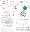
Fig. 1
Fig. 1. The renal mitochondrial protein acetylome identified PDHA1 hyperacetylation after CLP.
A Flow chart showing the mitochondrial protein acetylome in the mouse kidney 8 h after CLP (n = 3). B Volcano plot showing the differentially modified proteins. C The number of differentially modified proteins. D Subcellular localization of the differentially modified proteins. E Biological processes of the differentially modified proteins. F Differentially modified proteins involved in cellular respiration, the tricarboxylic acid cycle, and oxidative phosphorylation. G Differential PDHA1 acetylated lysine sites. H, I Mass spectrometry spectrum of the acetylated PDHA1 K267 and K385 sites. J Protein multiple sequence alignment analysis of the PDHA1 267 and 385 sites.
Fig. 2
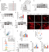
Fig. 2. SIRT3 downregulation mediates PDHA1 hyperacetylation and inactivation, leading to lactate overproduction.
A, B Changes in PDHA1 acetylation and PDH activity within 24 h after CLP in mice (n = 4). C, D Changes in PDHA1 acetylation and PDH activity in HK-2 cells within 24 h after LPS stimulation (n = 4). E, F Effects of SIRT3-specific inhibitor 3-TYP (5 mg/kg) treatment on CLP-induced PDHA1 hyperacetylation and PDH activity in mice (n = 5). G, H Effects of SIRT3 overexpression on LPS-induced PDHA1 hyperacetylation and PDH activity in HK-2 cells (n = 4). I, J Effects of SIRT3 knockdown on LPS-induced PDHA1 hyperacetylation and PDH activity in HK-2 cells (n = 4). K co-IP showing the interaction of SIRT3 with PDHA1 in HK-2 cells. L, M Effects of PDHA1 K385R mutation on PDHA1 acetylation and PDH activity in HK-2 cells (n = 4). N, O Effects of SIRT3 overexpression and SIRT3 knockdown on LPS-induced lactate production in HK-2 cells, respectively (n = 4). P Effects of PDHA1 K385R mutation on LPS-induced lactate production in HK-2 cells (n = 8). Data are mean ± SD; *P, #P < 0.05.
Fig. 3
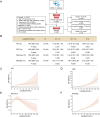
Fig. 3. High blood lactate levels are associated with a high incidence of AKI and poor prognosis in septic patients.
A Flow chart showing the retrospective analysis of sepsis patients with different lactate levels in the MIMIC-IV database. B The ORs (95% CIs) for the occurrence, RRT requirements, and recovery of AKI and HRs (95% CIs) for mortality across the groups. Regression models were adjusted for confounding variables selected from univariate analysis, including age, gender, ethnicity, admission type, liver disease, white blood cell, baseline creatinine, volume, and SOFA score. CF Concentration-response relationship between lactate and outcomes. Restricted cubic spline curves were used to evaluate the association between concentration of lactate and the occurrence, RRT requirements, recovery, as well as in-hospital mortality of AKI in septic patients.
Fig. 4
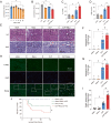
Fig. 4. High lactate levels directly contribute to SAKI aggravation.
A Effects of different concentrations (0 ~ 50 mM) of lactate on the viability of HK-2 cells (n = 6). B Lactate (10 mM) treatment significantly promoted the LPS-induced decrease in cell viability (n = 4). C, D Lactate (1 g/kg) pretreatment significantly increased CLP-induced increases in SCr and BUN in mice (n = 4). EG Haematoxylin-eosin (HE) staining (upper panel) and periodic acid-Schiff (PAS) staining (lower panel) of the kidney following CLP-induced sepsis (n = 25). Black arrow: renal tubular vacuoles, structural disorder; white arrow: brush border loss; white triangle: tubular lumen dilation; red arrow: renal tubular basement membrane rupture. Scale bars, 50 μm. H, I Cell apoptosis in the kidney was examined by TUNEL staining (n = 8). Scale bars, 50 μm. J The survival rates in the sham, sham+lactate, CLP, and CLP+lactate groups. Data are mean ± SD; *P, #P < 0.05.
Fig. 5
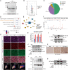
Fig. 5. The lactylome revealed that renal Fis1 lactylation was elevated after sepsis.
A Changes in Pan-Kla in mouse kidneys within 24 h after CLP. B Volcano plot showing the differentially lactylated proteins in the kidney 8 h after CLP (n = 3). C The number and intracellular localization of differentially lactylated proteins. D Biological processes of the differentially lactylated proteins. E Differentially lactylated proteins involved in the regulation of mitochondrion organization. F Mass spectrometry spectrum of the lactylated Fis1 K20 sites. G, H Fis1, Pan-Kla, and mitochondrial marker TOM20 immunofluorescent triple staining showing the co-localization of Fis1 and Pan-Kla. The images were analyzed by the Colocalization Finder plugin of ImageJ software (n = 6). Fis1 and PanKla were considered co-localized when the overlap coefficient was >0.6 and Pearson’s correlation coefficient was >0.5. Scale bars, 20 μm. I, J Changes in renal Fis1 lactylation following CLP or lactate (1 g/kg) administration in mice. K Immunohistochemistry showing the changes in Pan-Kla in mouse renal tissue after CLP and lactate administration. L Changes in Pan-Kla in HK-2 cells after lactate stimulation. M Changes in Fis1 lactylation after LPS or lactate stimulation in HK-2 cells. Data are mean ± SD; *P, #P < 0.05.
Fig. 6
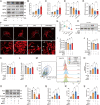
Fig. 6. Lactate aggravates SAKI by promoting excessive mitochondrial fission.
AE Effects of lactate on the protein expression of Fis1, DRP1, MFN1, and MFN2 in HK-2 cells (n = 6). FG The mitochondrial morphology of HK-2 cells was observed under confocal microscope by MitoTracker staining. The images were analyzed by the Mitochondrial Network Analysis (MiNA) plugin of ImageJ software (n = 17). Scale bars, 5 μm. H Lactate promoted the LPS-induced Fis1-DRP1 interaction in HK-2 cells. (I-J) Fis1 knockdown attenuated the reduction in cell viability and ATP depletion induced by lactate+LPS stimulation in HK-2 cells (n = 5). KL Mdivi-1 (10 μM) administration attenuated the reduction in cell viability and ATP depletion induced by lactate+LPS stimulation in HK-2 cells (n = 5). M, N Flow cytometry assessment after MitoSOX staining showed that Mdivi-1 reduced the LPS+lactate-induced mtROS elevation in HK-2 cells (n = 6). OQ The effects of Mdivi-1 on the expression of the antiapoptotic protein Bcl-2 and the proapoptotic protein Bak in HK-2 cells (n = 6). R, S Mdivi-1 reduced CLP+lactate-induced elevation of SCr and BUN in mice (n = 5). Data are mean ± SD; *P, #P, &P < 0.05.
Fig. 7
Fig. 7. Fis1 K20 is the key site by which lactate mediates Fis1 lactylation and exacerbates SAKI.
A Protein multiple sequence alignment analysis of Fis1 20 sites. B Verification of the specificity of the Fis1 K20la antibody by dot blot experiment. C Immunoblotting with the Fis1 K20la antibody after Fis1 IP in HK-2 cells. D, E Recognition of Fis1 lactylation in HK-2 whole-cell lysates by the Fis1 K20la antibody (n = 6). F Fis1 K20R mutation eliminated Fis1 K20la in HK-2 cells. G Fis1 K20R mutation inhibited the Fis1 interaction with DRP1 in HK-2 cells. H, I Effects of Fis1 WT and K20R plasmid overexpression on the mitochondrial network (n = 16). Scale bars, 5 μm. J, K Fis1 K20R mutation inhibited Fis1 WT-mediated mtROS overproduction (n = 6). LN Fis1 K20R mutation inhibited the Fis1 WT-mediated Bcl-2 expression decrease and Bak expression increase (n = 6). Data are mean ± SD; *P, #P, &P < 0.05.
Fig. 8
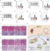
Fig. 8. The effects of reducing lactate and Fis1 lactylation on SAKI.
A, B Effects of DCA (5 mM) treatment on Fis1 K20la level in LPS-stimulated HK-2 cells (n = 6). C, D Effects of SIRT3 overexpression on Fis1 K20la level in LPS-stimulated HK-2 cells (n = 6). E, F Effects of 3-TYP (50 μM) and GSK (5 μM) treatment on Fis1 K20la level in LPS-stimulated HK-2 cells (n = 4). G, H Effects of intraperitoneal injection of DCA (25 mg/kg) on SCr and BUN in CLP mice (n = 3). I Effects of intraperitoneal injection of DCA on renal pathological damage in CLP mice. Black arrow: nuclei of renal tubular cells shed to the lumen; White arrow: renal tubular vacuoles, structural disorder; White triangle: brush border loss; Red arrow: renal tubular basement membrane rupture. Scale bars, 50 μm. J Effects of intraperitoneal injection of 3-TYP (5 mg/kg) and GSK (20 mg/kg) on renal pathological damage in CLP mice. Scale bars, 50 μm. K Schematic of PDHA1 hyperacetylation-mediated lactate overproduction promoting SAKI via Fis1 lactylation. Data are mean ± SD; *P, #P < 0.05.

References (V体育ios版)

    1. Bellomo R, Kellum JA, Ronco C, Wald R, Martensson J, Maiden M, et al. Acute kidney injury in sepsis. Intensive Care Med. 2017;43:816–28. doi: 10.1007/s00134-017-4755-7. - "VSports注册入口" DOI - PubMed
    1. Peerapornratana S, Manrique-Caballero CL, Gómez H, Kellum JA. Acute kidney injury from sepsis: current concepts, epidemiology, pathophysiology, prevention and treatment. Kidney Int. 2019;96:1083–99. doi: 10.1016/j.kint.2019.05.026. - DOI - PMC - PubMed
    1. Zeng S, Lin J, Zhang W. Research progress of sepsis-induced acute kidney injury. Zhonghua Wei Zhong Bing Ji Jiu Yi Xue. 2018;30:703–7. - PubMed
    1. Honore PM, Jacobs R, Hendrickx I, Bagshaw SM, Joannes-Boyau O, Boer W, et al. Prevention and treatment of sepsis-induced acute kidney injury: an update. Ann Intensive Care. 2015;5:51–51. doi: 10.1186/s13613-015-0095-3. - VSports注册入口 - DOI - PMC - PubMed
    1. Park S, Jeon JH, Min BK, Ha CM, Thoudam T, Park BY, et al. Role of the pyruvate dehydrogenase complex in metabolic remodeling: differential pyruvate dehydrogenase complex functions in metabolism. Diabetes Metab J. 2018;42:270–81. doi: 10.4093/dmj.2018.0101. - DOI (VSports app下载) - PMC - PubMed

Publication types (V体育官网)

MeSH terms (V体育2025版)

"VSports" Substances