"V体育ios版" Skip to main page content
U.S. flag

An official website of the United States government

Dot gov

The . gov means it’s official VSports app下载. Federal government websites often end in . gov or . mil. Before sharing sensitive information, make sure you’re on a federal government site. .

Https

The site is secure. The https:// ensures that you are connecting to the official website and that any information you provide is encrypted and transmitted securely V体育官网. .

. 2023 Apr 20:2023:3951940.
doi: 10.1155/2023/3951940. eCollection 2023.

Prognostic and Immunological Significance of the Molecular Subtypes and Risk Signatures Based on Cuproptosis in Hepatocellular Carcinoma

Affiliations

Prognostic and Immunological Significance of the Molecular Subtypes and Risk Signatures Based on Cuproptosis in Hepatocellular Carcinoma (V体育官网入口)

Xiaolong Tang et al. Mediators Inflamm. .

Abstract

Background: Hepatocellular carcinoma (HCC) remains a challenging medical problem. Cuproptosis is a novel form of cell death that plays a crucial role in tumorigenesis, angiogenesis, and metastasis. However, it remains unclear whether cuproptosis-related genes (CRGs) influence the outcomes and immune microenvironment of HCC patients VSports手机版. .

Method: From The Cancer Genome Atlas (TCGA) and International Cancer Genome Consortium (ICGC) databases, we obtained the mRNA expression file and related clinical information of HCC patients. We selected 19 CRGs as candidate genes for this study according to previous literature. We performed a differential expression analysis of the 19 CRGs between malignant and precancerous tissue V体育安卓版. Based on the 19 CRGs, we enrolled cluster analysis to identify cuproptosis-related subtypes of HCC patients. A prognostic risk signature was created utilizing univariate Cox regression and least absolute shrinkage and selection operator (LASSO) regression analyses. We employed independent and stratification survival analyses to investigate the predictive value of this model. The functional enrichment features, mutation signatures, immune profile, and response to immunotherapy of HCC patients were also investigated according to the two molecular subtypes and the prognostic signature. .

Results: We found that 17 CRGs significantly differed in HCC versus normal samples. Cluster analysis showed two distinct molecular subtypes of cuproptosis. Cluster 1 is preferentially related to poor prognosis, high activity of immune response signaling, high mutant frequency of TP53, and distinct immune cell infiltration versus cluster 2 V体育ios版. Through univariate and LASSO Cox regression analyses, we created a cuproptosis-related prognostic risk signature containing LIPT1, DLAT, MTF1, GLS, and CDKN2A. High-risk HCC patients were shown to have a worse prognosis. The risk signature was proved to be an independent predictor of prognosis in both the TCGA and ICGC datasets, according to multivariate analysis. The signature also performed well in different stratification of clinical features. The immune cells, which included regulatory T cells (Treg), B cells, macrophages, mast cells, NK cells, and aDCs, as well as immune functions containing cytolytic activity, MHC class I, and type II IFN response, were remarkably distinct between the high-risk and low-risk groups. The tumor immune dysfunction and exclusion (TIDE) score suggested that high-risk patients had a higher response rate to immune checkpoint inhibitors than low-risk patients. .

Conclusion: This research discovered the potential prognostic and immunological significance of cuproptosis in HCC, improved the understanding of cuproptosis, and may deliver new directions for developing more efficacious therapeutic techniques for HCC patients VSports最新版本. .


Conflict of interest statement

No potential conflicts of interest were disclosed.

VSports最新版本 - Figures

Figure 1
Figure 1
Identification of cuproptosis-related DEGs and exploration of the relationship between each CRG in HCC based on the TCGA database. (a) Cuproptosis-related DEGs expression patterns between HCC and normal tissue. The color legend represents the log2 (FPKM) value. (b) Pearson's correlation analysis of each CRG based on the HCC samples. (c) PPI network plot displayed the relationship between each CRG. Red and green nodes indicate up and downregulated genes, respectively. (d) The number of adjacent nodes between each CRG in the PPI network. p < 0.05, ∗∗p < 0.01, ∗∗∗p < 0.001.
Figure 2
Figure 2
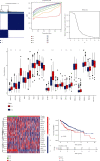
Consensus clustering of cuproptosis-associated subtypes and survival analysis in the TCGA. (a) Heatmap represented the consensus clustering solution (k = 2) for 19 CRGs among 502 HCC samples. (b, c) The consensus clustering delta area showed the cumulative distribution function area for k = 2 to 9. (d) Boxplots represented gene expression profiles for 19 genes in the two clusters. (e) An expression heatmap showed 19 genes grouped into two clusters. The color legend represents the log2 (FPKM) value. Red highlighted the high expression, and blue highlighted the low expression. (f) Kaplan–Meier curves of OS in different clusters. p < 0.05, ∗∗p < 0.01, ∗∗∗p < 0.001.
Figure 3
Figure 3
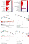
Results of functional enrichment analysis. (a) A list of the top 10 enriched GO terms. Topics contained biological processes (BP), cellular components (CC), and molecular functions (MF). (b) The top 30 most significant enriched KEGG pathways. (c) The top 5 GSEA-GO enrichment in cluster 1. (d) The top 5 GSEA-GO enrichment in cluster 2. (e) The top 5 GSEA-KEGG enrichment in cluster 1. (f) The top 5 GSEA-KEGG enrichment in cluster 2.
Figure 4
Figure 4
The somatic mutations landscape of two cuproptosis-related clusters. The top ten mutated genes in cluster 1 (a) and cluster 2 (b) were visualized using a waterfall plot.
Figure 5
Figure 5
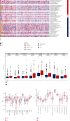
The immune landscape of two cuproptosis-related clusters in HCC. (a) The immune infiltration heatmap between the two clusters using TIMER, CIBERSORT, CIBERSORT-ABS, QUANTISEQ, MCPcounter, XCELL, and EPIC algorithms. (b) The gene expression levels of immune checkpoints for the two clusters. (c) The ssGSEA for examining subpopulation associations in immune cells. (d) The ssGSEA for examining subpopulation associations in immune functions. p < 0.05, ∗∗p < 0.01, ∗∗∗p < 0.001.
Figure 6
Figure 6
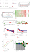
Formation of the risk score signature utilizing five CRGs in the TCGA. (a) Univariate Cox regression analysis selected six CRGs. (b, c) Detection of five prognostic CRGs using the LASSO Cox regression analysis. (d) Heatmaps of the five prognostic CRGs according to the distribution of risk scores. The color legend represents the log2 (FPKM) value. (e) The distribution of risk scores. (f) Patients' survival status according to the distribution of risk scores. (g) Kaplan-Meier survival analysis compared the OS between the high-risk and low-risk groups. (h) Kaplan-Meier survival analysis compared the PFS between the high-risk and low-risk groups. (i) The ROC curves for 1, 3, and 5 years of the risk model. (j) Mutation landscape of the five CRGs of the risk model.
Figure 7
Figure 7
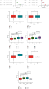
Exploration of the independent prognostic value and clinical feature of the risk score in HCC. (a, b) Through univariate and multivariate Cox regression analysis, the risk score was found to be an independent prognostic element for HCC patients. (c–i) The relationship between the risk score and different clinical parameters of HCC.
Figure 8
Figure 8
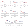
Kaplan-Meier analysis of the risk score in different stratifications according to clinicopathological characteristics. (a–h) HCC patients with varying clinical features (age, gender, stage, and grade) were analyzed using the Kaplan-Meier method according to the risk score.
Figure 9
Figure 9
Verification of the five CRGs signature in the ICGC cohort. (a) Heatmaps of five prognostic CRGs in the ICGC database according to the risk score distribution. The color legend represents the log2 (FPKM) value. (b) The risk scores distribution. (c) The survival status of each patient is according to the risk score distribution. (d) Kaplan-Meier curves for the OS of HCC patients. (e, f) The independent survival analysis of the risk scores and clinical traits through univariate and multivariate Cox regression analysis.
Figure 10
Figure 10
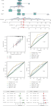
Predicting survival rates for HCC patients after one year, three years, and five years using the nomogram. (a) The nomogram model was formed to predict the survival rates of HCC patients in the TCGA cohorts. (b) Calibration curves of the nomogram. (c–e) The ROC curve explored the prognostic performance of the nomogram model. (f, g) Univariate and multivariate Cox analysis of the nomogram and clinical traits.
Figure 11
Figure 11
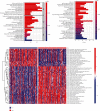
Functional enrichment analysis was performed according to the risk score. (a) A list of the top 10 significantly enriched GO terms. (b) A list of the top 30 most significantly enriched KEGG pathways. (c) The pathway activities scored by GSVA differently for high-risk and low-risk individuals.
Figure 12
Figure 12
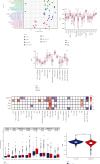
The immune landscape of cuproptosis-related risk score in HCC. (a) The forest plot displayed the connection between risk score and immune cell infiltration through TIMER, CIBERSORT, CIBERSORT-ABS, QUANTISEQ, MCPcounter, XCELL, and EPIC algorithms. (b, c) The bar graphs showed the difference in immune cell subpopulations and related functions between high-risk and low-risk groups. (d) The heatmap displayed the relationship of immune cell subpopulations and related functions with the five prognostic genes. (e) Differences in immune checkpoint expression between high-risk and low-risk groups. (f) The violin plots presented the TIDE scores between high-risk and low-risk groups. p < 0.05, ∗∗p < 0.01, ∗∗∗p < 0.001.

References

    1. Sung H., Ferlay J., Siegel R. L., et al. Global cancer statistics 2020: GLOBOCAN estimates of incidence and mortality worldwide for 36 cancers in 185 countries. CA: a Cancer Journal for Clinicians . 2021;71(3):209–249. doi: 10.3322/caac.21660. - DOI - PubMed
    1. Yang J. D., Hainaut P., Gores G. J., Amadou A., Plymoth A., Roberts L. R. A global view of hepatocellular carcinoma: trends, risk, prevention and management. Nature Reviews. Gastroenterology & Hepatology . 2019;16(10):589–604. doi: 10.1038/s41575-019-0186-y. - DOI - PMC - PubMed
    1. Villanueva A. Hepatocellular carcinoma. The New England Journal of Medicine . 2019;380(15):1450–1462. doi: 10.1056/NEJMra1713263. - DOI - PubMed
    1. Llovet J. M., Ricci S., Mazzaferro V., et al. Sorafenib in advanced hepatocellular carcinoma. The New England Journal of Medicine . 2008;359(4):378–390. doi: 10.1056/NEJMoa0708857. - DOI (V体育ios版) - PubMed
    1. Huang A., Yang X.-R., Chung W.-Y., Dennison A. R., Zhou J. Targeted therapy for hepatocellular carcinoma. Signal Transduction and Targeted Therapy . 2020;5(1):p. 146. doi: 10.1038/s41392-020-00264-x. - DOI - PMC - PubMed

MeSH terms