"V体育安卓版" Skip to main page content
U.S. flag

An official website of the United States government

Dot gov

The . gov means it’s official. Federal government websites often end in . gov or . mil VSports app下载. Before sharing sensitive information, make sure you’re on a federal government site. .

Https

The site is secure. The https:// ensures that you are connecting to the official website and that any information you provide is encrypted and transmitted securely V体育官网. .

. 2023 Mar 7;120(10):e2216722120.
doi: 10.1073/pnas.2216722120. Epub 2023 Feb 27.

VSports - FDX1-dependent and independent mechanisms of elesclomol-mediated intracellular copper delivery

Affiliations

"VSports最新版本" FDX1-dependent and independent mechanisms of elesclomol-mediated intracellular copper delivery

Mohammad Zulkifli et al. Proc Natl Acad Sci U S A. .

Abstract

Recent studies have uncovered the therapeutic potential of elesclomol (ES), a copper-ionophore, for copper deficiency disorders. However, we currently do not understand the mechanism by which copper brought into cells as ES-Cu(II) is released and delivered to cuproenzymes present in different subcellular compartments. Here, we have utilized a combination of genetic, biochemical, and cell-biological approaches to demonstrate that intracellular release of copper from ES occurs inside and outside of mitochondria. The mitochondrial matrix reductase, FDX1, catalyzes the reduction of ES-Cu(II) to Cu(I), releasing it into mitochondria where it is bioavailable for the metalation of mitochondrial cuproenzyme- cytochrome c oxidase. Consistently, ES fails to rescue cytochrome c oxidase abundance and activity in copper-deficient cells lacking FDX1 VSports手机版. In the absence of FDX1, the ES-dependent increase in cellular copper is attenuated but not abolished. Thus, ES-mediated copper delivery to nonmitochondrial cuproproteins continues even in the absence of FDX1, suggesting alternate mechanism(s) of copper release. Importantly, we demonstrate that this mechanism of copper transport by ES is distinct from other clinically used copper-transporting drugs. Our study uncovers a unique mode of intracellular copper delivery by ES and may further aid in repurposing this anticancer drug for copper deficiency disorders. .

Keywords: FDX1; copper; cytochrome c oxidase; elesclomol; mitochondria V体育安卓版. .


Conflict of interest statement (V体育安卓版)

The authors have organizational affiliations to disclose: Vishal M. Gohil serves as a consultant to Engrail Therapeutics for their effort in developing elesclomol–copper as a therapeutic agent for Menkes disease. The authors have patent filings to disclose: S. S V体育ios版. and V. M. G. are inventors on the patent US 2021/0290571 A1 submitted by Texas A&M University entitled “Compositions for the Treatment of Copper Deficiency and Methods of Use. ”.

Figures

Fig. 1.
Fig. 1.
Loss of FDX1 prevents ES-mediated rescue of mitochondrial respiration. (A) Western blot-based detection of FDX1 in the mitochondria isolated from wild-type (WT) and Ctr1−/− cells transduced with either empty vector or two different sgRNAs targeting Fdx1. ATP5A was used as a loading control. A representative blot from two independent trials is shown. (BD) Cells were cultured in the high-glucose media in the presence or absence of 1 nM ES, and cell numbers are plotted over time. Data are expressed as mean ± SD, n = 3. (E) Western blot analysis of COX1 and ATP5A in indicated cells treated with or without 1 nM ES for 72 h. A representative blot from two independent trials is shown. (F) The OCR of WT, Ctr1−/− and Ctr1−/−Fdx1−/− cells treated with or without 1 nM ES for 72 h. Oligomycin, CCCP, and antimycin A were used to measure adenosine triphosphate (ATP)-coupled respiration, maximum respiratory capacity, and mitochondria-specific respiration. Data are shown as mean ± SEM, n = 3.
Fig. 2.
Fig. 2.
Reduced FDX1 releases ES-bound Cu in vitro. (A) A schematic representation of in vitro assay to determine FDX1-mediated release of Cu from ES. (B) UV/Vis spectra of 50 µM human FDX1Red /FDX1Ox ± 50 µM ES–Cu(II) in Tris buffer (10 mM Tris, 50 mM NaCl, pH 7.5). (C) UV/Vis spectra of the BCS–Cu(I) complex in the presence of increasing concentrations (0 to 55 µM) of FDX1Red or (D) FDX1Ox protein. FDX1Ox/Red proteins were mixed with 1 mM BCS in Tris buffer, and 20 µM ES–Cu(II) was added to start the reaction. The final dimethyl sulfoxide (DMSO) concentration in the solution was 20% after injection.
Fig. 3.
Fig. 3.
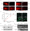
Uptake kinetics and intracellular distribution of Cu from the ES–Cu complex. (A and B) X-ray fluorescence microscopy elemental maps for phosphorus (P) and Cu in (A) Ctr1−/− and (B) Ctr1−/−Fdx1−/− cells treated with DMSO or 100 nM ES–Cu for 30 min. Elemental concentration ranges are represented by false coloring from the darkest (lowest concentration) to the brightest (highest concentration). Images are representative of two independent experiments. (C) Relative Cu accumulation in Ctr1−/− and Ctr1−/−Fdx1−/− cells treated with 100 nM ES–Cu for the indicated time intervals. Cu levels were normalized to 0 h. Data are expressed as mean ± SD, n = 3. (D) Immunofluorescence analysis of the steady-state localization and trafficking of endogenous ATP7A (green) in Ctr1−/− and Ctr1−/−Fdx1−/− cells treated with DMSO or 5 nM ES–Cu for 3 h. DAPI was used to stain the nuclei (blue). (E) SDS–PAGE/western blot analysis of CCS protein levels in the indicated cell types treated without or with 1 or 5 nM ES–Cu. β-actin was used as a loading control. (F) In-gel SOD1 activity in the indicated cell types treated with or without 1 or 5 nM ES–Cu. Coomassie stain of the same gel is used as a loading control.
Fig. 4.
Fig. 4.
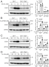
FDX1 is specific for Cu release from ES–Cu complex. Western blot analysis of COX1 protein levels in the indicated cells treated with or without (A) 1 or 5 nM ES–Cu, (B) 1 or 5 nM Cu-ATSM, (C) 1 or 5 nM CuCl2+ disulfiram, and (D) 1 or 5 µM Cu–histidine for 72 h. ATP5A was used as a loading control. The relative abundance of COX1 was quantified by densitometry analysis using ImageJ software. Data were normalized to protein levels in untreated Ctr1−/− cells and expressed as mean ± SD (n = 3). Statistical significance was assessed by one-way ANOVA with Tukey’s multiple comparison test using GraphPad Prism 9 software. ns: not significant; each dot on the bar chart represents individual data point.
Fig. 5.
Fig. 5.
A model depicting ES-mediated intracellular Cu distribution. ES binds Cu(II) in the extracellular environment forming an electrically neutral, square planar ES–Cu complex that can pass through the lipid membranes and enter the mitochondria where Cu(I) is released from ES–Cu by the action of FDX1 protein. The released Cu(I) is now bioavailable for the metalation of cytochrome c oxidase (solid arrow) and to a certain extent to other cuproenzymes present outside the mitochondria (broken arrow). Cu is also released from ES–Cu outside of the mitochondria where it is bioavailable to cytosolic cuproproteins—CCS and SOD1 (solid arrows) and other subcellular compartment.

References

    1. Garza N. M., Swaminathan A. B., Maremanda K. P., Zulkifli M., Gohil V. M., Mitochondrial copper in human genetic disorders. Trends Endocrinol. Metab. 34, 21–33 (2023). - PMC - PubMed
    1. Kaler S. G., Inborn errors of copper metabolism. Handb. Clin. Neurol. 113, 1745–1754 (2013). - V体育安卓版 - PMC - PubMed
    1. Papadopoulou L. C., et al. , Fatal infantile cardioencephalomyopathy with COX deficiency and mutations in SCO2, a COX assembly gene. Nat. Genet. 23, 333–337 (1999). - PubMed
    1. Valnot I., et al. , Mutations of the SCO1 gene in mitochondrial cytochrome c oxidase deficiency with neonatal-onset hepatic failure and encephalopathy. Am. J. Hum. Genet. 67, 1104–1109 (2000). - PMC - PubMed
    1. Baertling F., et al. , Mutations in COA6 cause cytochrome c oxidase deficiency and neonatal hypertrophic cardiomyopathy. Hum. Mutat. 36, 34–38 (2015). - PubMed

V体育官网入口 - Publication types

LinkOut - more resources