Skip to main page content
U.S. flag

An official website of the United States government

Dot gov

The . gov means it’s official VSports app下载. Federal government websites often end in . gov or . mil. Before sharing sensitive information, make sure you’re on a federal government site. .

Https

The site is secure. The https:// ensures that you are connecting to the official website and that any information you provide is encrypted and transmitted securely. V体育官网.

. 2022 Dec 4;11(23):3927.
doi: 10.3390/cells11233927.

VSports app下载 - Nrf2 Deficiency Exacerbated CLP-Induced Pulmonary Injury and Inflammation through Autophagy- and NF-κB/PPARγ-Mediated Macrophage Polarization

Affiliations

V体育平台登录 - Nrf2 Deficiency Exacerbated CLP-Induced Pulmonary Injury and Inflammation through Autophagy- and NF-κB/PPARγ-Mediated Macrophage Polarization

Jing Luo et al. Cells. .

Abstract

The balance between M1 and M2 macrophage polarization is involved in the regulation of pulmonary inflammation VSports手机版. Nuclear factor erythroid-derived 2-like 2 (Nfe2l2, also known as Nrf2), a nuclear transcription factor, is reported to play protective roles in acute lung injury (ALI) and inflammation, and increasing evidence indicates that the protective effects of Nrf2 are closely related to autophagy. This study aimed to explore whether Nrf2 is involved in sepsis-induced acute pulmonary injury and inflammation and in the role of macrophage polarization in the process. In the present study, sepsis patients, an Nrf2 knockout mouse that underwent cecal ligation and puncture (CLP), and lipopolysaccharide (LPS)-treated macrophage cell lines were employed to investigate the potential functions of Nrf2 in sepsis-induced lung injury and the underlying mechanisms. Clinical studies showed that the NRF2 mRNA level was inversely correlated with pulmonary inflammation and disease severity in patients with sepsis. Analyses in a CLP-treated Nrf2 knockout mouse model indicated that an Nrf2 deficiency promoted a CLP-induced increase in M1 macrophage polarization and apoptosis and inhibited CLP-induced upregulation of the autophagy level in lung tissues. Experiments in RAW264. 7 cells revealed that Nrf2 overexpression inhibited M1 macrophage polarization but promoted M2 macrophage polarization by improving the autophagy, and Nrf2 overexpression promoted PPARγ but inhibited NF-κB nuclear translocation. In conclusion, these results indicate that Nrf2 plays a protective role in sepsis-induced pulmonary injury and inflammation through the regulation of autophagy- and NF-κB/PPARγ-mediated macrophage polarization. .

Keywords: Nrf2; acute lung injury; autophagy; macrophage polarization; pulmonary inflammation V体育安卓版. .


Conflict of interest statement (V体育安卓版)

The authors declare no conflict of interest.

Figures

Figure 1
Figure 1
Patients with sepsis show a negative correlation between NRF2 mRNA level in peripheral blood and the severity of disease. (A) QRT-PCR detected the NRF2 mRNA level in the blood of 30 healthy controls and 30 sepsis patients at the acute and recovery stages. Paired t-test was used for data comparison between acute and recovery sepsis patients, and the NRF2 mRNA level was standardized to human housekeeping gene ACTB. (B) Correlation analysis of the APACHE II score and NRF2 mRNA level in the blood of acute sepsis patients. (C,D) Correlation analysis of TNF-α and IL-6 contents in BALFs and NRF2 mRNA level in the blood of acute sepsis patients. ** p < 0.01, *** p < 0.001.
Figure 2
Figure 2
Nrf2 deficiency exacerbates sepsis-induced ALI and promotes inflammation in a CLP mouse model; n = 6 mice per group. (A) Histological analyses of the lung tissues in WT and Nrf2−/− mice in both the sham and CLP groups. (B) Representative lung injury scores in (A). (C,D) Western blot analyses of Nrf2 protein in WT and Nrf2−/− mice. (E,F) The expressions of inflammatory cytokines including IL-1β and TNF-α in BALFs were measured with ELISA. ** p < 0.01, *** p < 0.001.
Figure 3
Figure 3
Transcriptome sequencing analyses of lung tissues in CLP-treated WT and Nrf2−/− mice. (A) Hierarchical clustered heatmap of DEGs in lung tissues of CLP-treated WT mice (WT+CLP) and CLP-treated Nrf2−/− mice (Nrf2−/− + CLP); n = 6 mice per group. (B) KEGG enrichment analysis identified the most significantly altered signaling pathways after Nrf2 deletion; n = 3 in each group. (C) Construction of Nrf2-related PPI regulatory network based on DEGs. (D) Heatmap of DEGs in CLP-treated WT and Nrf2−/− mice. (E) Network of KEGG pathway based on the similarity of gene expression profiles. (F) Volcano plot showing DEGs of CLP-treated WT and Nrf2−/− mice. The red markers represent genes that were significantly upregulated, and the green markers represent genes that were significantly downregulated, while insignificantly altered genes are highlighted in gray; log2 fold change > 1, Q value < 0.05.
Figure 4
Figure 4
Nrf2 deficiency promotes CLP-induced M1 macrophage polarization and apoptosis in lung tissues. n = 6 mice per group. (A,B) Immunofluorescence staining of iNOS and F4/80 in the lung tissues of each group. (C,D) TUNEL staining presented apoptotic cells in the lung tissues of WT and Nrf2−/− mice before or after CLP treatment. (E,F) Western blot analyses of apoptosis-associated proteins such as Bcl-2, Bax, and Cleaved—caspase3 in the lung tissues of each group. ** p < 0.01, *** p < 0.001.
Figure 5
Figure 5
Nrf2 deficiency inhibits CLP-induced upregulation of autophagy level in lung tissues; n = 6 mice per group. (A,B) Immunofluorescence staining of LC3 and F4/80 in the lung tissues of WT and Nrf2−/− mice before or after CLP treatment. (C,D) Western blot analyses of autophagy-associated proteins including LC3 I, LC3 II, Beclin1, and p62 in lung tissues of each group. ** p < 0.01, *** p < 0.001. (E) Transmission electron microscopy assay detected the numbers of double-membrane autophagosomes and autolysosomes (red arrows in the pictures) in lung tissues of each group.
Figure 6
Figure 6
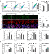
Nrf2 deficiency promotes M1 macrophage polarization and inhibits M2 macrophage polarization through autophagy modulation; n = 3 experiments per group. (A,B) Flow cytometry analyses of M1 macrophage polarization in BMDMs. (C,D) Immunofluorescence staining of CD206 and F4/80 in BMDMs. White arrows in the pictures indicate polarized macrophages. (E) Isolated BMDMs were treated with LPS/IFN-γ (15 ng/mL and 50 ng/mL, respectively) for 24 h to induce M1 macrophage polarization, and 0.1 μM RAPA was added simultaneously for autophagy activation. The levels of M1 macrophage markers such as iNOS, IL-6, IL-1β, and TNF-α were measured with qRT-PCR. (F) Isolated BMDMs were treated with IL-4/IL-13 (25 mg/mL) for 24 h to induce M2 macrophage polarization, and 0.1 μM RAPA was added simultaneously for autophagy activation. The levels of M2 macrophage markers such as Arg1, Fizz1, Ym1, and IL-10 were determined by qRT-PCR. * p < 0.05, ** p < 0.01, *** p < 0.001.
Figure 7
Figure 7
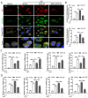
Nrf2 overexpression promotes LPS-induced upregulation of autophagy in vitro; n = 3 experiments per group. (A,B) Immunofluorescence staining of LAMP2 and LC3 in RAW264.7 cells. (C,D) Western blot analyses of the levels of LC3 I, LC3 II, Beclin1, and p62 proteins in RAW264.7 cells of each group. ** p < 0.01, *** p < 0.001.
Figure 8
Figure 8
Nrf2 overexpression inhibits M1 macrophage polarization and improves M2 macrophage polarization by promoting autophagy in vitro. Macrophage polarization induction was performed in RAW264.7 cells; n = 3 experiments per group. (A,B) Cells were first transfected with 2 μg of Nrf2 plasmid or vector for 48 h and treated with 1 μg/mL LPS for 24 h. Then, mRFP-GFP-LC3B plasmids were transfected, and 24 h later confocal images were taken to detect the numbers of autophagosomes (yellow dots) and autolysosomes (red dots). White arrows indicate autophagosomes in merged images. (C) RAW264.7 cells were treated with LPS/IFN-γ (15 ng/mL and 50 ng/mL, respectively) for 24 h to induce M1 macrophage polarization, and 2 mM 3-MA was added simultaneously for autophagy inhibition. The levels of M1 macrophage markers including iNOS, IL-6, IL-1β, and TNF-α were measured with qRT-PCR. (D) RAW264.7 cells were treated with IL-4/IL-13 (25 mg/mL) for 24 h to induce M2 macrophage polarization, and 2 mM 3-MA was added simultaneously for autophagy inhibition. The levels of M2 macrophage markers including Arg1, Fizz1, Ym1, and IL-10 were measured with qRT-PCR. ** p < 0.01, *** p < 0.001.
Figure 9
Figure 9
Nrf2 overexpression in vitro promotes PPARγ but inhibits NF-κB nuclear translocation; n = 3 experiments per group. (A,B) Immunofluorescence staining of NF-κB and PPARγ in RAW264.7 cells in each group. (C,D) Western blot analyses of Nrf2, NF-κB, and PPARγ levels in both cytoplasm and nucleus. ** p < 0.01, *** p < 0.001.
Figure 10
Figure 10
Hypothetical schema for the roles of Nrf2 in sepsis-induced lung injury. Nrf2 plays a protective role in sepsis-induced lung injury and inflammation through the regulation of autophagy-and NF-κB/PPARγ-mediated macrophage polarization.

"VSports注册入口" References

    1. Singer M., Deutschman C.S., Seymour C.W., Shankar-Hari M., Annane D., Bauer M., Bellomo R., Bernard G.R., Chiche J.-D., Coopersmith C.M., et al. The Third International Consensus Definitions for Sepsis and Septic Shock (Sepsis-3) JAMA. 2016;315:801–810. doi: 10.1001/jama.2016.0287. - DOI - PMC - PubMed
    1. Shashaty M.G.S., Reilly J.P., Faust H.E., Forker C.M., Ittner C.A.G., Zhang P.X., Hotz M.J., Fitzgerald D., Yang W., Anderson B.J., et al. Plasma receptor interacting protein kinase-3 levels are associated with acute respiratory distress syndrome in sepsis and trauma: A cohort study. Crit. Care. 2019;23:235. doi: 10.1186/s13054-019-2482-x. - DOI - PMC - PubMed
    1. Hwang J.-S., Kim K.-H., Park J., Kim S.-M., Cho H., Lee Y., Han I.-O. Glucosamine improves survival in a mouse model of sepsis and attenuates sepsis-induced lung injury and inflammation. J. Biol. Chem. 2019;294:608–622. doi: 10.1074/jbc.RA118.004638. - DOI - PMC - PubMed
    1. Xiong S., Hong Z., Huang L.S., Tsukasaki Y., Nepal S., Di A., Wu W., Ye Z., Gao X., Rao G.N., et al. IL-1β suppression of VE-cadherin transcription underlies sepsis-induced inflammatory lung injury. J. Clin. Investig. 2020;130:3684–3698. doi: 10.1172/JCI136908. - DOI - PMC - PubMed
    1. Liu Y., Zhou J., Luo Y., Li J., Shang L., Zhou F., Yang S. Honokiol alleviates LPS-induced acute lung injury by inhibiting NLRP3 inflammasome-mediated pyroptosis via Nrf2 activation in vitro and in vivo. Chin. Med. 2021;16:127. doi: 10.1186/s13020-021-00541-z. - "V体育官网" DOI - PMC - PubMed