VSports手机版 - Skip to main page content
U.S. flag

An official website of the United States government

Dot gov

The VSports app下载. gov means it’s official. Federal government websites often end in . gov or . mil. Before sharing sensitive information, make sure you’re on a federal government site. .

Https

The site is secure V体育官网. The https:// ensures that you are connecting to the official website and that any information you provide is encrypted and transmitted securely. .

. 2023 Jan;42(2):83-98.
doi: 10.1038/s41388-022-02537-x. Epub 2022 Nov 12.

CST1 inhibits ferroptosis and promotes gastric cancer metastasis by regulating GPX4 protein stability via OTUB1

Affiliations

CST1 inhibits ferroptosis and promotes gastric cancer metastasis by regulating GPX4 protein stability via OTUB1

Dongbao Li et al. Oncogene. 2023 Jan.

Abstract

Metastasis is an important factor contributing to poor prognosis in patients with gastric cancer; yet, the molecular mechanism leading to this cell behavior is still not well understood. In this study, we explored the role of cysteine protease inhibitor SN (Cystatin SN, CST1) in promoting gastric cancer metastasis. We hypothesized that CST1 could regulate gastric cancer progression by regulating GPX4 and ferroptosis. Whole transcriptome sequencing suggested that the expression of CST1 was significantly increased in metastatic cancer, and high CST1 expression was correlated with a worse prognosis. Our data further confirmed that the overexpression of CST1 may significantly promote the migration and invasion of gastric cancer cells in vitro and enhance liver, lung, and peritoneal metastasis of gastric cancer in nude mice. Meanwhile, high expression of CST1 promoted the epithelial-mesenchymal transition (EMT) of gastric cancer cells. Mechanistically, a co-immunoprecipitation experiment combined with mass spectrometry analysis confirmed that CST1 could interact with GPX4, a key protein regulating ferroptosis. CST1 relieves GPX4 ubiquitination modification by recruiting OTUB1, improving GPX4 protein stability and reducing intracellular reactive oxygen species (ROS), thereby inhibiting ferroptosis and, in turn, promoting gastric cancer metastasis. Moreover, clinical data suggested that CST1 is significantly increased in peripheral blood and ascites of gastric cancer patients with metastasis; multivariate Cox regression model analysis showed that CST1 was an independent risk factor for the prognosis of gastric cancer patients. Overall, our results elucidated a critical pathway through which high CST1 expression protects gastric cancer cells from undergoing ferroptosis, thus promoting its progression and metastasis VSports手机版. CST1 may be used as a new oncological marker and potential therapeutic target for gastric cancer metastasis. .


Conflict of interest statement (V体育官网入口)

The authors declare no competing interests.

"VSports在线直播" Figures

Fig. 1
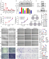
Fig. 1. CST1 is up-regulated in primary and metastatic GC tissues and is associated with a poor prognosis.
Heatmap mainly showing expression levels of 20 up-regulated and 20 down-regulated different expressed genes in primary GC (A) and metastatic GC (C) vs. adjacent normal tissues (>1.5-fold). Volcano plots with differentially expressed genes in primary GC (B) and metastatic GC (D) vs. adjacent normal tissues. E Venn diagram representation of 768 overlapped up-regulated genes. F Histogram was shown the overlapped highest 20 up-regulated genes in these two clusters. G, H Higher expression of CST1 was found in GC samples than the matched normal tissues (based on GSE54129 and GSE66229 database). I, J Kaplan–Meier plots of overall survival and progression-free survival for GC samples from the KM Ploter database. K RT-qPCR analysis of CST1 mRNA expression in 100 pairs of GC patient samples. Data are shown as the mean ± SD of triplicate independent sets of experiments; statistical significance was assessed by paired t-test, *<0.05, n = 100. L Western blot analysis was performed using an antibody against CST1 in 5 pairs of GC patients’ samples (upper panel); protein band intensities were measured by ImageJ software and normalized to GAPDH (lower panel). M IHC staining was performed using an antibody against CST1 and representative photographs of CST1 in GC patients. Scale bar: 100 μm. N IHC stain scoring of CST1 in 185 GC tissues and 185 normal tissues, statistical significance was assessed by unpaired t-test, ****<0.0001.
Fig. 2
Fig. 2. CST1 promotes GC cell metastasis but not proliferation in vitro.
A, B RT-qPCR and Western blot analysis showing the expression of CST1 in different GC cell lines. Total GAPDH was used as a loading control. C Western blot analysis of HGC-27 and MKN45 stably transfected with CST1 overexpression/knockdown lentiviruses and control lentiviruses. Total GAPDH was used as a loading control. D CCK8 assay analyzed the proliferation of HGC-27-Vector/HGC-27-CST1 and MKN45-shNC/MKN45-sh1-CST1/MKN45-sh2-CST1 stable cell lines. Data are shown as the mean ± SD of triplicate independent sets of experiments; statistical significance was assessed by paired t-test. E A colony formation assay. Left panel: representative images, right panel: quantification analysis. Data from independent experiments are presented as the mean ± SD. Statistical was assessed by unpaired t-test, ns means no significance. F Wound healing analysis for assessing migration of HGC-27-Vector/HGC-27-CST1 and MKN45-shNC/MKN45-sh1-CST1/MKN45-sh2-CST1 at 0, 24, and 48 h. Representative images (left panel) and quantification (right panel) are shown as indicated. Data from independent experiments are presented as the mean ± SD. Statistical significance was assessed by an unpaired t-test. ***p < 0.001. Scale bar: 100 μm. G Transwell migration and Matrigel invasion assays were performed to assess migration and invasion ability of CST1-overexpression and knockdown stable cell lines. Representative images (left panel) and quantification (right panel) are shown as indicated. Data from independent experiments are presented as the mean ± SD. Statistical significance was assessed by an unpaired t-test. ***p < 0.001. Scale bar: 100 μm.
Fig. 3
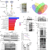
Fig. 3. CST1 interacts with GPX4 to improve the stability of GPX4 protein.
A Schematic of CST1 interactor discovery. The potential interactors were presented at higher levels in the CST1 experimental group than in the IgG control group. Three replicates of IP-MS were conducted. B Silver staining of IP cell lysates. C Venn diagram of three times of LC-MS results showing 63 co-upregulated proteins. D KEGG pathways analysis of 63 co-upregulated proteins. E Immunoprecipitation of the CST1 protein by an anti-GPX4 antibody in MKN45 cells. IgG was used as a negative control. F Immunoprecipitation of the GPX4 protein by an anti-Flag antibody in HEK293 cells transfected with pcDNA3.1-FLAG-CST1. PcDNA3.1-vector was used as a negative control. G Western blot showing GPX4 expression in HGC-27 and MKN45 stable cell lines; total GAPDH was used as a loading control. H, I Degradation of the GPX4 protein was measured after the treatment of 200 µg/ml CHX at the indicated time points in HEK293T, which transfected with Flag-tagged CST1 expression plasmids and Myc-tagged GPX4 expression plasmids. J, K Western blot analysis of GPX4 expression after treatment with 20 µM MG132 for 4 h in MKN45 stable cell lines. Data were expressed as a fold-change relative to control. L Analysis of GPX4 ubiquitination was performed by immunoprecipitation using an anti-Myc antibody, followed by immunoblot with anti-HA antibody and anti-Myc antibody in HEK293T cells transfected with the indicated constructs.
Fig. 4
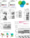
Fig. 4. CST1 relieves GPX4 ubiquitination through deubiquitinase OTUB1.
A GSEA analysis of our previous transcriptome sequencing data and GEO data set (GSE66229) enriched the deubiquitination pathway. B Predicting the deubiquitinase DUBs that interact with GPX4 through the online database (BioGRID, IntAct) and Venn analysis with all known DUBs. Intersection proteins include OTUB1 and OTUD5. C Co-IP assay on HEK293T cells transfected with Flag-tagged OTUB1 and OTUD5 plasmids. D WB detection of Flag, HA, Myc tagged proteins after IP in cells co-transfected with Myc-tagged CST1 and Flag-tagged OTUB1. E Endogenous Co-IP of GPX4 in MKN45-shNC/MKN45-sh1-CST1/MKN45-sh2-CST1. WB detection of IP proteins; OTUB1 binding to GPX4 was significantly reduced. F HGC-27-Vector/HGC-27-CST1 cells were transiently transfected with OTUB1 siRNA. The ubiquitination assay showed that the level of ubiquitin bound by GPX4 increased with the decrease of OTUB1, and the ubiquitination level of GPX4 was more obvious in HGC27 cells overexpressing CST1. G HGC-27-Vector/HGC-27-CST1 cells were transiently transfected with Flag-OTUB1-Con/WT/D88A and HA-GPX4 plasmids, WB detection showed that the GPX4 protein in the OTUB1-WT group was more stable, while the GPX4 protein in the OTUB1-D88A group was reduced. H Predicted binding complex models of CST1, OTUB1 and GPX4 by using the Cluspro online protein docking tool. I Schematic diagram of the construction of full-length and truncated CST1 proteins. J HEK293T cells were transfected with CST1-FL/N/C-Myc plasmids and OTUB1-Flag, GPX4-HA plasmid, respectively. Myc protein was immunoprecipitated, WB showed that CST1-FL and CST1-N truncated protein can bind to OTUB1, while CST1-FL and CST1-C truncated proteins can bind to GPX4. The SDS-PAGE used for separating CST fragments was 15%. (GSEA Gene set enrichment analysis, Con control/empty plasmid, WT Wild-type plasmid, D88A the D88 site of OTUB1 is point mutated to A plasmid).
Fig. 5
Fig. 5. CST1 reduces intracellular ROS and inhibits ferroptosis through GPX4.
A Treatment of HGC-27-Vector/HGC-27-CST1 with the ferroptosis inducer erastin (10 μM) inhibited the decrease of HGC-27-CST1 cell viability relative to the DMSO-treated group (****<0.0001). B Treating MKN45-shNC/MKN45-sh1-CST1/MKN45-sh2-CST1 cells with erastin (10 μM) decreased the viability more significantly than the DMSO-treated group (****<0.0001). After ferroptosis inhibitor liproxstatin-1 treatment, the viability of MKN45-sh cells increased. C, D The level of ROS in HGC-27-Vector/HGC-27-CST1cells detected by fluorescence microscopy. The results showed that in the erastin-treated group, CST1 reduced the level of intracellular ROS (****<0.0001). E, F In MKN45-shNC/MKN45-sh1-CST1/MKN45-sh2-CST1 cells, intracellular ROS was significantly increased after erastin treatment (****<0.0001), while ferroptosis inhibitor liproxstatin-1 reversed this process. G MDA content in HGC-27-CST1 cells decreased, and this difference was more significant after erastin treatment (****<0.0001). H MDA content in MKN45-sh cells significantly increased, and the difference was more significant after erastin treatment. After liproxstain-1 treatment, the MDA content decreased again. I The GSH content in HGC-27-CST1 cells increased. J GSH content in MKN45-sh cells decreased, and the difference was more significant after erastin treatment and increased after liproxstatin-1 treatment (****<0.0001).
Fig. 6
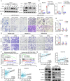
Fig. 6. CST1 mediates GPX4 protein stability to promote migration and invasion in epithelial-mesenchymal transition manner in GC cells.
A Western blot analysis of GPX4 in HCG27 and MKN45 stable cell lines transfected with GPX4-siRNA or exogenous overexpressing GPX4. B, C Transwell migration and invasion assays were performed in HGC-27-Vector/HGC-27-CST1 treated with GPX4-siRNA and control as indicated. D, E Transwell migration and invasion assays were performed in MKN45-shNC/MKN45-sh1-CST1/MKN45-sh2-CST1 treated with transient transfection of GPX4 exogenous overexpression or vector control as indicated. Data are presented as the mean ± SD. Statistical significance was assessed by an unpaired t-test. ****p < 0.0001. F Gene set enrichment analysis of CST1 related to invasiveness and degradation of the extracellular matrix. G, H Correlation analysis of CST1 and FN1/Snail in GSE54129 and GSE66229 as illustrated in the dot plot (Person’s correlation test). I Western blot analysis of E-cadherin, Vimetin, FN1 expression in the indicated cells.
Fig. 7
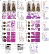
Fig. 7. CST1 promotes distant metastasis in vivo.
A Left: Images of peritoneal metastasis in nude BALB/c mice after injection of HGC-27-Vector/HGC-27-CST1/HGC-27-GPX4#sh cells into their abdominal cavity; images taken 60 days after injection. Right: statistical significance of the peritoneal nodules number and the ascites volume assessed by paired t-test, *p < 0.05, **p < 0.01. B Left: CST1-silenced MKN45 peritoneum xenograft tumors 30 days after injection. Right: statistical significance of the peritoneal nodules number and the ascites volume assessed by paired t-test, **p < 0.01, ****p < 0.0001. C Left: corresponding images of the lungs after injection of HGC-27-Vector/HGC-27-CST1/HGC-27-GPX4#sh cells by tail vein; images taken 3 months after injection. Right: statistical significance of the metastasis nodules number assessed by paired t-test, **p < 0.01. D Left: lung metastasis model of MKN45-shNC/MKN45-sh1-CST1/MKN45-sh2-CST1 8 weeks after tail vain injection. Right: statistical significance of the metastasis nodules number assessed by paired t-test, ***p < 0.001. E, F Left: HGC-27-Vector/HGC-27-CST1 and MKN45-shNC/MKN45-sh1-CST1/MKN45-sh2-CST1 cells were injected into tail vein; metastatic tumors in the livers were assessed 2 and 3 months after injection. Right: statistical significance of the metastasis nodules number assessed by paired t-test, **p < 0.01, ***p < 0.001. G Western blot analysis indicated metastasis tumors for GPX4 and CST1. H The content of MDA in the peritoneal metastatic tumor tissue of nude mice was detected, and the difference was statistically significant (***p < 0.001, ****p < 0.0001). HE-stained sections were magnified ×0.66 and ×5, respectively.
Fig. 8
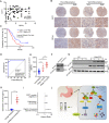
Fig. 8. High CST1 and GPX4 expression levels correlate with tumor aggressiveness and poor clinical outcome in GC patients.
A IHC for 95 GC patients’ tissue. Pearson correlation analysis of the expression of CST1 and GPX4. B Representative graph of CST1 and GPX4 expression in IHC according to the degree of differentiation of gastric cancer tissues. C Survival analysis; the survival time of 95 gastric cancer patients was analyzed; patients with high CST1 expression had a shorter survival (p = 0.0036). D ROC curve analysis of the sensitivity and specificity of CST1 in the diagnosis of gastric cancer patients (AUC = 0.9311, p < 0.0001). E Serum ELISA assay in 50 normal people, 45 stage I–II GC patients, and 30 stage III–IV GC patients revealed that CST1 was highly expressed in GC patients’ serum compared with normal people and highly expressed consistent with patient’s clinicopathological stage (p < 0.0001). F, G Western blot detecting intracellular and extracellular CST1 in HEK293T cells with the FLAG-CST1 expression vector. H Peritoneal lavage fluid of 50 GC patients without metastasis and 30 GC patients with malignant ascites by ELISA. Expression of CST1 in malignant ascites was significantly higher than without metastasis patients (p < 0.0001). I Multivariate Cox regression model analysis of the relationship between CST1 expression and prognosis of gastric cancer. J Mechanistic diagram showing the role of CST1 in gastric cancer cells.

V体育官网入口 - References

    1. Sung H, Ferlay J, Siegel RL, Laversanne M, Soerjomataram I, Jemal A, et al. Global cancer statistics 2020: GLOBOCAN estimates of incidence and mortality worldwide for 36 cancers in 185 countries. CA Cancer J Clin. 2021;71:209–49. doi: 10.3322/caac.21660. - DOI - PubMed
    1. Smyth EC, Moehler M. Late-line treatment in metastatic gastric cancer: today and tomorrow. Ther Adv Med Oncol. 2019;11:1758835919867522. doi: 10.1177/1758835919867522. - DOI (VSports最新版本) - PMC - PubMed
    1. Abrahamson M, Alvarez-Fernandez M, Nathanson CM. Cystatins. Biochem Soc Symp. 2003;70:179–99. doi: 10.1042/bss0700179. - "VSports app下载" DOI - PubMed
    1. de Sousa-Pereira P, Amado F, Abrantes J, Ferreira R, Esteves PJ, Vitorino R. An evolutionary perspective of mammal salivary peptide families: cystatins, histatins, statherin and PRPs. Arch Oral Biol. 2013;58:451–8. doi: 10.1016/j.archoralbio.2012.12.011. - DOI - PubMed
    1. Hiltke TR, Lee TC, Bobek LA. Structure/function analysis of human cystatin SN and comparison of the cysteine proteinase inhibitory profiles of human cystatins C and SN. J Dent Res. 1999;78:1401–9. doi: 10.1177/00220345990780080501. - DOI - PubMed

Publication types