<kbd dir="kkRSvnu"></kbd> Skip to main page content
U.S. flag

An official website of the United States government

Dot gov

The . gov means it’s official. Federal government websites often end in . gov or . mil. Before sharing sensitive information, make sure you’re on a federal government site. VSports app下载.

Https

The site is secure V体育官网. The https:// ensures that you are connecting to the official website and that any information you provide is encrypted and transmitted securely. .

. 2022 Sep 30;22(1):297.
doi: 10.1186/s12935-022-02703-x.

"V体育安卓版" Mucosal-associated invariant T cells predict increased acute graft-versus-host-disease incidence in patients receiving allogeneic hematopoietic stem cell transplantation

Affiliations

Mucosal-associated invariant T cells predict increased acute graft-versus-host-disease incidence in patients receiving allogeneic hematopoietic stem cell transplantation

Zhao Wang et al. Cancer Cell Int. .

Abstract

Background: Mucosal-associated invariant T (MAIT) cells are innate-like T cells, some studies have reported that the number of circulating MAIT cells reduced in patients with acute graft-versus-host-disease (aGVHD) development VSports手机版. However, the role of donor MAIT cells on aGVHD development and subsequent functional change still remain unclear. .

Methods: The study recruited 86 patients with hematological malignancies who underwent allogeneic hematopoietic cell transplantation (HCT) from May 1, 2018 to June 30, 2019 V体育安卓版. MAIT cells, their subset, and cytokine levels were measured by flow cytometry. Gray's test was used to assess the impact of graft MAIT cell proportion and number on aGVHD incidence. The Cox proportional hazard model was used in the multivariate analysis. The comparison for continuous variables was assessed using Mann-Whitney analysis. RNA-sequencing was performed to investigate the possible molecular pathway changes. .

Results: Our study showed that the proportion of MAIT cells in grafts was not different from normal controls, but the CD4/8 subsets were altered. Taking the median of the proportion and number of MAIT cells in the graft as the threshold, the results showed that the incidence of grade B-D aGVHD in patients with MAIT cell proportion ≥ 3. 03% was significantly higher than that in patients with MAIT cell proportion < 3. 03% (56. 3%, 95% CI 37. 1-71. 2 versus 23. 1%, 95% CI 13. 8-46 V体育ios版. 2; P = 0. 038). The number of MAIT cells in the graft was not associated with aGVHD development (P = 0. 173), however, when the graft contained more CD4 positive, CD8 positive, and CD4/CD8 double-positive MAIT cells, the incidence of aGVHD was significantly increased (P = 0. 019, P = 0. 035 and P = 0. 027, respectively). Besides, reduced frequencies and counts of circulating MAIT cells were observed in patients with aGVHD when compared to patients without aGVHD, accompanied by enhanced production of Tumor necrosis factor-α, Interferon-γ and upregulated programmed death-1, CXC Chemokine Receptor-6 (CXCR6) and CD38 expression. Gene set enrichment analysis of MAIT cell RNA-seq data showed interferon-α response pathway upregulated in aGVHD patients when compared with patients without aGVHD and healthy controls. .

Conclusions: Our study shows that MAIT cells in grafts and peripheral blood are both closely related to the aGVHD development post allogeneic HCT VSports最新版本. Interferon-α response pathway perhaps is a critical regulation mechanism for the MAIT cell involvement in aGVHD development. .

Keywords: Cytokines; Graft versus host disease; Hematopoietic cell transplantation; MAIT cell. V体育平台登录.


Conflict of interest statement

The authors declare that they have no competing interests.

Figures
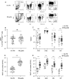
Fig. 1
Fig. 1
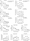
Mucosal-associated-invariant T (MAIT) cells mobilization characteristics in PB-grafts. A Gating strategy and a representative example of flow cytometric analysis of one peripheral blood graft (PB-graft) sample and one healthy subject peripheral blood (HC-PB) sample. B The frequency of MAIT cells in PB-grafts (n = 69) and HC-PB (n = 48). C Proportion of CD4, CD8, DN, DP subsets in PB-grafts (n = 66) and healthy controls (n = 40). D The number of MAIT cells in PB-grafts (n = 69) and HC-PB (n = 48). C number of CD4, CD8, DN, DP subsets in PB-grafts (n = 66) and healthy controls (n = 40). *P-value < 0.05; **P-value < 0.01; ***P-value < 0.001; NS: no significance
Fig. 2
Fig. 2
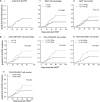
Factors that affect the reconstitution of MAIT cells post allogeneic HCT. A Mucosal-associated-invariant T cell frequencies and counts in peripheral blood from recipients receiving HLA-matched sibling donor transplantation (n = 45) and alternative donor transplantation (n = 41). B MAIT cell frequencies and counts from recipients receiving transplantation with ATG (n = 64) and without ATG (n = 22) in the conditioning regimes. C MAIT cell frequencies and counts from recipients receiving HLA-matched sibling donor transplantation with ATG (n = 23) and without ATG (n = 22) in the conditioning regimes. D MAIT cell frequencies and counts from recipients existing with or without bacterial infection (n = 63 and 23, respectively); fungal infection (n = 17 and 69, respectively) and viral infection (n = 34 and 52, respectively). Mann–Whitney test was used to assess significant differences between groups. Mean ± standard error is shown. *P-value < 0.05; **P-value < 0.01; ***P-value < 0.001
Fig. 3
Fig. 3
MAIT cell proportion and number in grafts predicted grade (BD) aGVHD development. (AI) Cumulative incidence of grades BD aGVHD in the entire cohort (n = 69); grades BD aGVHD incidence according to (AII) MAIT cell proportion; (AIII) MAIT cell number; (BI) CD4-CD8 + MAIT cell proportion; (BII) CD4 + CD8-MAIT cell number; (BIII) CD4 + CD8 + MAIT cell number and (BIV) CD4-CD8 + MAIT cell number in PB-grafts. Gray’s test was used to compare the aGVHD cumulative incidence between groups
Fig. 4
Fig. 4
Reduced circulating MAIT cells proportion and number in aGVHD recipients. A, B MAIT cell early reconstitution in the entire cohort (n = 86). C, D circulating MAIT cell proportion and number in recipients with grades BD aGVHD in comparison with grades 0‒A aGVHD post allogeneic HCT. Mean ± standard error is shown. Mann–Whitney test was used to assess significant differences between groups. *P-value < 0.05; **P-value < 0.01; ***P-value < 0.001
Fig. 5
Fig. 5
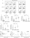
Functional activation of MAIT cells in patients with aGVHD. Representative gating for PD-1, CXCR6, CD38 expression, and IFN-γ, TNF-α production on MAIT cells from PB-grafts, patients without aGVHD and patients with aGVHD development. B Expression of PD-1, CXCR6, CD38 on MAIT cells from PB-grafts (n = 26), patients without aGVHD (n = 22) and patients with aGVHD (n = 23). C Production of IFN-γ, TNF-α in MAIT cells from PB-grafts (n = 16), patients without aGVHD (n = 16) and patients with aGVHD (n = 14). D Spearman rank correlation between circulating MAIT cell frequency and PD-1, IFN-γ and TNF-α expression on MAIT cells from recipients receiving allogeneic HCT. Mean ± standard error is shown. Mann‒Whitney test was used to assess significant differences between groups. *P-value < 0.05; **P-value < 0.01; ***P-value < 0.001; NS: no significance
Fig. 6
Fig. 6
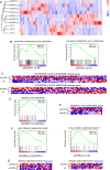
Differential gene expression analysis for MAIT cells from patients and healthy controls. A Heatmap of total differential gene expressions. B Gene set enrichment analysis (GSEA) for IFN-α response pathway enrichment in MAIT cells from aGVHD patients and non-aGVHD patients when compared to those fron healthy controls (i–ii); Moserle IFN-αresponse pathway enrichment in patients with aGVHD when compared to that in patients without aGVHD (iii). C Heatmap presenting the expression of IFN-α-response hallmark genes and Moserle IFN-α-response hallmark genes. D GSEA for primary-immunodeficiency gene set in patients without aGVHD when compared with that in patients with aGVHD and healthy controls. E Heatmap presenting the expression of primary-immunodeficiency hallmark genes

"VSports注册入口" References

    1. Jagasia M, Arora M, Flowers ME, Chao NJ, McCarthy PL, Cutler CS, et al. Risk factors for acute GVHD and survival after hematopoietic cell transplantation. Blood. 2012;119(1):296–307. doi: 10.1182/blood-2011-06-364265. - DOI - PMC - PubMed
    1. Paczesny S, Hanauer D, Sun Y, Reddy P. New perspectives on the biology of acute GVHD. Bone Marrow Transplant. 2010;45(1):1–11. doi: 10.1038/bmt.2009.328. - DOI - PMC - PubMed
    1. Dusseaux M, Martin E, Serriari N, Peguillet I, Premel V, Louis D, et al. Human MAIT cells are xenobiotic–resistant, tissue-targeted, CD161hi IL-17–secreting T cells. Blood. 2011;117(4):1250–1259. doi: 10.1182/blood-2010-08-303339. - "VSports手机版" DOI - PubMed
    1. McWilliam HE, Birkinshaw RW, Villadangos JA, McCluskey J, Rossjohn J. MR1 presentation of vitamin B-based metabolite ligands. Curr Opin Immunol. 2015;34:28–34. doi: 10.1016/j.coi.2014.12.004. - DOI (V体育2025版) - PubMed
    1. Treiner E, Duban L, Bahram S, Radosavljevic M, Wanner V, Tilloy F, et al. Selection of evolutionarily conserved mucosal–associated invariant T cells by MR1. Nature. 2003;422(6928):164–169. doi: 10.1038/nature01433. - "V体育安卓版" DOI - PubMed