<tt lang="BvUPii"></tt> VSports手机版 - Skip to main page content
U.S. flag

An official website of the United States government

Dot gov

The VSports app下载. gov means it’s official. Federal government websites often end in . gov or . mil. Before sharing sensitive information, make sure you’re on a federal government site. .

Https

The site is secure V体育官网. The https:// ensures that you are connecting to the official website and that any information you provide is encrypted and transmitted securely. .

. 2022 Jun 24;13(1):3615.
doi: 10.1038/s41467-022-31413-1.

Coordination of metal center biogenesis in human cytochrome c oxidase

Affiliations

Coordination of metal center biogenesis in human cytochrome c oxidase

Eva Nývltová et al. Nat Commun. .

Abstract (V体育安卓版)

Mitochondrial cytochrome c oxidase (CcO) or respiratory chain complex IV is a heme aa3-copper oxygen reductase containing metal centers essential for holo-complex biogenesis and enzymatic function that are assembled by subunit-specific metallochaperones. The enzyme has two copper sites located in the catalytic core subunits. The COX1 subunit harbors the CuB site that tightly associates with heme a3 while the COX2 subunit contains the binuclear CuA site. Here, we report that in human cells the CcO copper chaperones form macromolecular assemblies and cooperate with several twin CX9C proteins to control heme a biosynthesis and coordinate copper transfer sequentially to the CuA and CuB sites. These data on CcO illustrate a mechanism that regulates the biogenesis of macromolecular enzymatic assemblies with several catalytic metal redox centers and prevents the accumulation of cytotoxic reactive assembly intermediates VSports手机版. .


Conflict of interest statement

The authors declare no competing interests.

Figures
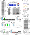
Fig. 1
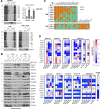
Fig. 1. COX11 is dispensable for CcO biogenesis in human mitochondria.
A Steady-state levels of CcO subunits and assembly factors in COX11-KO cells. B Heat map showing the levels of CcO subunits and assembly factors normalized by ACTIN levels in COX11-KO, COX19-KO, and PET191-KO cells, presented as % of WT The map represents the average of three independent experiments. Two-sided unpaired t-test, *p < 0.05. C BN-PAGE analysis of ETC complexes (CIII2, CIV, CIV2) and supercomplexes (III2 + CIV and I + III2 + CIVn) in COX11-KO cells reconstituted or not with WT COX11. CIV subassemblies (sub CIV) are labeled as S1-S3. S1 contains COX1-COX14-COA3-CMC1 and S2-S3 contain COX1-COX14-COA3-COX4-COX5a with or without assembly factors such as SURF1 or MITRAC7. S4 is a CIV subcomplex formed by the off-pathway joining of the COX1 and COX3 assembly modules when COX2 is absent or in limited amounts. D, GJ CIV (CcO) activity measured spectrophotometrically in the indicated cell lines. In G, H, J, the cells were incubated in the presence or absence of 1 mM CuCl2 or 1 nM elesclomol (ES) + 1 mM CuCl2 for 7 days. The bar graphs represent the average ± SD of three independent experiments. Black dots represent individual data points. Two-sided unpaired t-test, ****p < 0.0001. E Endogenous cell respiration measured polarographically in COX11-KO, COX19-KO, or PET191-KO cells reconstituted or not with the corresponding WT gene. The bar graphs represent the average ± SD of three independent experiments. Black dots represent individual data points. Two-sided unpaired t-test, ****p < 0.0001. F Total mitochondrial cytochrome spectra. Cytochromes c and c1 peak at 550 nm and cytochrome b peaks at 560 nm. The precise wavelength at which the absorbance of a + a3 cytochromes peak is annotated. The height of the peaks was calculated using the Quant mode of the UV-Probe software (Shimadzu) and expressed as the a + a3/b ratio. In the lower graph, the data from three independent experiments were plotted with bars representing the mean ± SD. Black dots represent individual data points. Two-sided unpaired t-test. GJ CcO activity measured spectrophotometrically in the indicated cell lines. In G, J, the cells were incubated in the presence or absence of 1.5 mM Cu-His for 7–10 days with addition or not of elesclomol (ES) as indicated in G. The bar graphs represent the average ± SD of three independent experiments. Black dots represent individual data points. Two-sided unpaired t-test, ****p < 0.0001. ns, no significant. K Steady-state levels of CcO subunits and assembly factors in COX11-KO cells overexpressing CcO assembly factors. The bar graphs represent the average ± SD of three independent experiments. Black dots represent individual data points. Two-sided unpaired t-test, ****p < 0.0001. ns, no significant. Figures A, C, K are representative of three independent repetitions with similar results. Source data for A, B, and DK, are provided as a Source Data file.
Fig. 2
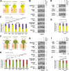
Fig. 2. COX11 redox state and copper binding are regulated by PET191 and COX19.
A Reverse thiol trapping approach to detect the native cysteine residues inaccessible to a cell-permeable alkylating compound (2-iodoacetamide, IAA) that upon denaturation with SDS (sodium dodecyl sulfate) and full reduction with TCEP (tris(2-carboxyethyl)phosphine) are bound to AMS (4-acetamido-4’-maleimidylstilbene-2,2’- disulfonic acid), which adds ~540 Da per thiol. Three potential scenarios are depicted to account for copper (Cu)-bound COX11 dimer and oxidized or reduced COX11 monomers. BD Reverse COX11 thiol trapping in the indicated cell lines. The bar graphs represent the average ± SD of four independent experiments. Two-sided unpaired t-test, ****p < 0.0001. Figures BD are representative of three independent repetitions with similar results. Source data for BD are provided as a Source Data file.
Fig. 3
Fig. 3. CcO subunits and metallochaperones undergo stable and transient interactions to drive and coordinate metal center assembly.
A Co-immunoprecipitation (co-IP) of endogenous COX11 with newly synthesized mitochondrial polypeptides, identified in the right-hand side. The graph shows the densitometry (average ± SD) of three independent experiments. Black dots represent individual data points. Two-sided unpaired t-test, ****p < 0.0001. The ratio “α-COX11 IP signal vs. control background signal” for two parameters, COX1/ATP6 and COX2/ATP6, is presented. B Co-IP of COX11-FLAG with CcO subunits and assembly factors in the presence or absence of the crosslinker DSP. Ex, extract; Un, unbound; IP, immunoprecipitate. C Stable interactions among CcO assembly factors determined by IP followed by either immunoblotting (WB) or mass spectrometry (MS). D Transient interactions among CcO assembly factors determined by IP in the presence of the crosslinker DSP (dithiobis(succinimidyl propionate)) or the vehicle DMSO (dimethyl sulfoxide) followed by immunoblotting. The heat maps show the average quantification of three independent experiments. Two-sided unpaired t-test. *p < 0.05. Figures A, B are representative of three independent repetitions with similar results. Source data for A, B, D are provided as a Source Data file.
Fig. 4
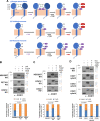
Fig. 4. SCO1 and SCO2 redox state and copper binding are regulated by COX11, PET191, and COX19.
Potential redox and copper (Cu) binding states of SCO1 (A) or SCO2 (F). B, D, G, I Reverse SCO1 or SCO2 thiol trapping in the indicated cell lines. C, E, H, J The graphs show the quantification of six (for SCO1) or five (for SCO2) independent experiments as in B, D, G, I, respectively. The bars represent the mean ± SD. Two-sided unpaired t-test, ****p < 0.0001. ns, no significant. Figures B, D, G, I are representative of six (B, D) or five (G, I) independent repetitions with similar results. Source data for BD and HJ are provided as a Source Data file.
Fig. 5
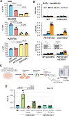
Fig. 5. COX11- and PET191-containing metallochaperone modules coordinate copper and heme center assembly to minimize accumulation of pro-oxidant deleterious CcO assembly intermediates.
A ROS generation in the indicated cell lines measured using superoxide-sensitive dyes dihydroethidium (DHE) and MitoSOX and the H2O2-sensitive dichlorodihydrofluorescein diacetate (CM-H2DCFDA). The bar graphs are quantification (mean ± SD) of three independent experiments. Black dots represent individual data points. Two-sided unpaired t-test, ****p < 0.0001. B H2O2 sensitivity measured as the release of lactate dehydrogenase (LDH) to the growth medium. In the bottom graph, the inset shows immunoblot analysis of COX10 steady-state levels in WT and PET191-KO HEK293T cells following 10 days of treatment with siRNA-COX10 or non-targeting (NT) control. ACTIN was used as the loading control. The bar graphs are quantification (mean ± SD) of nine independent experiments. Black dots represent individual data points. Two-sided unpaired t-test, ****p < 0.0001. Black stars are for comparisons with WT, and red starts for comparison with PET191-KO. Immunoblots in this panel are representative of three independent repetitions with similar results. Source data for the immunoblots are provided as a Source Data file. C Experimental workflow for copper measurements. Mitochondria isolated from WT or KO cell lines expressing FLAG-tagged COX11 or PET191 were solubilized in native conditions (see details in the Methods section), then the extracts used for FLAG-immunoprecipitation (IP), the IPed proteins eluted, and analyzed their metal content by inductively coupled plasma MS (ICP-MS). The panel was created with Biorender.com. D Copper content in COX11- and PET191-containing metallochaperone modules measured by ICP-MS. Mitochondria were purified from COX11-KO or PET191-KO cells expressing the indicated FLAG-tagged proteins. The proteins were immunoprecipitated using anti-FLAG conjugated beads, and their metal content was assessed by ICP-MS. The data is expressed as total Cu normalized by [34S]. The bar graphs are quantification (mean ± SD) of three (COX11-KO + COX11-C219C), six (COX11-KO + COX11 and PET191-KO + PET191 or + PET191-C30A,C41A) or nine (WT, COX11-KO + PET191 or + PET191-C30A,C41A) independent samples. Black dots represent individual data points. Two-sided unpaired t-test, ****p < 0.0001. Source data for A, B, D are provided as a Source Data file.
Fig. 6
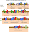
Fig. 6. Model of CcO assembly depicting the coordination of metal center biogenesis.
The role of metallochaperone modules and the several regulatory checkpoints are indicated. See the text for details.

References

    1. Gray HB, Winkler JR. Electron transfer in proteins. Annu. Rev. Biochem. 1996;65:537–561. doi: 10.1146/annurev.bi.65.070196.002541. - DOI - PubMed
    1. Timon-Gomez A, et al. Mitochondrial cytochrome c oxidase biogenesis: Recent developments. Semin. Cell Dev. Biol. 2018;76:163–178. doi: 10.1016/j.semcdb.2017.08.055. - DOI - PMC - PubMed
    1. Khalimonchuk O, Bird A, Winge DR. Evidence for a pro-oxidant intermediate in the assembly of cytochrome oxidase. J. Biol. Chem. 2007;282:17442–17449. doi: 10.1074/jbc.M702379200. - DOI - PubMed
    1. Bode M, et al. Inaccurately assembled cytochrome c oxidase can lead to oxidative stress-induced growth arrest. Antioxid. Redox Signal. 2013;18:1597–1612. doi: 10.1089/ars.2012.4685. - DOI - PMC - PubMed
    1. Yoshikawa S, et al. Redox-coupled crystal structural changes in bovine heart cytochrome c oxidase. Science. 1998;280:1723–1729. doi: 10.1126/science.280.5370.1723. - DOI (V体育2025版) - PubMed

Publication types