<code lang="Pu3y8p"></code> Skip to main page content (VSports手机版)
U.S. flag

An official website of the United States government

Dot gov

The VSports app下载. gov means it’s official. Federal government websites often end in . gov or . mil. Before sharing sensitive information, make sure you’re on a federal government site. .

Https

The site is secure. The https:// ensures that you are connecting to the official website and that any information you provide is encrypted and transmitted securely. V体育官网.

. 2022 Jun 24;8(25):eabm6504.
doi: 10.1126/sciadv.abm6504. Epub 2022 Jun 24.

VSports在线直播 - Human gingival mesenchymal stem cells retain their growth and immunomodulatory characteristics independent of donor age

Affiliations

Human gingival mesenchymal stem cells retain their growth and immunomodulatory characteristics independent of donor age (V体育官网入口)

Jay R Dave (V体育官网) et al. Sci Adv. .

Abstract

Aging has been reported to deteriorate the quantity and quality of mesenchymal stem cells (MSCs), which affect their therapeutic use in regenerative medicine VSports手机版. A dearth of age-related stem cell research further restricts their clinical applications. The present study explores the possibility of using MSCs derived from human gingival tissues (GMSCs) for studying their ex vivo growth characteristics and differentiation potential with respect to donor age. GMSCs displayed decreased in vitro adipogenesis and in vitro and in vivo osteogenesis with age, but in vitro neurogenesis remained unaffected. An increased expression of p53 and SIRT1 with donor age was correlated to their ability of eliminating tumorigenic events through apoptosis or autophagy, respectively. Irrespective of donor age, GMSCs displayed effective immunoregulation and regenerative potential in a mouse model of LPS-induced acute lung injury. Thus, we suggest the potential of GMSCs for designing cell-based immunomodulatory therapeutic approaches and their further extrapolation for acute inflammatory conditions such as acute respiratory distress syndrome and COVID-19. .


Figures
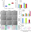
Fig. 1.
Fig. 1.. In vitro morphological and growth characteristics of GMSCs.
(A) Age distribution of donors categorized into group A (13 to 31 years; mean age ± SD, 23.29 ± 6.26 years; n = 14), group B (37 to 55 years; mean age ± SD, 45.69 ± 6.49 years; n = 13), and group C (59 to 80 years; mean age ± SD, 65.36 ± 6.57 years; n = 14). (B) GMSCs from all age groups displayed almost similar average rate of proliferation between passages 8 and 11. (C to K) GMSCs from groups A, B, and C displayed spindle-shaped fibroblast morphology in the in vitro cultures (magnification, ×10; scale bars, 100 μm) at passages 0, 8, and 12. (L to N) The size of colonies in group A GMSCs is larger than that in group C (eosin staining, pink) (magnification, x4; scale bars, 500 μm). (O) Group A, B, and C GMSCs displayed similar PDT at passages 9 and 11, but the doubling time of group C GMSCs significantly increased at passage 13. (P) No significant difference in average population doublings is observed between passages 9 and 13 (*P < 0.05, all pairwise multiple comparison). (Q) GMSCs from all the groups showed similar efficiency of colony formation.
Fig. 2.
Fig. 2.. Stem cell and senescence markers.
(A and B) Group A, B, and C GMSCs expressed MSC surface markers (CD44, CD90, CD73, and CD105) with negligible expression of hematopoietic stem cell markers (CD45 and CD34). (C to E) Representative images of SA-β-gal+ve cells (blue-green color of SA-β-gal–stained cells, highlighted in the figure; magnification, ×10; scale bars, 100 μm) between passages 8 and 12. (F and G) GMSCs from group C showed significantly higher population of senescing cells in both early (average of passages 4 to 6) and late passage (average of passages 9 to 12) (*P < 0.05, all pairwise multiple comparison). (H) Group C GMSCs displayed enhancement in the expression of p53 and SIRT1. The gene expression profiles of group B and C GMSCs were normalized with group A.
Fig. 3.
Fig. 3.. Cell cycle regulators, growth factor receptors, and wound healing ability.
(A) Group C GMSCs displayed an up-regulation in expression of CDKN2A as compared to groups A and B. No significant difference in levels of CDKN1A was observed. (B) As compared to groups A and B, group C GMSCs exhibited an increased expression of growth factor receptors (PDGFR, FGFR, and EGFR). The gene expression profiles of group B and C GMSCs were normalized with group A. (C to L) Group B GMSCs displayed a significantly higher rate of migration as compared to groups A and C. There was very low but consistent difference in the rate of wound closure in all groups of GMSCs (magnification, ×10; scale bars, 100 μm) (at time point 10 hours, for group B versus group C, *P < 0.05; at time point 14 hours, for group A versus group B, *P < 0.05 and for group B versus group C, ***P < 0.001; at time point 24 hours, for group A versus group B and group B versus group C, ***P < 0.001; at time point 30 hours, for group A versus group B, **P < 0.01 and for group B versus group C, ***P < 0.001; at time point 48 hours, for group A versus group B, *P < 0.05).
Fig. 4.
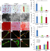
Fig. 4.. Multilineage differentiation potentials.
(A to C) Accumulation of oil globules by GMSCs of all groups (magnification, ×40; scale bars, 50 μm; oil globules accumulated inside the cells are stained with Oil Red O, which gives red color). (D) The number of oil globules declined with donor age (*P < 0.05, group A versus group C). (E to G) The osteogenic differentiation potential of GMSCs also showed a decline with donor age, in terms of decrease in bone nodule formation (magnification, ×10; scale bars, 100 μm; bone nodules appear as brownish black deposits on cell monolayer). (H) ALP expression was maintained in all groups of osteogenic GMSCs. (I to K) ARS was used to stain the mineralization of matrix in terms of calcium phosphate synthesis (ARS, red; magnification, ×10; scale bars, 100 μm), (L) which showed a significant decline in groups B and C as compared to group A (*P < 0.05, group A versus group B and group A versus group C). (M to O) GMSCs differentiated into neurons after induction for 21 days. Upon neurogenic induction, GMSCs from all the groups displayed similar expression of β-III tubulin (green, phalloidin; red, DAPI; blue, nuclei) and (P to R) nestin (red, phalloidin; green, DAPI; blue, nuclei), indicating that GMSCs have a skewed potential toward neuroectodermal differentiation (magnification, ×63; scale bars, 50 μm). (S) The graph displays average intensities of β-III tubulin and nestin. The average intensity was calculated after analyzing four different locations in a given sample (at ×40 magnification; fig. S1).
Fig. 5.
Fig. 5.. Ectopic bone formation.
Subcutaneous implantation of GMSC-seeded scaffold in SCID mice for 12 weeks resulted in osteogenic differentiation of GMSCs, as indicated by immunostaining for matrix proteins, (A to D) osteocalcin (green), and (F to I) collagen type I (red, pseudo-color) [magnification, ×60 (inset); scale bars, 100 μm; magnification, ×10; scale bars, 50 μm; nuclei, DAPI (blue)]. (E) Intensities of osteocalcin (***P < 0.001, only scaffold versus group B; **P < 0.01, group B versus group C; *P < 0.05, group A versus group B) and (J) collagen type I (*P < 0.05, only scaffold versus group A) were calculated from average intensities of ten random locations on retrieved implants, which indicated a decline in contribution of aging osteogenic GMSCs toward matrix mineralization. (K to N) Group C GMSCs displayed lower accumulation of calcium (blue) and phosphate (green) in implants as compared to implants seeded with group A and B GMSCs (magnification, ×1000; scale bars, 60 μm). (O) Quantitative analysis of Ca and P indicates significantly lower levels in scaffolds seeded with group C GMSCs (***P < 0.001; only scaffold versus group A; only scaffold versus group B; group A versus group C and group B versus group C). (A, F, and K) Acellular scaffolds (only scaffold) showed negligible levels of matrix proteins and mineralization.
Fig. 6.
Fig. 6.. In vitro and in vivo immunoregulation by GMSCs.
(A) In vitro coculture of GMSCs with PHA-activated PBMNCs in a ratio of 1:7.5 demonstrated a significant suppression of PBMNC proliferation by all groups of GMSCs. Group A displayed maximum reduction at 96 hours (***P < 0.001, stimulated PBMNCs versus groups A, B, and C). (B) Mice with LPS administration displayed significantly high infiltration of neutrophils in BAL and lung tissue as compared to sham control. Intravenous administration of group A, B, and C GMSCs displayed reduction in the number of infiltrated neutrophils (*P < 0.05, sham versus LPS-induced ALI; LPS-induced ALI versus group A MSCs and LPS-induced ALI versus group C MSCs). (C to E) Induction of ALI by intranasal administration of LPS showed elevated gene levels of pro-inflammatory cytokines, TNF-α, TGF-β, and IFN-γ, in the cellular components of BAL as compared to sham control. Administration of group A, B, and C GMSCs has been found to reduce the expression levels of these pro-inflammatory cytokines. (F and G) Histological analysis of lung sections displayed indications of ALI in mice administered with LPS as compared to sham control. (H to J) A significant reduction in all the damage parameters was observed in mice administered with GMSCs from groups A, B, and C, resulting in improvement of overall lung health (magnification, ×20; scale bars, 50 μm; hematoxylin and eosin staining). Histological parameters studied (i) infiltration of neutrophils in alveoli and (ii) interstitial spaces; (iii) hyaline membrane formed due to fibrin polymerization that leaks into the interstitial/alveolar space; (iv) presence of proteinaceous debris in the alveolar space (such as fibrin strands); (v) thickening of alveolar septa; (vi) desquamation of alveolar epithelium; and (vii) thrombosis.

References

    1. Asumda F. Z., Age-associated changes in the ecological niche: Implications for mesenchymal stem cell aging. Stem Cell Res. Ther. 4, 47 (2013). - PMC (VSports在线直播) - PubMed
    1. De Mora J. F., Juan A. D., The decay of stem cell nourishment at the niche. Rejuvenation Res. 16, 487–494 (2013). - PMC - PubMed
    1. Ma D., Ma Z., Zhang X., Wang W., Yang Z., Zhang M., Wu G., Lu W., Deng Z., Jin Y., Effect of age and extrinsic microenvironment on the proliferation and osteogenic differentiation of rat dental pulp stem cells in vitro. J. Endod. 35, 1546–1553 (2009). - PubMed
    1. Iohara K., Murakami M., Nakata K., Nakashima M., Age-dependent decline in dental pulp regeneration after pulpectomy in dogs. Exp. Gerontol. 52, 39–45 (2014). - PubMed
    1. Alzer H., Kalbouneh H., Alsoleihat F., Abu Shahin N., Ryalat S., Alsalem M., Alahmad H., Tahtamouni L., Age of the donor affects the nature of in vitro cultured human dental pulp stem cells. Saudi Dent. J. 33, 524–532 (2021). - PMC - PubMed