Skip to main page content
U.S. flag

An official website of the United States government

Dot gov

The . gov means it’s official. Federal government websites often end in . gov or . mil. Before sharing sensitive information, make sure you’re on a federal government site. VSports app下载.

Https

The site is secure V体育官网. The https:// ensures that you are connecting to the official website and that any information you provide is encrypted and transmitted securely. .

. 2022 May 25;14(646):eabj2829.
doi: 10.1126/scitranslmed.abj2829. Epub 2022 May 25.

"VSports手机版" MAIT and Vδ2 unconventional T cells are supported by a diverse intestinal microbiome and correlate with favorable patient outcome after allogeneic HCT

Affiliations

MAIT and Vδ2 unconventional T cells are supported by a diverse intestinal microbiome and correlate with favorable patient outcome after allogeneic HCT (VSports在线直播)

Hana Andrlová et al. Sci Transl Med. .

Abstract

Microbial diversity is associated with improved outcomes in recipients of allogeneic hematopoietic cell transplantation (allo-HCT), but the mechanism underlying this observation is unclear. In a cohort of 174 patients who underwent allo-HCT, we demonstrate that a diverse intestinal microbiome early after allo-HCT is associated with an increased number of innate-like mucosal-associated invariant T (MAIT) cells, which are in turn associated with improved overall survival and less acute graft-versus-host disease (aGVHD). Immune profiling of conventional and unconventional immune cell subsets revealed that the prevalence of Vδ2 cells, the major circulating subpopulation of γδ T cells, closely correlated with the frequency of MAIT cells and was associated with less aGVHD VSports手机版. Analysis of these populations using both single-cell transcriptomics and flow cytometry suggested a shift toward activated phenotypes and a gain of cytotoxic and effector functions after transplantation. A diverse intestinal microbiome with the capacity to produce activating ligands for MAIT and Vδ2 cells appeared to be necessary for the maintenance of these populations after allo-HCT. These data suggest an immunological link between intestinal microbial diversity, microbe-derived ligands, and maintenance of unconventional T cells. .


Figures
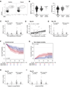
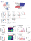
Figure 1:
Figure 1:. MAIT cells are supported by a diverse intestinal microbiome after HCT and predict favorable patient outcomes
A) FACS plots of MAIT cells (MR1+/CD161+) in a healthy volunteer and a representative allo-HCT patient. B) MAIT cell frequencies among CD3+ cells and absolute counts on day 30 (+/−10) and day 100 (+/−20) after HCT (frequencies: day 30 n=147, day 100 n=72; healthy n=15; absolute counts: day 30 n=137, day 100 n=70, healthy control n=15). C) Recipients of BM or PBSC grafts were classified into > median (higher) or ≤ median (lower) α-diversity and MAIT frequency at day 30 was compared. (Wilcoxon rank-sum test, BM n=15 ≤ median, n=14 > median, p=0.32; PBSC n=59 ≤ median, n=59 > median, p=0.014). D) Stool α-diversity at day 7 to day 21 as a continuous variable correlated with MAIT frequency at day 30 (Pearson method; n = 118, R=0.25, p=0.0063). Each dot represents an individual patient sample. E) Patients were divided as in C), data shown for day 100 MAIT frequency (Wilcoxon rank-sum test, BM, n=8 ≤ median α-diversity, n=8 > median, p=0.84; PBSC, n=28 ≤ median α-diversity, n=28 > median, p=0.43). F) Kaplan-Meier survival analysis of PBSC-graft recipients stratified by MAIT cell frequency (log-rank test, n=118, p=0.047). G) Cumulative incidence of NRM in PBSC-graft recipients with > median or ≤ median MAIT cell frequency (Gray’s test, n=118, p=0.031). H) Recipients of either BM or PBSC grafts were classified by GVHD status (grade 2–4, Y=Yes, grade 2–4 GVHD present, N=No grade 2–4 GVHD). MAIT cell frequency at day 30 shown (Wilcoxon rank-sum test, BM, n=15 grade 0–1 GVHD, n=11, grade 2–4, p=0.92; PBSC, n=53 grade 0–1 GVHD, n=36 with grade 2–4, p=0.0047). I) BM or PBSC recipients were classified by the presence of lower GI GVHD (Y=Yes, lower GI GVHD present or N=No, no lower GI GVHD); MAIT cell frequencies are shown (BM, n=23 without lower GI GVHD, n=3 with lower GI GVHD, p=0.52; PBSC, n=73 without lower GI GVHD, n=16 with any stage lower GI GVHD, p=0.031). H+I) Patients with blood samples collected before or on the day of GVHD onset were included. C+E, H+I) Each dot represents a single patient, boxes represent median with interquartile range.
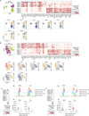
Figure 2:
Figure 2:. Deep immune profiling reveals higher frequency of the Vδ2 subset of γδ T cells in the samples with higher MAIT frequency after HCT
A) UMAP clusters in patients with day 30 MAIT frequency higher (blue) or lower than/equal to (red) the population median. Populations different between the two groups are marked by a circle and a rectangle. B) Gating strategy for both the conventional and unconventional T cell populations, which were used to define the UMAP clusters in A. Concatenated CD3+ cells from the same 76 samples from panel A were used to establish the gating scheme. DN = double negative, DP = double positive, SCM = stem cell memory, CM = central memory, TM = transitional memory, EM = effector memory, TE = terminal effector. C) UMAP clustering color coded to identify the gated populations. Clusters that were different between samples with higher and lower MAIT frequency are outlined: MAIT cells (rectangle) and Vδ2 cells (circle). D) Vδ2 cell frequency as a function of higher or lower MAIT frequency (Wilcoxon rank-sum test, n= 76, p=0.004). Each dot represents a single patient, boxes represent median with interquartile range. E) Heatmap of 20 populations identified by self-organizing maps (FlowSOM) algorithm, demonstrating mean fluorescence intensity (MFI) of 19 identifying surface markers. F) Frequency of each FlowSOM population in each patient sample represented by 3000 concatenated CD3+ events/sample. Each dot represents a single patient, lines represent median with interquartile range. Mann-Whitney test was used to calculate differences between the two groups for each population and FDR correction was performed for multiple hypothesis testing, total n=76, *p<0.05, **p < 0.01. G) UMAP projection of 20 populations of CD3+ cells identified by the FlowSOM clustering algorithm. Dots indicate clusters that were different between samples with higher and lower MAIT frequency.
Figure 3:
Figure 3:. The frequency of the Vδ2 subset of γδ T cell is associated with higher fecal α-diversity in peri-engraftment stool samples and lower rates of overall and lower intestinal acute GVHD
A) MAIT and Vδ2 cell frequency analyzed as continuous variables in day 30 blood samples from recipients of PBSC grafts (Pearson correlation, n=118, R=0.38, p=2.8e-05). B) Vδ2 cell frequencies among CD3+ cells and absolute counts on day 30 (+/−10) and day 100 (+/−20) after HCT in PBSC graft recipients (frequencies: day 30 n=118, day 100 n=56; healthy n=15; absolute counts: day 30 n=110, day 100 n=54, healthy control n=15). C) PBSC graft recipients were classified as in Fig 1C into > median (higher) and ≤ median (lower) α-diversity and Vδ2 cell frequency in each subgroup at day 30 was compared (Wilcoxon rank-sum test, n = 118, p=0.0026). D) Fecal α-diversity between day 7 and day 21 as a continuous variable correlated with Vδ2 frequency (Pearson correlation, n = 118, R=0.22, p=0.017). E) PBSC graft recipients were classified by GVHD grade (grade 2–4, Y=Yes, grade 2–4 GVHD present, N=No grade 2–4 GVHD) and Vδ2 frequencies compared in each group (Wilcoxon rank-sum test, n=53 grade 0–1 GVHD, n=36 grade 2–4, p=0.013). F) Patients were classified by lower GI GVHD status (Y=Yes, lower GI GVHD present or N=No, no lower GI GVHD) and Vδ2 frequencies were compared in each group (Wilcoxon rank-sum test, n=73 without lower GI GVHD, n=16 with any stage lower GI GVHD, p=0.0099). E+F) Patients with blood samples collected before or on the day of GVHD onset were included. A and D) Each dot represents a single patient sample. C+ E+ F) Each dot represents a single patient, and boxes represent median with interquartile range.
Figure 4:
Figure 4:. Distinct microbial taxa and metabolic pathways support MAIT and Vδ2 cell populations
A) Differences in peri-engraftment stool microbiota composition between PBSC recipients with MAIT cell frequency > median (higher) or ≤ median (lower) of all patients depicted using LEfSE (n=425 samples collected on day 7 to day 21 post-HCT from 118 patients; median relative abundance per patient at genus level). B) Dominant taxa driving the difference between the two groups (LDA>4, p<0.01). C) Taxa dominant in the higher MAIT cell group from the LEfSE analysis plotted as a relative abundance in patients with higher versus lower MAIT cells as defined above. D) Taxa dominant in the lower MAIT cell group identified by the LEfSE analysis plotted as a relative abundance in patients stratified by MAIT cell frequency. C and D) Wilcoxon rank-sum test was performed to compare taxa abundances between groups, FDR correction was performed for multiple hypothesis testing. E) Predicted gene abundance of the riboflavin biosynthesis pathway in PBSC recipients as a function of MAIT cell frequency (Wilcoxon rank-sum test, n=118, p=0.098). F) Heatmap representing microbial gene level abundance of key riboflavin biosynthesis enzymes in patients with higher and lower MAIT cell frequency. G) Differences in predicted microbial gene abundance of specific genes encoding key riboflavin biosynthesis enzymes. H) Metabolomic analysis was performed on stool samples collected from patients with previously defined higher or lower MAIT cells. Riboflavin concentrations measured as peak area normalized to internal standard, median value was used when more than one sample per patient was analyzed (Wilcoxon rank-sum test, n=35, p=0.066). I) Predicted microbial gene level abundance of the 2-C-methyl-D-erythritol 4-phosphate/1-deoxy-D-xylulose 5-phosphate (MEP) biosynthesis pathway in PBSC recipients with > median or ≤ median Vδ2 cell frequency using PICRUSt2 analysis (Wilcoxon rank-sum test, n=118, p=0.00054). J) Heatmap representing microbial gene level abundance of key MEP biosynthesis enzymes in PBSC recipients with higher and lower Vδ2 cell frequency. K) Differences in microbial gene level abundance of key MEP biosynthesis enzymes. L) HMBPP peak area was measured in the same patient cohort as in H. Patients were divided into those with detectable or not detectable HMBPP and Vδ2 frequencies are shown (Wilcoxon rank-sum test, n=35, p=7.6e-05). E-G and I-K) Total n=425 samples collected d7 to d21 post-transplantation from 118 PBSC allograft recipients were used, plotted is median predicted pathway/enzyme gene abundance in all samples available in this time window per patient. Each dot represents a single patient, and boxes represent median with interquartile range. C-E, G-I, K,L) Each dot represents a single patient, boxes/lines represent median with interquartile range. G,K) Mann-Whitney test was used to compare the groups for each enzyme and FDR correction was performed for multiple hypothesis testing, n=118, **p < 0.01, ***p<0.001, ns = not significant.
Figure 5:
Figure 5:. MAIT and Vδ2+ T cell subsets acquire effector cytotoxic transcriptional signature post-transplantation and are mostly Th/Tc1 polarized
A) Upper left: UMAP clustering of MAIT cells from patients (n=5) and healthy donors (n=5). Upper right: Dotplot demonstrating the most differentially regulated genes between patients and healthy donors. Lower: UMAP plots of the most differentially regulated genes between healthy and patient MAIT cells. B) Upper left: UMAP clustering of Vδ2 cells from patients and healthy donors. Upper right: Dotplot quantifying the most differentially regulated genes between patients and healthy donors. Lower: UMAP plots of most most differentially regulated genes between healthy and patient Vδ2 cells. C) Gene expression of T-bet and RORγt in patients (n=5) and healthy controls (n=5). D, E) Representative histograms and quantification of RORγt and T-bet expression in MAIT cells from patients with previously defined above median MAIT cell frequency (n=14) and healthy controls (n=3) by flow cytometry. Geometric MFI = geometric mean fluorescence intensity. F) Gene expression of T-bet and RORγt in Vδ2 cells in patients (n=5) and healthy controls (n=5). G, H) Representative histograms of RORγt and T-bet expression in Vδ2 populations from PBMC samples of patients with previously defined above-medianVδ2 cell frequency (n=22) and healthy controls (n=3) by flow cytometry. E+H) Each dot represents a single patient, lines represent median with interquartile range. Mann-Whitney test was used to compare the groups, *p<0.05.

V体育平台登录 - References

    1. Godfrey DI, Uldrich AP, McCluskey J, Rossjohn J, Moody DB, The burgeoning family of unconventional T cells. Nature Immunology 16, 1114–1123 (2015). - PubMed
    1. Treiner E, Duban L, Bahram S, Radosavljevic M, Wanner V, Tilloy F, Affaticati P, Gilfillan S, Lantz O, Selection of evolutionarily conserved mucosal-associated invariant T cells by MR1. Nature 422, 164–169 (2003). - PubMed
    1. Godfrey DI, MacDonald HR, Kronenberg M, Smyth MJ, Kaer LV, NKT cells: what’s in a name? Nat Rev Immunol 4, 231–237 (2004). - PubMed
    1. Hintz M, Reichenberg A, Altincicek B, Bahr U, Gschwind RM, Kollas A-K, Beck E, Wiesner J, Eberl M, Jomaa H, Identification of E-4-hydroxy-3-methyl-but-2-enyl pyrophosphate as a major activator for human γδ T cells in Escherichia coli. FEBS Letters 509, 317–322 (2001). - PubMed
    1. Sandstrom A, Peigné C-M, Léger A, Crooks James E., Konczak F, Gesnel M-C, Breathnach R, Bonneville M, Scotet E, Adams Erin J., The Intracellular B30.2 Domain of Butyrophilin 3A1 Binds Phosphoantigens to Mediate Activation of Human Vγ9Vδ2 T Cells. Immunity 40, 490–500 (2014). - PMC - PubMed

"VSports在线直播" Publication types

"V体育2025版" Substances