Skip to main page content
U.S. flag

An official website of the United States government

Dot gov

The . gov means it’s official. Federal government websites often end in . gov or . mil. Before sharing sensitive information, make sure you’re on a federal government site. VSports app下载.

Https

The site is secure V体育官网. The https:// ensures that you are connecting to the official website and that any information you provide is encrypted and transmitted securely. .

. 2022 Mar 15;22(1):67.
doi: 10.1186/s12902-022-00968-x.

Circ_0000064 promotes high glucose-induced renal tubular epithelial cells injury to facilitate diabetic nephropathy progression through miR-532-3p/ROCK1 axis (V体育ios版)

Affiliations

Circ_0000064 promotes high glucose-induced renal tubular epithelial cells injury to facilitate diabetic nephropathy progression through miR-532-3p/ROCK1 axis

"VSports最新版本" Huanlan Wang et al. BMC Endocr Disord. .

Abstract

Background: Circular RNA (circRNA) has been shown to mediate diabetic nephropathy (DN) development by regulating renal tubular epithelial cells (RTECs) injury VSports手机版. However, the role and mechanism of circ_0000064 in high glucose (HG)-induced RTECs injury have not been fully elucidated. .

Methods: Human RTECs (HK-2) were exposed to HG to induce cell injury. Cell oxidative stress was assessed by detecting the levels of oxidative stress-markers. Moreover, cell proliferation and apoptosis were determined by CCK8 assay, EDU assay and flow cytometry V体育安卓版. The protein levels of proliferation markers, apoptosis markers and Rho-associated coiled-coil-containing kinase 1 (ROCK1) were measured using western blot analysis. Furthermore, quantitative real-time PCR was performed to assess the expression of circ_0000064, microRNA (miR)-532-3p and ROCK1. The interaction between miR-532-3p and circ_0000064 or ROCK1 was confirmed by dual-luciferase reporter assay and RNA pull-down assay. .

Results: Our results revealed that HG treatment could promote HK-2 cells oxidative stress, apoptosis, fibrosis, and inhibit proliferation. Circ_0000064 expression was increased in the serum of DN patients and HG-induced HK-2 cells, and silenced circ_0000064 could relieve HG-induced HK-2 cells injury. MiR-532-3p could be sponged by circ_0000064, and its overexpression also alleviated HG-induced HK-2 cells injury. Besides, the regulation of circ_0000064 knockdown on HG-induced HK-2 cells injury could be reversed by miR-532-3p inhibitor V体育ios版. Additionally, ROCK1 was a target of miR-532-3p, and its expression was inhibited by circ_0000064 knockdown. The inhibition effect of circ_0000064 knockdown on HG-induced HK-2 cells injury also could be reversed by overexpressing ROCK1. .

Conclusion: In summary, circ_0000064 knockdown might alleviate HG-induced HK-2 cells injury via regulating the miR-532-3p/ROCK1 axis, which provided a new perspective for DN treatment VSports最新版本. .

Keywords: Circ_0000064; Diabetic nephropathy; High glucose; ROCK1; miR-532-3p. V体育平台登录.


Conflict of interest statement

The authors declare that there are no conflicts of interest.

Figures

Fig. 1
Fig. 1
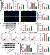
HG could induce HK-2 cells injury. HK-2 cells were cultured under HG, NG or mannitol condition. A-B Cell oxidative stress was assessed by determining the levels of SOD and MDA. CCK8 assay (C) and EDU assay (D) were performed to assess cell proliferation. E-G The protein expression of PCNA and cyclin D1 was measured by WB analysis. H Cell apoptosis rate was evaluated by flow cytometry. I-K WB analysis was used to examine the protein expression of Bax and Bcl2. ***P < 0.001
Fig. 2
Fig. 2
Circ_0000064 was overexpressed in DN patients and HG-induced HK-2 cells. A The basic information of circ_0000064 was shown. B The expression of circ_0000064 in the serum of DN patients with microalbuminuria or macroalbuminuria and normal healthy volunteers was measured by qRT-PCR. C QRT-PCR was performed to detect circ_0000064 expression in HK-2 cells under HG, NG or mannitol condition. ***P < 0.001
Fig. 3
Fig. 3
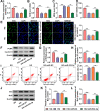
Circ_0000064 knockdown alleviated HG-induced HK-2 cells injury. HK-2 cells were transfected with si-NC or si-circ_0000064 and then treated with HG. A QRT-PCR was used to measure circ_0000064 expression. B-C The levels of SOD and MDA were measured to assess cell oxidative stress. CCK8 assay (D) and EDU assay (E) were used to detect cell proliferation. F-H WB analysis was utilized to determine the protein expression of PCNA and cyclin D1. I Flow cytometry was used to assess cell apoptosis rate. J-L The protein expression of Bax and Bcl2 was detected by WB analysis. **P < 0.01, ***P < 0.001
Fig. 4
Fig. 4
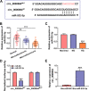
MiR-532-3p could be sponged by circ_0000064. A The binding sites and mutant sites between circ_0000064 and miR-532-3p were shown. B The expression of miR-532-3p was detected by qRT-PCR in the serum of DN patients with microalbuminuria or macroalbuminuria and normal healthy volunteers. C QRT-PCR was used to measure the miR-532-3p expression in HK-2 cells under HG, NG or mannitol condition. Dual-luciferase reporter assay (D) and RNA pull-down assay (E) were used to assess the interaction between circ_0000064 and miR-532-3p. ***P < 0.001
Fig. 5
Fig. 5
MiR-532-3p relieved HG-induced HK-2 cells injury. HK-2 cells were transfected with miR-NC or miR-532-3p mimic and then treated with HG. A The expression of miR-532-3p was detected by qRT-PCR. B-C Cell oxidative stress was analyzed by detecting the levels of SOD and MDA. Cell proliferation was assessed using CCK8 assay (D) and EDU assay (E). F-H The protein expression of PCNA and cyclin D1 was examined using WB analysis. I Cell apoptosis rate was analyzed using flow cytometry. J-L The protein expression of Bax and Bcl2 was determined using WB analysis. **P < 0.01, ***P < 0.001
Fig. 6
Fig. 6
Circ_0000064 regulated HG-induced HK-2 cells injury by sponging miR-532-3p. A QRT-PCR analysis was performed to measure miR-532-3p expression in HG-induced HK-2 cells transfected with anti-miR-NC or anti-miR-532-3p. B-L HK-2 cells were transfected with si-NC, si-circ_0000064, si-circ_0000064 + anti-miR-NC or si-circ_0000064 + anti-miR-532-3p, and then treated with HG. B-C The levels of SOD and MDA were determined to measure cell oxidative stress. Cell proliferation was analyzed using CCK8 assay (D) and EDU assay (E). F-H WB analysis was used to detect the protein expression of PCNA and cyclin D1. I Cell apoptosis rate was analyzed by flow cytometry. J-L The protein expression of Bax and Bcl2 was examined by WB analysis. *P < 0.05, **P < 0.01, ***P < 0.001
Fig. 7
Fig. 7
MiR-532-3p targeted ROCK1. A The binding sites and mutant sites between miR-532-3p and ROCK1 3’UTR were shown. B-C The mRNA and protein expression of ROCK1 in the serum of DN patients with microalbuminuria or macroalbuminuria and normal healthy volunteers was examined by qRT-PCR and WB analysis. D WB analysis was used to measure the protein expression of ROCK1 in HK-2 cells under HG, NG or mannitol condition. E Dual-luciferase reporter assay was used to confirm the interaction between miR-532-3p and ROCK1. F ROCK1 protein expression was detected by WB analysis in HK-2 cells transfected with anti-miR-NC or anti-miR-532-3p. G WB analysis was used to measure ROCK1 protein expression in HK-2 cells transfected with si-NC, si-circ_0000064, si-circ_0000064 + anti-miR-NC or si-circ_0000064 + anti-miR-532-3p. **P < 0.01, ***P < 0.001
Fig. 8
Fig. 8
ROCK1 partially reversed the regulation of si-circ_0000064 on HG-induced HK-2 cells injury. A WB analysis was used to detect ROCK1 protein expression in HG-induced HK-2 cells transfected with pcDNA3.1 or ROCK1 overexpression vector. B-L HK-2 cells were transfected with si-NC, si-circ_0000064, si-circ_0000064 + pcDNA3.1 or si-circ_0000064 + ROCK1, and then treated with HG. B-C Cell oxidative stress was evaluated by measuring the levels of SOD and MDA. Cell proliferation was detected by CCK8 assay (D) and EDU assay (E). (F-H) The protein expression of PCNA and cyclin D1 was detected using WB analysis. I Cell apoptosis rate was determined using flow cytometry. J-L WB analysis was performed to analyze the protein expression of Bax and Bcl2. *P < 0.05, **P < 0.01, ***P < 0.001

References

    1. Schernthaner G, Schernthaner GH. Diabetic nephropathy: new approaches for improving glycemic control and reducing risk. J Nephrol. 2013;26(6):975–985. doi: 10.5301/jn.5000281. - DOI - PubMed
    1. Dronavalli S, Duka I, Bakris GL. The pathogenesis of diabetic nephropathy. Nat Clin Pract Endocrinol Metab. 2008;4(8):444–452. doi: 10.1038/ncpendmet0894. - "VSports" DOI - PubMed
    1. Usuelli V, La Rocca E. Novel therapeutic approaches for diabetic nephropathy and retinopathy. Pharmacol Res. 2015;98:39–44. doi: 10.1016/j.phrs.2014.10.003. - V体育2025版 - DOI - PubMed
    1. Andresdottir G, Jensen ML, Carstensen B, Parving HH, Hovind P, Hansen TW, Rossing P. Improved prognosis of diabetic nephropathy in type 1 diabetes. Kidney Int. 2015;87(2):417–426. doi: 10.1038/ki.2014.206. - DOI - PubMed
    1. Liu L, Wang Y, Yan R, Liang L, Zhou X, Liu H, Zhang X, Mao Y, Peng W, Xiao Y, et al. BMP-7 inhibits renal fibrosis in diabetic nephropathy via miR-21 downregulation. Life Sci. 2019;238:116957. doi: 10.1016/j.lfs.2019.116957. - DOI - PubMed