Skip to main page content (VSports在线直播)
U.S. flag

An official website of the United States government

Dot gov

The . gov means it’s official. Federal government websites often end in . gov or VSports app下载. mil. Before sharing sensitive information, make sure you’re on a federal government site. .

Https

The site is secure. The https:// ensures that you are connecting to the official website and that any information you provide is encrypted and transmitted securely V体育官网. .

. 2022 Feb 21:13:821275.
doi: 10.3389/fgene.2022.821275. eCollection 2022.

VSports注册入口 - The Landscape of Featured Metabolism-Related Genes and Imbalanced Immune Cell Subsets in Sepsis

Affiliations

The Landscape of Featured Metabolism-Related Genes and Imbalanced Immune Cell Subsets in Sepsis

Han She et al. Front Genet. .

"V体育ios版" Abstract

Sepsis is a heterogeneous disease state triggered by an uncontrolled inflammatory host response with high mortality and morbidity in severely ill patients. Unfortunately, the treatment effectiveness varies among sepsis patients and the underlying mechanisms have yet to be elucidated. The present aim is to explore featured metabolism-related genes that may become the biomarkers in patients with sepsis. In this study, differentially expressed genes (DEGs) between sepsis and non-sepsis in whole blood samples were identified using two previously published datasets (GSE95233 and GSE54514) VSports手机版. A total of 66 common DEGs were determined, namely, 52 upregulated and 14 downregulated DEGs. The Gene Set Enrichment Analysis (GSEA) results indicated that these DEGs participated in several metabolic processes including carbohydrate derivative, lipid, organic acid synthesis oxidation reduction, and small-molecule biosynthesis in patients with sepsis. Subsequently, a total of 8 hub genes were screened in the module with the highest score from the Cytoscape plugin cytoHubba. Further study showed that these hub DEGs may be robust markers for sepsis with high area under receiver operating characteristic curve (AUROC). The diagnostic values of these hub genes were further validated in myocardial tissues of septic rats and normal controls by untargeted metabolomics analysis using liquid chromatography-mass spectrometry (LC-MS). Immune cell infiltration analysis revealed that different infiltration patterns were mainly characterized by B cells, T cells, NK cells, monocytes, macrophages, dendritics, eosinophils, and neutrophils between sepsis patients and normal controls. This study indicates that metabolic hub genes may be hopeful biomarkers for prognosis prediction and precise treatment in sepsis patients. .

Keywords: bioinformatics; biomarkers; immune cell infiltration; metabolomics; sepsis. V体育安卓版.


Conflict of interest statement

The authors declare that the research was conducted in the absence of any commercial or financial relationships that could be construed as a potential conflict of interest.

"VSports注册入口" Figures

FIGURE 1
FIGURE 1
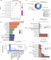
Metabolism-related genes are among DEGs found in sepsis patient versus healthy control whole blood. (A) Principal components analysis (PCA) score plot of GSE95233 and GSE54514. Red represents control patients, and blue represents sepsis patients. (B) Heatmap of GSE95233 and GSE54514. (C) Volcano plot of GSE95233 and GSE54514. Gray dots indicate downregulated DEGs while red dots indicate upregulated DEGs. Statistically significant DEGs were identified as those with Student’s t-test p-values < 0.05. (D) Venn diagram of DEGs identified from the two GEO datasets (-UP: upregulated DEGs, -DOWN: downregulated DEGs). (E) Co-expression network of the differentially expressed metabolism-related genes identified from GSE95233. Red depicts high gene expression and blue depicts low gene expression. (F) Heatmap of the 66 DEGs between patients with sepsis and healthy controls in the training cohort GSE95233.
FIGURE 2
FIGURE 2
Metabolism-related pathways are involved in the pathophysiology of sepsis. (A) Kyoto Encyclopedia of Genes and Genomes (KEGG). (B) Gene ontology (GO) results of biological process (BP), cellular component (CC), and molecular function (MF). (C) Cellular component (CC). (D) Molecular function (MF). (E) Biological process (BP). (F) Biological pathway.
FIGURE 3
FIGURE 3
Metabolic changes were identified by metabolomics profiling in hearts of septic rats and sham controls. (A) Principal components analysis (PCA) score plot for metabolomics analysis in septic rats and normal control. Orange represents sepsis rats, and blue represents normal control. (B) Volcano plot, (C) circle diagram, and (D) heatmap analyzed by TBtools showing the significantly changed metabolites in septic rats and normal control. (E) Bubble plot of the metabolic pathway enrichment analysis identified in septic rats and normal control. The different color depths of circles represent the p-value of pathway enrichment analysis.
FIGURE 4
FIGURE 4
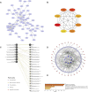
Hub genes were identified by the PPI network complex of metabolic DEGs. (A) Sixty-six DEGs with 66 nodes and 119 edges were displayed using STRING. (B) The 8 most important hub genes were screened using the Cytoscape software plugin cytoHubba. The PPI network data from STRING was further analyzed by Cytoscape and hub genes identification was performed by cytoHubba. (C) Gene–gene interaction networks and functions of 8 hub genes in GeneMANIA. (D)The landscape of metabolic network of hub genes. (E) Enriched pathways of hub genes in Metascape.
FIGURE 5
FIGURE 5
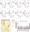
The expressions of the hub genes were different between sepsis patients and healthy controls in the training dataset GSE95233. (A–H) Validation of expression of metabolic-related hub genes in patients with sepsis and normal controls in GSE95233. (I) Hierarchical clustering analysis demonstrates identified metabolic-related gene expression patterns of heart tissues between septic rat and normal groups in the training cohort. (J) Relative expression of identified hub genes in myocardial tissues was compared between septic rats and control rats using quantitative real-time PCR. Differences between two groups were analyzed using the t-test. *p < 0.05, **p < 0.01, ***p < 0.001.
FIGURE 6
FIGURE 6
The eight hub genes had potential diagnostic roles in sepsis. (A) ROC of ADCY3. (B) ENTPD1. (C) NME1. (D) NME6. (E) POLD4. (F) POLE4. (G) POLR2J. (H) POLR2L.
FIGURE 7
FIGURE 7
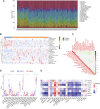
The expressions of the hub genes were correlated with immune cell infiltration in GSE95233. (A) Composition and distribution of inferred immune cell infiltration subsets in each sample. (B) Heatmap of visualizing the differentially infiltrated immune cells between sepsis and healthy control. The horizontal bar indicated the clustering information of samples that were divided into two major clusters. Vertical bars discriminated between upregulated (red) and downregulated (blue) genes. (C) Correlation matrix displaying the Pearson’s correlation values for each comparison between the immune cells. The intensity of the color indicated the strength of the correlation between two immune cells. Red indicates positive correlations and green indicates negative correlations. (D) Boxplot of immune-cell proportion comparisons between sepsis patients and healthy controls (the blue and red boxplots stand for control and sepsis, respectively). Abbreviation: CIBERSORT: Cell type identification by estimating relative subsets of RNA transcripts. (E) Pearson’s correlation analysis between infiltrating immune cells and identified hub genes. Red nodes indicate positive correlation while blue nodes indicate negative correlation. *p < 0.05, **p < 0.01, ***p < 0.001.

References

    1. Abdelmohsen K., Srikantan S., Tominaga K., Kang M.-J., Yaniv Y., Martindale J. L., et al. (2012). Growth Inhibition by miR-519 via Multiple P21-Inducing Pathways. Mol. Cel Biol 32 (13), 2530–2548. 10.1128/mcb.00510-12 - DOI - PMC - PubMed
    1. Bastid J., Cottalorda-Regairaz A., Alberici G., Bonnefoy N., Eliaou J.-F., Bensussan A. (2013). ENTPD1/CD39 Is a Promising Therapeutic Target in Oncology. Oncogene 32 (14), 1743–1751. 10.1038/onc.2012.269 - DOI - PubMed
    1. Bellelli R., Belan O., Pye V. E., Clement C., Maslen S. L., Skehel J. M., et al. (2018). POLE3-POLE4 Is a Histone H3-H4 Chaperone that Maintains Chromatin Integrity during DNA Replication. Mol. Cel 72 (1), 112–e5. 10.1016/j.molcel.2018.08.043 - DOI - PMC - PubMed
    1. Chen C., Chen H., Zhang Y., Thomas H. R., Frank M. H., He Y., et al. (2020). TBtools: An Integrative Toolkit Developed for Interactive Analyses of Big Biological Data. Mol. Plant 13 (8), 1194–1202. 10.1016/j.molp.2020.06.009 - VSports app下载 - DOI - PubMed
    1. Cho J. H., Ju W. S., Seo S. Y., Kim B. H., Kim J.-S., Kim J.-G., et al. (2021). The Potential Role of Human NME1 in Neuronal Differentiation of Porcine Mesenchymal Stem Cells: Application of NB-hNME1 as a Human NME1 Suppressor. Ijms 22 (22), 12194. 10.3390/ijms222212194 - VSports最新版本 - DOI - PMC - PubMed

LinkOut - more resources (V体育ios版)