Skip to main page content
U.S. flag

An official website of the United States government

Dot gov

The . gov means it’s official VSports app下载. Federal government websites often end in . gov or . mil. Before sharing sensitive information, make sure you’re on a federal government site. .

Https

The site is secure. The https:// ensures that you are connecting to the official website and that any information you provide is encrypted and transmitted securely V体育官网. .

. 2022 Jan 31;11(2):298.
doi: 10.3390/antiox11020298.

Vulnerability of Triple-Negative Breast Cancer to Saponin Formosanin C-Induced Ferroptosis

Affiliations

VSports在线直播 - Vulnerability of Triple-Negative Breast Cancer to Saponin Formosanin C-Induced Ferroptosis

Hsin-Chih Chen et al. Antioxidants (Basel). .

"V体育ios版" Abstract

Targeting ferritin via autophagy (ferritinophagy) to induce ferroptosis, an iron- and reactive oxygen species (ROS)-dependent cell death, provides novel strategies for cancer therapy VSports手机版. Using a ferroptosis-specific inhibitor and iron chelator, the vulnerability of triple-negative breast cancer (TNBC) MDA-MB-231 cells to ferroptosis was identified and compared to that of luminal A MCF-7 cells. Saponin formosanin C (FC) was revealed as a potent ferroptosis inducer characterized by superior induction in cytosolic and lipid ROS formation as well as GPX4 depletion in MDA-MB-231 cells. The FC-induced ferroptosis was paralleled by downregulation of ferroportin and xCT expressions. Immunoprecipitation and electron microscopy demonstrated the involvement of ferritinophagy in FC-treated MDA-MB-231 cells. The association of FC with ferroptosis was strengthened by the results that observed an enriched pathway with differentially expressed genes from FC-treated cells. FC sensitized cisplatin-induced ferroptosis in MDA-MB-231 cells. Through integrated analysis of differentially expressed genes and pathways using the METABRIC patients' database, we confirmed that autophagy and ferroptosis were discrepant between TNBC and luminal A and that TNBC was hypersensitive to ferroptosis. Our data suggest a therapeutic strategy by ferroptosis against TNBC, an aggressive subtype with a poor prognosis. .

Keywords: breast cancer; ferritinophagy; ferroptosis potential index; formosanin C; gene database. V体育安卓版.


Conflict of interest statement

The authors declare no conflict of interest.

Figures

Figure 1
Figure 1
FC-induced ferroptosis is more effective in MDA-MB-231 cells. (A) Effect of various types of compounds on the growth of MCF-7 and MDA-MB-231 cells. Cells received treatments of erastin (10 μM), RSL3 (5 μM), cisplatin (10 μM), lapatinib (0.5 μM), curcumin (20 μM), formosanin C (FC; 10 μM), garcinielliptone FC (20 μM), justicidin A (10 μM), lupeol (100 μM), pterostilbene (100 μM), and resveratrol (100 μM) separately in the presence and absence of ferroptosis inhibitor ferrostatin-1 (Fer-1; 5 μM) for 24 h. Cell growth were analyzed by sulforhodamine B assay. * Compared to without Fer-1, p < 0.05; Student’s t-test. # p < 0.05; Student’s t-test. n.s., not significant. (B) Effect of iron chelator on the growth of the cells received FC. Cells received treatments of FC in the presence and absence of deferoxamine (100 μM) for 24 h. The ferroptosis inducer, RSL3 (5 μM), was used as a positive control. * Compared to without deferoxamine, p < 0.05; Student’s t-test. DFO denotes deferoxamine. (C) Iron-enhanced cell growth inhibition of FC. Cells received treatments of RSL3 (1 μM) or FC (5 μM) in the presence and absence of ferric ammonium citrate for 24 h. * Compared to 0 μM of ferric ammonium citrate, p < 0.05; Student’s t-test. # Compared to MCF-7 cells under the same experimental conditions, p < 0.05; Student’s t-test.
Figure 1
Figure 1
FC-induced ferroptosis is more effective in MDA-MB-231 cells. (A) Effect of various types of compounds on the growth of MCF-7 and MDA-MB-231 cells. Cells received treatments of erastin (10 μM), RSL3 (5 μM), cisplatin (10 μM), lapatinib (0.5 μM), curcumin (20 μM), formosanin C (FC; 10 μM), garcinielliptone FC (20 μM), justicidin A (10 μM), lupeol (100 μM), pterostilbene (100 μM), and resveratrol (100 μM) separately in the presence and absence of ferroptosis inhibitor ferrostatin-1 (Fer-1; 5 μM) for 24 h. Cell growth were analyzed by sulforhodamine B assay. * Compared to without Fer-1, p < 0.05; Student’s t-test. # p < 0.05; Student’s t-test. n.s., not significant. (B) Effect of iron chelator on the growth of the cells received FC. Cells received treatments of FC in the presence and absence of deferoxamine (100 μM) for 24 h. The ferroptosis inducer, RSL3 (5 μM), was used as a positive control. * Compared to without deferoxamine, p < 0.05; Student’s t-test. DFO denotes deferoxamine. (C) Iron-enhanced cell growth inhibition of FC. Cells received treatments of RSL3 (1 μM) or FC (5 μM) in the presence and absence of ferric ammonium citrate for 24 h. * Compared to 0 μM of ferric ammonium citrate, p < 0.05; Student’s t-test. # Compared to MCF-7 cells under the same experimental conditions, p < 0.05; Student’s t-test.
Figure 2
Figure 2
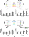
FC increases ROS generation and iron accumulation. (A) FC elevated cytosolic ROS. (B) FC elevated lipid ROS. Cells received treatments of FC in the presence and absence of ferroptosis inhibitor ferrostatin-1 (Fer-1, 5 μM) for 24 h. Cytosolic and lipid ROS were detected using flow cytometry after staining with H2DCFDA and C11-BODIPY, respectively. The higher the intensity (the peak shifts to the right) of H2DCFDA or C11-BODIPY fluorescence is, the richer the cytosolic or lipid ROS are, respectively. * Compared to without Fer-1, p < 0.05; Student‘s t-test. # Compared to the corresponding vehicle, p < 0.05; Student’s t-test. (C) FC-reduced GPX4 protein level. The GPX4 protein levels were detected with enzyme-linked immunosorbent assay kit for GPX4. Cells received treatments of FC for 24 h. # Compared to the corresponding vehicle, p < 0.05; Student’s t-test. (D) FC increased intracellular iron accumulation. Cells received treatments of FC in the presence and absence of iron chelator deferoxamine (100 μM) for 24 h. Ferroptosis inducer, RSL3 (5 μM), was used as a positive control. Intracellular iron accumulation was detected using flow cytometry after Phen green SK staining. * Compared to without deferoxamine, p < 0.05; Student‘s t-test. # Compared to the corresponding vehicle, p < 0.05; Student’s t-test. DFO denotes deferoxamine.
Figure 2
Figure 2
FC increases ROS generation and iron accumulation. (A) FC elevated cytosolic ROS. (B) FC elevated lipid ROS. Cells received treatments of FC in the presence and absence of ferroptosis inhibitor ferrostatin-1 (Fer-1, 5 μM) for 24 h. Cytosolic and lipid ROS were detected using flow cytometry after staining with H2DCFDA and C11-BODIPY, respectively. The higher the intensity (the peak shifts to the right) of H2DCFDA or C11-BODIPY fluorescence is, the richer the cytosolic or lipid ROS are, respectively. * Compared to without Fer-1, p < 0.05; Student‘s t-test. # Compared to the corresponding vehicle, p < 0.05; Student’s t-test. (C) FC-reduced GPX4 protein level. The GPX4 protein levels were detected with enzyme-linked immunosorbent assay kit for GPX4. Cells received treatments of FC for 24 h. # Compared to the corresponding vehicle, p < 0.05; Student’s t-test. (D) FC increased intracellular iron accumulation. Cells received treatments of FC in the presence and absence of iron chelator deferoxamine (100 μM) for 24 h. Ferroptosis inducer, RSL3 (5 μM), was used as a positive control. Intracellular iron accumulation was detected using flow cytometry after Phen green SK staining. * Compared to without deferoxamine, p < 0.05; Student‘s t-test. # Compared to the corresponding vehicle, p < 0.05; Student’s t-test. DFO denotes deferoxamine.
Figure 3
Figure 3
FC changes expressions of proteins related to antioxidant system and iron metabolism. Cells received treatments of FC for 24 and 48 h. Whole cell lysates were prepared and subjected to Western blot analysis using anti-ferroportin, anti-xCT, anti-FTH1, and anti-LC3 antibodies. β-actin antibody was used as an internal control. FPN denotes ferroportin. The intensity of each protein expression band was quantified (n = 3). * and ** Compared to the corresponding control (0 μM of FC), p < 0.05 and p < 0.01, respectively; Student‘s t-test.
Figure 4
Figure 4
Ferritinophagy in FC-treated MDA-MB-231 cells. (A) The interaction of LC3 and FTH1. Cells received treatments of FC for 24 h. The cell lysates were immunoprecipitated using anti-LC3 antibody, and the immune complexes were subjected to Western blot using anti-LC3 and anti-FTH1 antibody, separately. The intensity of each protein expression band was quantified (n = 3). * and ** compared to the corresponding control (0 μM of FC), p < 0.05 and p < 0.01, respectively; Student‘s t-test. (B) Mitochondrial morphology. The arrow indicates the differences in the morphology of mitochondria between control and FC-treated cells. (C) Co-localization of gold-stained ferritin and NCOA4. Cells received treatments of FC (10 μM) for 24 h. Autophagosomes/autolysosomes and gold particles were quantified. *** denote p < 0.001; Student’s t-test. The arrow and triangle indicate NCOA4 (12 nm) and FTH1 (20 nm), respectively. The images were taken by electron microscopy.
Figure 4
Figure 4
Ferritinophagy in FC-treated MDA-MB-231 cells. (A) The interaction of LC3 and FTH1. Cells received treatments of FC for 24 h. The cell lysates were immunoprecipitated using anti-LC3 antibody, and the immune complexes were subjected to Western blot using anti-LC3 and anti-FTH1 antibody, separately. The intensity of each protein expression band was quantified (n = 3). * and ** compared to the corresponding control (0 μM of FC), p < 0.05 and p < 0.01, respectively; Student‘s t-test. (B) Mitochondrial morphology. The arrow indicates the differences in the morphology of mitochondria between control and FC-treated cells. (C) Co-localization of gold-stained ferritin and NCOA4. Cells received treatments of FC (10 μM) for 24 h. Autophagosomes/autolysosomes and gold particles were quantified. *** denote p < 0.001; Student’s t-test. The arrow and triangle indicate NCOA4 (12 nm) and FTH1 (20 nm), respectively. The images were taken by electron microscopy.
Figure 5
Figure 5
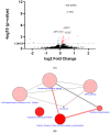
Biological function of FC via bioinformatics analysis. (A) FC-altered mRNA expression. Cells received treatments of FC (0.5 μM) for 6 h to avoid secondary responses under a higher treatment concentration for a longer period time. RNA sequencing was carried out to investigate the transcriptomic alteration of FC. Volcano plot displays the log2 fold change (FC/vehicle) and -log10 (p-value) of the genes altered by FC. DEGs were defined as genes with p < 0.05. Red and blue dots denote upregulated (log2 fold change > 0) DEGs and downregulated (log2 fold change < 0) DEGs, respectively. Black dots denote non-DEGs. The names of the top 5 DEGs are shown. (B) The over-represented pathways analyzed using the DEGs between FC treatment and vehicle via CPDB. The size of each dot designates the entity number of genes in the pathway. The intensity of dot color denotes the p-value. The darker the color is, the smaller the p-value is. The line between two dots was analyzed by the function of these two pathways to show the number of genes overlapping said pathways. The breadth of the line indicates the strength of the correlation between two dots.
Figure 6
Figure 6
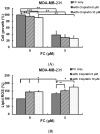
Effect of FC on cisplatin-treated cells. (A) Combination of FC and anti-cancer drug cisplatin increased cell growth inhibition. (B) Combination of FC and cisplatin increased lipid ROS formation. Cells received treatments of FC in the presence and absence of cisplatin for 24 h. Cell growth was analyzed by sulforhodamine B assay. Lipid ROS were detected using flow cytometry after staining with C11-BODIPY. *, **, and *** denote p < 0.05, p < 0.01, and p < 0.001; Student’s t-test.
Figure 7
Figure 7
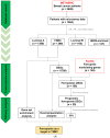
Ferroptosis is shown to account for discrepancy between TNBC and luminal A. (A) Flow chart of in silico data collection and analyses. (B) Nottingham prognostic index obtained from METABRIC cohort. Differences among groups were analyzed by one-way ANOVA and Tukey’s multiple comparison test. Medians with different subscript letters are significantly different, at p < 0.05. (C) The over-represented pathways analyzed using the DEGs between patients with TNBC and luminal A via CPDB. The size of each dot designates the entity number of genes in the pathway. The intensity of dot color denotes the p-value. The darker the color is, the smaller the p-value is. The line between two dots was analyzed by the function of these two pathways to show the number of genes overlapping said pathways. The breadth of the line indicates the strength of the correlation between two dots. (D) Identification of ferroptosis as a potential regulator on TNBC using GSEA. The enrichment score is normalized to account for the size of the gene set, demonstrating significant enrichment (p < 0.05). NES denotes normalized enrichment score. (E) Genes associated with ferroptosis and prognosis of patients with TNBC and luminal A. DEGs (FDR < 0.05) between patients with TNBC and luminal A in the METABRIC cohort were intersected with experimentally validated ferroptosis driver and suppressor genes. The common genes (red boxes) were selected for survival analyses (left panel). Ferroptosis-related prognostic DEGs were shown and the values represented mean of the log2 gene expression (right panel). (F) The FPI between patients with TNBC and luminal A. Sample-wise enrichment scores of the prognostic ferroptosis driver DEG set and suppressor DEG set were independently generated using GSVA algorithm. The enrichment score of the prognostic ferroptosis driver DEG set minus that of the prognostic ferroptosis suppressor DEG set was defined as FPI. The horizontal dashed line indicates FPI = 0, which means the potential of ferroptosis is neutral. The lower and upper extents of the box represent the 25th and 75th percentiles, respectively. The parallel line in the box represents the median. The lower and upper extreme of the whisker represents minimum and maximum, respectively (B) and (F). NPI denotes Nottingham prognostic index.
Figure 7
Figure 7
Ferroptosis is shown to account for discrepancy between TNBC and luminal A. (A) Flow chart of in silico data collection and analyses. (B) Nottingham prognostic index obtained from METABRIC cohort. Differences among groups were analyzed by one-way ANOVA and Tukey’s multiple comparison test. Medians with different subscript letters are significantly different, at p < 0.05. (C) The over-represented pathways analyzed using the DEGs between patients with TNBC and luminal A via CPDB. The size of each dot designates the entity number of genes in the pathway. The intensity of dot color denotes the p-value. The darker the color is, the smaller the p-value is. The line between two dots was analyzed by the function of these two pathways to show the number of genes overlapping said pathways. The breadth of the line indicates the strength of the correlation between two dots. (D) Identification of ferroptosis as a potential regulator on TNBC using GSEA. The enrichment score is normalized to account for the size of the gene set, demonstrating significant enrichment (p < 0.05). NES denotes normalized enrichment score. (E) Genes associated with ferroptosis and prognosis of patients with TNBC and luminal A. DEGs (FDR < 0.05) between patients with TNBC and luminal A in the METABRIC cohort were intersected with experimentally validated ferroptosis driver and suppressor genes. The common genes (red boxes) were selected for survival analyses (left panel). Ferroptosis-related prognostic DEGs were shown and the values represented mean of the log2 gene expression (right panel). (F) The FPI between patients with TNBC and luminal A. Sample-wise enrichment scores of the prognostic ferroptosis driver DEG set and suppressor DEG set were independently generated using GSVA algorithm. The enrichment score of the prognostic ferroptosis driver DEG set minus that of the prognostic ferroptosis suppressor DEG set was defined as FPI. The horizontal dashed line indicates FPI = 0, which means the potential of ferroptosis is neutral. The lower and upper extents of the box represent the 25th and 75th percentiles, respectively. The parallel line in the box represents the median. The lower and upper extreme of the whisker represents minimum and maximum, respectively (B) and (F). NPI denotes Nottingham prognostic index.

VSports手机版 - References

    1. Sioshansi S., Huber K.E., Wazer D.E. The implications of breast cancer molecular phenotype for radiation oncology. Front. Oncol. 2011;1:12. doi: 10.3389/fonc.2011.00012. - DOI - PMC - PubMed
    1. Szymiczek A., Lone A., Akbari M.R. Molecular intrinsic versus clinical subtyping in breast cancer: A comprehensive review. Clin. Genet. 2021;99:613–637. doi: 10.1111/cge.13900. - DOI - PubMed
    1. Nunez Abad M., Calabuig-Farinas S., Lobo de Mena M., Jose Godes Sanz de Bremond M., Garcia Gonzalez C., Torres Martinez S., Garcia-Garcia J.A., Iranzo Gonzalez-Cruz V., Camps Herrero C. Update on systemic treatment in early triple negative breast cancer. Ther. Adv. Med. Oncol. 2021;13:1758835920986749. doi: 10.1177/1758835920986749. - DOI - PMC - PubMed
    1. Dwivedi S., Purohit P., Misra R., Lingeswaran M., Vishnoi J.R., Pareek P., Misra S., Sharma P. Single Cell Omics of Breast Cancer: An Update on Characterization and Diagnosis. Indian J. Clin. Biochem. 2019;34:3–18. doi: 10.1007/s12291-019-0811-0. - DOI (V体育官网) - PMC - PubMed
    1. Eiger D., Agostinetto E., Saude-Conde R., de Azambuja E. The Exciting New Field of HER2-Low Breast Cancer Treatment. Cancers. 2021;13:1015. doi: 10.3390/cancers13051015. - DOI - PMC - PubMed

V体育官网 - LinkOut - more resources