"VSports手机版" Skip to main page content
U.S. flag

An official website of the United States government

Dot gov

The . gov means it’s official VSports app下载. Federal government websites often end in . gov or . mil. Before sharing sensitive information, make sure you’re on a federal government site. .

Https

The site is secure. The https:// ensures that you are connecting to the official website and that any information you provide is encrypted and transmitted securely. V体育官网.

. 2021 Dec 2;20(1):156.
doi: 10.1186/s12943-021-01469-6.

LncRNA PKMYT1AR promotes cancer stem cell maintenance in non-small cell lung cancer via activating Wnt signaling pathway

Affiliations

"V体育官网入口" LncRNA PKMYT1AR promotes cancer stem cell maintenance in non-small cell lung cancer via activating Wnt signaling pathway

Yaomei He et al. Mol Cancer. .

Abstract

Background: Non-small cell lung cancer (NSCLC) is the most common type of human lung cancers, which has diverse pathological features VSports手机版. Although many signaling pathways and therapeutic targets have been defined to play important roles in NSCLC, limiting efficacies have been achieved. .

Methods: Bioinformatics methods were used to identify differential long non-coding RNA expression in NSCLC. Real-time RT-PCR experiments were used to examine the expression pattern of lncRNA PKMYT1AR, miR-485-5p. Both in vitro and in vivo functional assays were performed to investigate the functional role of PKMYT1AR/miR-485-5p/PKMYT1 axis on regulating cell proliferation, migration and tumor growth. Dual luciferase reporter assay, fluorescent in situ hybridization (FISH), immunoblot, co-immunoprecipitation experiments were used to verify the molecular mechanism V体育安卓版. .

Result: Here, we identify a human-specific long non-coding RNA (lncRNA, ENST00000595422), termed PKMYT1AR (PKMYT1 associated lncRNA), that is induced in NSCLC by Yin Yang 1 (YY1) factor, especially in cancerous cell lines (H358, H1975, H1299, H1650, A549 and SPC-A1) compared to that in normal human bronchial epithelium cell line (BEAS-2B). We show that PKMYT1AR high expression correlates with worse clinical outcome, and knockdown of PKMYT1AR inhibits tumor cell proliferation, migration and xenograft tumor formation abilities. Bioinformatic analysis and a luciferase assay demonstrate that PKMYT1AR directly interacts with miR-485-5p to attenuate the inhibitory role on its downstream oncogenic factor PKMYT1 (the protein kinase, membrane-associated tyrosine/threonine 1) in NSCLC. Furthermore, we uncover that miR-485-5p is downregulated in both cancerous cell lines and peripheral blood serum isolated from NSCLC patients compared to reciprocal control groups. Consistently, forced expression of miR-485-5p inhibits the proliferation and migration abilities of tumor cells V体育ios版. Moreover, we provide evidence showing that PKMYT1AR targeting antisense oligonucleotide (ASO) dramatically inhibit tumor growth in vivo. Mechanistic study shows that PKMYT1AR/ miR-485-5p /PKMYT1 axis promotes cancer stem cells (CSCs) maintenance in NSCLC via inhibiting β-TrCP1 mediated ubiquitin degradation of β-catenin proteins, which in turn causes enhanced tumorigenesis. .

Conclusions: Our findings reveal the critical role of PKMYT1AR/miR-485-5p /PKMYT1 axis during NSCLC progression, which could be used as novel therapeutic targets in the future VSports最新版本. .

Keywords: Cancer stem cells (CSCs); Non-small cell lung cancer; PKMYT1; PKMYT1AR; miR-485-5p. V体育平台登录.


Conflict of interest statement

The authors declare have no conflict of interest.

Figures
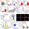
Fig. 1
Fig. 1
LncRNA PKMYT1AR is highly expressed in NSCLC. a LncRNA PKMYT1AR was identified by integrative analysis using GEO datasets, GSE81089 (Blue): Next Generation Sequencing (RNAseq) from NSCLC, GSE144520 (Red): whole-transcriptome sequencing of A549 cells and cisplatin-resistant A549/DPP cells, GSE157427 (Green): gene expression profile for lung cancer stem cells. b The relative expression level of PKMYT1AR in fresh paired tissues isolated from NSCLC patients using Real-time RT-PCR assay, n=24. c The expression of PKMYT1AR in paired NSCLC peripheral blood serum examined by the Real-time RT-PCR assay, n=30. d The relative expression level of PKMYT1AR in TCGA-LUAD (adenocarcinoma; Normal: 59; Tumor: 533) and TCGA-LUSC (squamous cell carcinoma; Normal: 49; Tumor: 502), respectively. e The relative expression pattern of PKMYT1AR in GSE81089 dataset (Normal: 19, Tumor: 197). f The relative expression level of PKMYT1AR in NSCLC cancerous cell lines, including H358, H1975, H1299, H1650, A549 and SPC-A1 examined by Real-time RT-PCR, compared to normal human bronchial epithelial cell line: BEAS-2B. g PKMYT1AR high expression correlates with worse survival rate. h The ROC curve for PKMYT1AR (AUC=0.719) in LUAD using TCGA dataset. i The subcellular localization of PKMYT1AR was predicted by LncLocater. j PKMYT1AR was majorly localized in the cytoplasm of A549 cells using nuclear and cytoplasmic RNA fractionation assay followed by Real-time RT-PCR. β-actin and U1 expressions were used as cytoplasmic and nuclear fraction controls, respectively. k The sub-cellular localization of PKMYT1AR was examined by FISH. The nuclei were stained with DAPI (blue), and the 18S RNA was used as cytoplasm-localized RNA control. Scale bar=25μm. l The YY1 binding motif within the PKMYT1AR promoter region was predicted using JASPAR. m The correlation between YY1 and PKMYT1AR were examined in TCGA-LUAD dataset. n Depletion of YY1 reduced PKMYT1AR expression in A549 cells examined by the Real-time RT-PCR assay. * P < 0.05, ** P < 0.01, *** P < 0.001. MYT1AR=PKMYT1AR
Fig. 2
Fig. 2
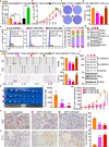
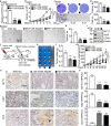
PKMYT1AR knockdown inhibits tumor cell growth and migration. a Establishment of PKMYT1AR overexpression and knockdown cell lines in A549 verified by Real-time RT-PCR. b-d PKMYT1AR knockdown dramatically inhibited A549 cell proliferation (b) and colony formation ability (c), (d) is the quantification data for (c). e-f. Effect of PKMYT1AR knockdown on the G0/G1 cell cycle transition was tested in A549 cells by PI staining and flow cytometry. f is the quantification data for (e). g PKMYT1AR knockdown regulated the expressions of cell cycle transition mediators, including CDK2, CDK6, cyclin D1, p21 and p27. Indicated cell extracts were probed with indicated antibodies. h-i Knockdown of PKMYT1AR inhibited A549 cell migration using wound healing (h) and transwell (i) assays. Quantification data were also indicated, and the OD570 values for trans-well assay were indicated below. Scale bar= 50 μm. j Indicated cell extracts were probed with indicated antibodies to examine the expression patterns of cell migration regulators, including E-cadherin, N-cadherin, Vimentin and Slug. k-m PKMYT1AR knockdown inhibited xenograft tumor formation in vivo. Representative xenograft tumor images (k), tumor masses (l) and tumor volumes (m) were shown. n-o Representative IHC staining of Ki67 (n) and Cleaved Caspase 3 (CC3, o) for indicated xenograft tumors. Quantification data were also indicated. Scale bar=50 μm. * P < 0.05, ** P < 0.01, *** P < 0.001. HPF=high power field, pCDH-Vec=pCDH lenti-viral plasmid vector control. ove=over-expression, sh#1=shRNA#1, sh#2=shRNA#2
Fig. 3
Fig. 3
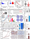
miR-485-5p inhibits tumor progression. a Correlation analysis between PKMYT1AR and miR-485-5p using TCGA-LAUD dataset. b The decreased expression of miR-485-5p in TCGA-LAUD dataset. c The prognostic value of miR-485-5p in TCGA-LAUD examined by Kaplan-Meier Plotter. d The expression of miR-485-5p in fresh peripheral blood serum samples isolated from normal and NSCLC patients, respectively, verified by Real-time RT-PCR. e The expression of PKMYT1AR after over-expression of miR-485-5p, miR-216a-5p and miR-6884-5p in A549 cells examined by Real-time RT-PCR. f A schematic picture of the wild-type (WT) and mutant (MUT) PKMYT1AR luciferase reporter plasmids. g The luciferase activities of the PKMYT1AR luciferase reporters (WT or MUT) were examined in HEK-293T cells with miR-485-5p mimics or mimic NC co-expression. h miR-485-5p regulated A549 cell proliferation assay. i Relative miR-485-5p expression examined by Real-time RT-PCR assay in indicated cells. j-k miR-485-5p regulated A549 colony formation assay (j), (k) is the quantification data for (j). l-m miR-485-5p regulated A549 cell migration examined by wound healing (l) and trans-well (m) assays. Quantification data were also presented, and the OD570 values for trans-well assay were indicated below. Scale bar=50 μm. n-p Representative xenograft tumor images (n), tumor masses (o) and tumor volumes (p) were shown for indicated groups. A549 cells were used. q-r Representative IHC staining of Ki67 (q) and CC3 (r) for indicated xenograft tumors. Quantification data were also indicated. Scale bar=50 μm. * P < 0.05, ** P < 0.01, *** P < 0.001. mimics=miR-485-5p mimics, miR-NC=NC=miRNA mimics control, Anti-Ctrl=miRNA inhibitor control, WT=PKMYT1AR wild-type luciferase reporter, MUT=PKMYT1AR mutant luciferase reporter
Fig. 4
Fig. 4
PKMYT1 is targeted by miR-485-5p and highly expressed in NSCLC. a Identifying PKMYT1 as the downstream target of miRNA-485-5p using various datasets (Green: StarBase; Pink: Targetscan; Yellow: miRWalk), and the top 50% of the target genes were selected. b The positive correlation between PKMYT1AR and PKMYT1 expressions in TCGA-LAUD dataset. c The negative correlation between miRNA-485-5p and PKMYT1 expressions in TCGA-LAUD dataset. d The prognostic value of PKMYT1 in TCGA-LAUD examined by Kaplan-Meier Plotter. e The relative expression level of PKMYT1 in TCGA-LUAD (Normal: 59; Tumor: 533) and TCGA-LUSC (Normal: 49; Tumor: 502), respectively. f The relative expression of PKMYT1 in GSE30219 datasetset (No-relapse:164; Relapse:114). g The ROC curve for PKMYT1 (AUC=0.954) in LUAD using TCGA dataset. h-i miR-485-5p mimics or inhibitor suppressed or promoted, respectively, PKMYT1 expression in A549 cells tested by Real-time RT-PCR (h) and immunoblot (i). j A schematic graph of the wild-type (WT) and mutant (MUT) PKMYT1 3’-UTR containing luciferase reporter plasmids. k The luciferase activities of the PKMYT1 3’-UTR containing luciferase reporters (WT or MUT) were examined in HEK-293T cells with miR-485-5p mimics or mimic NC co-expression. l-m IHC staining of PKMYT1 proteins in NSCLC cancerous tissues, m is the quantification data for (l). Scale bar=50 μm. n PKMYT1 high expression correlates with worse overall survival using IHC data from tissue microarray (l-m). * P < 0.05, ** P < 0.01, *** P < 0.001
Fig. 5
Fig. 5
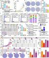
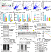
Depletion of PKMYT1 inhibits tumor growth. a-b Establishment of PKMYT1 knockdown in A549 cells, verified by Real-time RT-PCR (a) and immunoblot (b). c PKMYT1 knockdown dramatically inhibited A549 cell proliferation using growth curve assay. d PKMYT1 knockdown inhibited colony formation ability of A549 and SPC-A1 cells, respectively. Quantification data were also indicated. e-f Effect of PKMYT1 knockdown on the G0/G1 cell cycle transition was examined in A549 cells by PI staining and flow cytometry. f is the quantification data for (e). g PKMYT1 knockdown regulated the expressions of cell cycle transition mediators, including CDK2, CDK6, cyclin D1, p21 and p27. Indicated cell extracts were probed with indicated antibodies. h-i PKMYT1 regulated A549 cell migration examined by wound healing (h) and trans-well (i) assays. Quantification data were also presented, and the OD570 values for trans-well assay were indicated below. Scale bar=50 μm. j Indicated cell extracts were probed with indicated antibodies to examine the expression patterns of cell migration regulators, including E-cadherin, N-cadherin, Vimentin and Slug. k Validation of PKMYT1 over-expression by Real-time RT-PCR (top) and immunoblot (bottom) assays. Blue arrow head: exogenous HA-tagged PKMYT1; black arrow head: endogenous PKMYT1. l–r PKMYT1 forced expression overcame PKMYT1AR knockdown effect by cell growth curve (l), wound healing (m-n), trans-well (o, q) and colony formation assays (p, r), (n, q, r) were quantification data for reciprocal assays. Scale bar=50 μm. * P < 0.05, ** P < 0.01, *** P < 0.001. MYT1 ove=PKMYT1 over-expression
Fig. 6
Fig. 6
PKMYT1AR knockdown promotes tumor cell response to DDP. a-b The positive correlations between PKMYT1AR (a), or PKMYT1 (b), and stem cell maintenance related genes, including CD44, SOX2, OCT4 and Nanog, were examined using TCGA-LUAD dataset by Pearson’s correlation analysis. c-f Representative images of colony formation in indicated cells exposed to 0, 4, 6 Gy of X-ray irradiation. d, f were quantification data for indicated assays. g-j Representative images of colony formation in indicated cells exposed to 0, 4, 6 Gy of X-ray irradiation. h, j were quantification data for indicated assays. * P < 0.05, ** P < 0.01, *** P < 0.001
Fig. 7
Fig. 7
Depletion of PKMYT1AR inhibits cancer stem cell maintenance. a Relative mRNA expressions of PKMYT1AR and cancer stem cell marker genes, including CD44, OCT4, SOX2, Nanog in A549 cells, were examined by Real-time RT-PCR upon PKMYT1AR knockdown. b Tumor sphere formation abilities of indicated cells after PKMYT1AR knockdown were examined. Scale bar=50 μm. c Total extracts of indicated cells were probed with indicated antibodies by immunoblot. d Relative mRNA expressions of PKMYT1 and cancer stem cell marker genes in A549 cells, were examined by Real-time RT-PCR upon PKMYT1 knockdown. e Tumor sphere formation abilities of indicated cells after PKMYT1 knockdown were examined. Scale bar=50 μm. f Total cell extracts of indicated cells were probed with indicated antibodies by immunoblot. g Relative mRNA expressions of PKMYT1 and cancer stem cell marker genes in A549 cells, were examined by Real-time RT-PCR with miR-485-5p mimics or miR-NC co-expression. h-i Tumor sphere formation abilities of indicated cells after miR-485-5p mimics or miRNA controls co-transfection were examined (h) in A549 cells. i is the quantification data for (h). Scale bar=50 μm. j Total extracts of indicated cells were probed with indicated antibodies by immunoblot. k-l Rescue effect of PKMYT1AR over-expression on miR-485-5p mimics-mediated phenotype was examined by tumor sphere formation assay (k), l is the quantification data for (k). Scale bar=50 μm. m-n Rescue effect of PKMYT1 over-expression on miR-485-5p mimics-mediated phenotype was examined by tumor sphere formation assay (m), (n) is the quantification data for (k). o-p Rescue effect of PKMYT1 over-expression on PKMYT1AR depletion-mediated phenotype was examined by tumor sphere formation assay (o), (p) is the quantification data for (o). * P < 0.05, ** P < 0.01, *** P < 0.001
Fig. 8
Fig. 8
PKMYT1 activates Wnt signaling. a Wnt signaling pathway was enriched by GSEA analysis. b The correlations between PKMYT1 and Wnt signaling factors, including β-catenin, Axin2, c-Myc and Cyclin D1 in TCGA-LUAD , were examined using pearson’s correlation analysis. c Wnt signaling pathway activity was examined using TOPFlash reporter assay in indicated cells after Wnt3a or LiCl (100 μM) treatment, respectively. d Relative expression of indicated transcripts were examined by Real-time RT-PCR upon PKMYT1 knockdown. e Membrane-tethered form of cancer stem cell marker CD133 was examined in indicated cells by flow cytometry assay. Quantification result was indicated (right). f Total and phosphorylated forms of β-catenin proteins were examined by immunoblot with indicated antibodies in A549 cells. β-catenin Phosphorylation level was detected after normalization of endogenous β-catenin proteins, but not β-actin. g Total β-catenin proteins were reduced after PKMYT1 knockdown in A549 cells, which can be reversed by MG132 (20 μM) treatment. h PKMYT1 knockdown inhibited nuclear accumulation of β-catenin proteins in A549 cells by immunoblot. Lamin B: nuclear fraction; GAPDH: cytosol fraction. i-j Lysates of indicated cells treated by cycloheximide (CHX: 100 μg/mL) were examined by immunoblot with indicated antibodies. (j) is the quantification data for (i). k-l Examining the ubiquitination levels of β-catenin proteins upon PKMYT1 knockdown (k) or over-expression (l) by immunoblot using indicated antibodies under different co-transfection conditions. m Co-IP assay was used to detect the association among PKMYT1, β-TrCP1 and β-catenin in HEK-293T cells. Myc-tagged and HA-tagged PKMYT1 were used for different assays. * P < 0.05, ** P < 0.01, *** P < 0.001
Fig. 9
Fig. 9
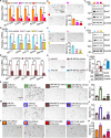
PKMYT1AR targeting ASOs impede tumor growth. a The relative expressions of PKMYT1AR transcripts were examined by Real-time RT-PCR after indicated ASO transfection. b Knockdown of PKMYT1AR inhibited cell proliferation examined by growth curve assays. c-d PKMYT1AR targeting ASOs repressed cell growth by colony formation assay. d is the quantification data for (c). e-f PKMYT1AR targeting ASOs repressed cell migration by trans-well assay (e). f is the quantification data for e. Scale bar=50 μm. g-h PKMYT1AR targeting ASOs inhibited cancer stem cell self-renewal ability by tumor-sphere formation assay (g). h is the quantification data for (g). Scale bar=50 μm. i PKMYT1AR targeting ASOs inhibited cancer stem cell marker genes expressions examined by immunoblot with indicated antibodies. j Schematic view of xenograft mouse model treated by indicated ASOs. k-m Representative xenograft tumor images (k), tumor masses (l) and tumor volumes (m) were shown for indicated groups treated by indicated ASOs. A549 cells were used. n-o Representative IHC staining of PKMYT1, Ki67 and CC3 (n) for indicated xenograft tumors. o is the quantification data for (n). Scale bar=50 μm. * P < 0.05, ** P < 0.01, *** P < 0.001. ASO NC=ASO control
Fig. 10
Fig. 10
A model demonstrating how PKMYT1AR/miR-485-5p/PKMYT1 axis activates Wnt signaling and cancer stem cell self-renewal ability

References

    1. Travis WD, Brambilla E, Nicholson AG, Yatabe Y, Austin JHM, Beasley MB, Chirieac LR, Dacic S, Duhig E, Flieder DB, et al. The 2015 World Health Organization classification of lung tumors: impact of genetic, clinical and radiologic advances since the 2004 classification. J Thorac Oncol. 2015;10:1243–1260. - PubMed
    1. Lo Russo G, Imbimbo M, Garassino MC. Is the chemotherapy era in advanced non-small cell lung cancer really over? Maybe not yet. Tumori. 2016;2016:223–225. - PubMed
    1. Miller KD, Siegel RL, Lin CC, Mariotto AB, Kramer JL, Rowland JH, Stein KD, Alteri R, Jemal A. Cancer treatment and survivorship statistics, 2016. CA Cancer J Clin. 2016;66:271–289. - PubMed
    1. Roth A, Diederichs S. Long noncoding RNAs in lung cancer. Long non-coding Rnas in human disease. Curr Top Microbiol Immunol. 2016;394:57–110. - "V体育安卓版" PubMed
    1. Kopp F, Mendell JT. Functional classification and experimental dissection of long noncoding RNAs. Cell. 2018;172:393–407. - PMC - PubMed

MeSH terms

Substances

LinkOut - more resources