Skip to main page content
U.S. flag

An official website of the United States government

Dot gov

The . gov means it’s official. Federal government websites often end in . gov or . mil. Before sharing sensitive information, make sure you’re on a federal government site. VSports app下载.

Https

The site is secure V体育官网. The https:// ensures that you are connecting to the official website and that any information you provide is encrypted and transmitted securely. .

. 2021 Oct 28:14:5581-5596.
doi: 10.2147/JIR.S327736. eCollection 2021.

Transthyretin and Receptor for Advanced Glycation End Product's Differential Levels Associated with the Pathogenesis of Rheumatoid Arthritis

Affiliations

Transthyretin and Receptor for Advanced Glycation End Product's Differential Levels Associated with the Pathogenesis of Rheumatoid Arthritis (V体育2025版)

Monu et al. J Inflamm Res. .

Abstract

Objective: Rheumatoid arthritis (RA) is a chronic autoimmune, inflammatory joint disease VSports手机版. The identification of multifaceted etiological changes at the protein level in RA remains an important need. We aimed to identify differential proteins (DPs) and gene profiles to uncover inflammatory indicators and their association to RA pathogenesis. .

Methods: 2-DE and SWATH-MS were used to identify DPs in RA and healthy control plasma. Fluorescence phenylboronate gel electrophoresis (Flu-PAGE) with mass spectrometry was used for protein glycation in RA plasma. Disease specificity of identified DPs was confirmed by ELISA and Western blot analysis V体育安卓版. The gene expressions of selected DPs were evaluated by qRT-PCR in PBMCs of RA, systemic lupus erythematosus (SLE), spondyloarthritis (SpA), and osteoarthritis (OA). The functional implication of glycated protein was determined by in- silico and validated by in vitro analysis in fibroblast-like synoviocytes. .

Results: A total of 150 DPs (127 increased and 23 decreased) were identified by 2-DE and SWATH-MS analysis in RA plasma compared to healthy control (HC). Nine proteins were identified as glycated by Flu-PAGE LC-MS/MS. Transthyretin (TTR), serotransferrin, and apolipoprotein-A1 (Apo-A1) were found to be differential and glycated. ELISA and Western blot results revealed the disease-specific increased expression of TTR and RAGE in RA. The qRT-PCR results signify the aberrant gene expression of TTR and RAGE, found to be associated with RA when compared with SLE, SpA, and OA PBMCs. TTR-RAGE interactions were predicted by in-silico and validated by in- vitro analysis using RA-FLS V体育ios版. The increased levels of pro-inflammatory cytokines IL-6, IL-1β, TNF-α, and differently expressed TTR and RAGE were confirmed in fibroblast-like synoviocytes under inflammatory conditions. .

Conclusion: Our findings showed that the level of TTR was increased in RA plasma, along with an altered glycation rate. TTR and RAGE aberrant gene expression in PBMCs are the key events associated with RA, and TNF-α activates the NF-KB pathways and promote TTR and RAGE differential expressions that may have pathogenic/inflammatory significance. VSports最新版本.

Keywords: advanced glycation end products; differential proteins; post-translational modifications; receptor for advanced glycation end products; rheumatoid arthritis; transthyretin. V体育平台登录.


Conflict of interest statement

The authors declare no conflict of interests.

"VSports" Figures

Figure 1
Figure 1
Differential proteins identification in RA plasma: (A) 2-DE analysis of DPs from RA plasma: A representative 2-DE gel image of RA and HC plasma pooled samples (n=5, each) demonstrates differential protein spot intensities. Spots no. 8 (TTR) densitometry analysis shows a substantial increase in RA compared to HC, p<0.0457. The experiment was repeated three times to assess the repeatability of individual spots between groups, the significance level indicate *p < 0.05. (B) SWATH-MS analysis of RA plasma: The distribution of differentially expressed proteins (upregulated and downregulated) in RA plasma is depicted in a pie chart.
Figure 2
Figure 2
Glycated proteins detection and Identification in RA plasma: (A) Flu-PAGE analysis for detecting glycated proteins in RA plasma: The representative gel image displays glycated proteins identified by Flu-PAGE using RA and HC albumin-depleted plasma pools (n=3 each). Left panel; The representative gel image of Flu-PAGE shows glycated proteins detected in Lane-1ʹ (RA plasma incubated with fluorescein-boronic acid-treated RA-FB), Lane 2ʹ (RA plasma incubated with fluorescein control), Lane-3ʹ (healthy plasma incubated with fluorescein-boronic acid-treated HC-FB) and Lane 4ʹ (healthy plasma incubated with fluorescein control). The arrows indicate glycated proteins fluorescent bands (A8-I8) identified by mass spectrometry (MS). Right panel; shows the silver-stained proteins band of the same Flu-PAGE and loading pattern of the samples (Lanes 1–4). (B) The Flu-PAGE % glycation variation in RA and HC plasma: The fluorescent band intensities represent the percentage (%) of glycation in pooled RA and HC plasma (n=3, each) of the Flu-PAGE obtained from (A). TTR (7.78%), serotransferrin (5.37%), and apolipoprotein-A1 (3.38%) are all determined by the percentage glycation difference measured using three replicates, and the data were analysed by normalizing fluorescence intensities of Flu-PAGE to total intensities of silver-stained same gel by densitometry analysis Image Lab (Bio-Rad).
Figure 3
Figure 3
Dot plots of differential proteins TTR, serotransferrin, apolipoprotein-A and RAGE specificity in RA validation by ELISA. The multiple comparisons between groups indicate the significant and non-significant difference between HC vs RA and OA. The plasma TTR (A), serotransferrin (B) Apo-A1 (C) and RAGE (D) were compared between RA, HC and OA (n=24, each), dot plot indicates individual protein abundance of each group found higher of TTR with statistical significance p<0.0039, and significant lower serotransferrin level p<0.0001, lower non-significant Apo-A1 level p<0.234 compared between groups respectively. Similarly, RAGE result statistical significance between groups indicates significant higher-level p<0.0017 between HC and RA, whereas, no significance (ns) level in comparison between HC and OA. The data are presented mean ± SEM. The Student’s t-test was used for statistical significance difference calculation between groups. (E) Correlation analysis between ACPA and TTR: The ACPA concentration shows a positive correlation with TTR with r = 0.1380 indicates the goodness of fit with p<0.0199. (F and G) The graph shows a positive correlation between TTR and DAS-28 (ESR) score with r = 0.4021 and p<0.0135, whereas, RAGE shows non-significant (ns) correlation with r = 0.3072 and p<0.1443 in RA patients. The significance level indicates *p < 0.05; **< 0.01, **< 0.001 and ****< 0.0001.
Figure 4
Figure 4
Dot plots and ROC curve of differential TTR and RAGE validation by ELISA. (A and B) The plasma TTR and RAGE protein were compared between RA (n=100) and HC (n=50). (C and D) The ROC plots indicating individual protein abundance of each group with (AUC: 0.732) for TTR, and (AUC: 0.703) for RAGE, respectively. Independent t-tests were used to determine statistical significance p<0.0001 for TTR and p<0.0003 for RAGE. (E) The dot plot indicates significant p<0.0060 with increased average levels 80.94598 pg/mL in RA than HC 18.37638 pg/mL for TTR and RAGE p<0.0241 significant increased 89.45769 pg/mL in RA compared to the 59.25481pg/mL HC concentration determined by commercially available ELISA kit in HC and RA (n=24 each). For details in the estimation plots the green dot shows the difference between the two means (HC and RA), and the green line shows the 95% confidence interval of that difference. The data are presented as mean ±SEM. (F) Correlation analysis between TTR and RAGE: The graph illustrates correlation analysis measured by Pearson with r = 0.1865 indicates the goodness of fit with p<0.0276 and RAGE concentration was found to be positively correlated with TTR using ELISA between groups. The significance level indicates *p < 0.05; **< 0.01, ***< 0.001 and ****< 0.0001.
Figure 5
Figure 5
Western blot analysis of differential proteins (DPs) TTR and RAGE in RA: Western blot analysis representing the differential level of TTR and RAGE in RA (n=10) pooled plasma compared to healthy controls (n=10). (A) The mean densitometric analysis of TTR shows a significant p<0.021 increased level with fold change 1.7-fold in RA compared to HC. (B) The mean densitometric analysis of RAGE shows a significantly increased level with fold change 1.4-fold in RA compared to HC, normalizing with total protein as a loading control. Data represent mean ± SEM, and statistical significance is determined by Student’s t-test, the significance level indicate *p < 0.05.
Figure 6
Figure 6
PBMCs differential gene expression of TTR, RAGE and pro-inflammatory cytokines (IL-6 and TNF-α) between HC and RA (n=10 each). (A and B) Increased significant gene expression of TTR (p<0.0048) and RAGE (p<0.0003) respectively in RA PBMCs compared to HC. (C and D) Increased significant gene expression of IL-6 (p<0.0004) and TNF-α (p<0.0018) respectively in RA PBMCs compared to HC. The estimation plot of each indicates the difference between the mean of HC and RA. The green dot shows the difference between the two means (HC, and RA), and the green line shows the 95% confidence interval of that difference. The data are presented as mean ±SEM. The significance level indicates **< 0.01, and ***< 0.001.
Figure 7
Figure 7
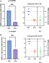
PBMC’s differential gene expression of TTR, RAGE between RA and OA. (A and B) Shows the increased significant gene expression of TTR (p<0.0058) and RAGE (p<0.0029) respectively in RA PBMCs compared to OA. The estimation plot of each groups shows a green dot indicates a difference between the mean of RA and OA, the green line represents the 95% confidence interval of that difference. The data are presented as mean ±SEM. The significance level indicates **< 0.01.
Figure 8
Figure 8
Differential gene expression analysis of TTR, RAGE, pro-inflammatory cytokine (IL-6, TNF-α) in RA, SLE, and SPA PBMCs by qRT-PCR. The altered gene expression was analyzed in RA (n=10), SLE, SpA (n=3 each) PBMCs compared to HC (n=10). (A) Shows the gene expression of TTR significantly increased in RA compared to SLE, SPA and OA PBMCs compared to HC, respectively. (B) Shows the increased gene expression of RAGE significantly in RA compared to SLE, SpA and OA. (C and D) Shows the increased pro-inflammatory cytokines IL-6 significantly in RA, SLE, SpA but non-significant (ns) in OA compare to HC respectively whereas TNF-α was found to be significantly increased in all compare to HC PBMCs. The data were normalized with β–actin and/or GAPDH as an internal loading control. Values are presented as the mean ± SEM. The significance level indicates *p < 0.05; **< 0.01, ***< 0.001 and ****< 0.0001, ns = non-significant, analyzed by one-way ANOVA.
Figure 9
Figure 9
Fold induction of pro-inflammatory cytokines (IL-6, and IL-1β) and NF-κB pathway subunits (Rel A/p65, p50, and TNF-α) mRNA expression analysis in synovial cells by qRT-PCR. The mRNA expression in SW982 cells induced with TNF-α (10ng/mL) for 1hr compared to control (un-induced). (A) Fold induction of IL-6 gene expression indicates a significantly increased level (p<0.0094). (B) The fold induction of IL-1β gene expression indicates a significant (p<0.0001) increased mRNA level. (C) Fold induction of p50gene expression shows a significant (p<0.0001) up-regulation. (D) Fold induction of Rel A/p65 gene expression was observed significant (p<0.0010) on TNF-α induction. (E) Fold induction of TNF-α gene expression shows a significant (p<0.0004) higher level. The data was normalized with GAPDH as an internal loading control and the values are presented as the mean ± SEM (n = 3). The significance level indicates **< 0.01, ***< 0.001 and ****< 0.0001 versus normal control or TNF-α induction analysed by Student’s t-test.
Figure 10
Figure 10
Fold induction of TTR and RAGE mRNA expression analysis in synovial cells by qRT-PCR. The mRNA expression in SW982 cells induced with TNF-α (10ng/mL) for 1hr, compared to control un-induced (A) Fold induction of TTR gene expression significantly (p<0.028) increased upon TNF-α induction compare to control. (B). Similarly, fold induction of RAGE gene expression indicates a significant (p<0.0011) increased level in TNF-α induced synoviocytes. The data normalized with GAPDH as an internal loading control and the values presented the mean ± SEM (n = 3). The significance level indicates *p < 0.05; **< 0.01 versus normal control and TNF-α treatment analysed by Student’s t-test.

References

    1. Smolen JS, Aletaha D, Barton A, et al. Rheumatoid arthritis. Nat Rev Dis Primers. 2018;4:18001. - PubMed
    1. WHO. Rheumatoid arthritis; 2018. Available from: www.who.int/chp/topics/rheumatic/en/. Accessed October 8, 2021.
    1. Guo Q, Wang Y, Xu D, Nossent J, Pavlos NJ, Xu J. Rheumatoid arthritis: pathological mechanisms and modern pharmacologic therapies. Bone Res. 2018;6:15. - V体育官网入口 - PMC - PubMed
    1. Zavala-Cerna MG, Martínez-García EA, Torres-Bugarín O, et al. The clinical significance of posttranslational modification of autoantigens. Clinic Rev Allerg Immunol. 2014;47:73–90. - PubMed
    1. Santos AL, Lindner AB. Protein posttranslational modifications: roles in aging and age-related disease. Oxid Med Cell Longev. 2017;2017:5716409. - PMC - PubMed

V体育2025版 - LinkOut - more resources