Skip to main page content
U.S. flag

An official website of the United States government

Dot gov

The . gov means it’s official. Federal government websites often end in . gov or . mil VSports app下载. Before sharing sensitive information, make sure you’re on a federal government site. .

Https

The site is secure V体育官网. The https:// ensures that you are connecting to the official website and that any information you provide is encrypted and transmitted securely. .

Review
. 2021 Aug 12;11(18):8813-8835.
doi: 10.7150/thno.62521. eCollection 2021.

Inflammation-related pyroptosis, a novel programmed cell death pathway, and its crosstalk with immune therapy in cancer treatment

Affiliations
Review

"V体育2025版" Inflammation-related pyroptosis, a novel programmed cell death pathway, and its crosstalk with immune therapy in cancer treatment

Sheng-Kai Hsu et al. Theranostics. .

"V体育安卓版" Abstract

In recent decades, chemotherapies targeting apoptosis have emerged and demonstrated remarkable achievements. However, emerging evidence has shown that chemoresistance is mediated by impairing or bypassing apoptotic cell death. Several novel types of programmed cell death, such as ferroptosis, necroptosis, and pyroptosis, have recently been reported to play significant roles in the modulation of cancer progression and are considered a promising strategy for cancer treatment. Thus, the switch between apoptosis and pyroptosis is also discussed. Cancer immunotherapy has gained increasing attention due to breakthroughs in immune checkpoint inhibitors; moreover, ferroptosis, necroptosis, and pyroptosis are highly correlated with the modulation of immunity in the tumor microenvironment VSports手机版. Compared with necroptosis and ferroptosis, pyroptosis is the primary mechanism for host defense and is crucial for bridging innate and adaptive immunity. Furthermore, recent evidence has demonstrated that pyroptosis exerts benefits on cancer immunotherapies, including immune checkpoint inhibitors (ICIs) and chimeric antigen receptor T-cell therapy (CAR-T). Hence, in this review, we elucidate the role of pyroptosis in cancer progression and the modulation of immunity. We also summarize the potential small molecules and nanomaterials that target pyroptotic cell death mechanisms and their therapeutic effects on cancer. .

Keywords: Nonapoptotic programmed cell death; cell death switch; immunotherapy; inflammasome; pyroptosis. V体育安卓版.


Conflict of interest statement (V体育官网入口)

Competing Interests: The authors have declared that no competing interest exists.

Figures

Figure 1
Figure 1
Morphological changes during pyroptotic cell death. The inflammasome is activated by PAMPs and DAMPs or chemotherapy, and this activation leads to the activation of caspase-1 or caspase-3, which eventually results in the cleavage of gasdermin D and gasdermin E to form N-terminal fragments, respectively. First, gasdermin-mediated pore formation (nonselective channel) promotes the release of inflammatory substances, including IL-1β and IL-18, due to the large inner diameters of gasdermin pores. Subsequently, the formation of these nonselective channels and the influx of water contribute to cell swelling, membrane blebbing with bubble-like protrusions (known as pyroptotic bodies), continuous cell swelling, and loss of plasma membrane integrity to culminate in rupture of the cell membrane. Finally, HMGB1 and ATP are secreted after cell membrane rupture, followed by pyroptotic cell death.
Figure 2
Figure 2
Molecular mechanisms and cellular physiology of pyroptosis. The molecular mechanism of pyroptosis is primarily divided into the canonical pathway (dependent on caspase-1) and the noncanonical pathway (dependent on caspase-4/5/11 or mediated by caspase-3 or caspase-8). (A) NLRP1, NLRP3, NLRC4, AIM2, and pyrin inflammasomes trigger pyroptosis by inducing the activation of caspase-1. Here, the NLRP3 inflammasome is shown as an example in the figure. The pathway is mainly initiated through the binding of DAMPs or PAMPs to PRRs. PRRs can then recruit the adaptor ASC and pro-caspase-1 to form inflammasomes (the NLRP1 and NLRC4 inflammasomes are exceptions in which ASC is not needed). Procaspase-1 is cleaved by inflammasomes to activate caspase-1, and activated caspase-1 leads to the maturation of IL-1β and IL-18 and the cleavage of GSDMD at 272FLTD275 to generate the N- and C-termini. The N-terminus forms oligomers and translocates to the cell membrane, which finally induces pyroptosis and the secretion of intracellular substances, such as IL-1β, IL-18, HMGB1, ATP, and LDH. Moreover, the released cytokines and DAMPs trigger inflammation and the subsequent immune response. (B) The caspase-4/-5/-11-dependent noncanonical pathway is triggered by LPS directly interacting with the CARD of pro-caspase-4/-5/-11 (caspase-4/-5 in humans; caspase-11 in mice). Subsequently, this interaction contributes to the activation of caspase and the cleavage of GSDMD to yield the N- and C-termini. The N-terminus forms oligomers and translocates to the cell membrane to induce pyroptosis. (C) The caspase-3-mediated noncanonical pathway is primarily initiated by chemotherapies, mitochondrial dysfunction, or the accumulation of generated ROS. This pathway shares a common upstream signaling pathway with apoptosis; nevertheless, the switch between apoptosis and pyroptosis depends on the cellular content of GSDME. Chemotherapy induces the translocation of BAX/BAK to the mitochondrial outer membrane to form pores, resulting in MOMP and cytochrome C release. Subsequently, this process even sequentially activates caspase-9 and caspase-3. Moreover, caspase-3 is also activated by death receptor signaling-induced caspase-8. Activated caspase-3 can cleave GSDME after Asp270 to generate the C- and N-termini. Similarly, the N-terminus forms oligomers and translocates to the cell membrane to induce pyroptosis. (D) GSDMC is upregulated by the PD-L1/p-STAT3 axis and specifically cleaved by death receptor signaling-induced caspase-8. The GSDMC N-terminus forms oligomers and translocates to the cell membrane to induce pyroptosis.
Figure 3
Figure 3
Crosstalk in immune responses induced by pyroptotic cell death. Cytokines (e.g., IL-1β and IL-18) and DAMPs (e.g., HMGB1 and ATP) can be released through pyroptotic cell death. IL-1β promotes antigen presentation between dendritic cells and T lymphocytes and drives the differentiation of naïve CD4+ T lymphocytes toward a Th17 phenotype. Castano et al. suggested that IL-1β can inhibit MET in metastatic breast cancer cells by inducing the expression of ZEB1. IL-18 interacts with IL-18 receptors on immune cells, such as NK cells and Th1 cells, further inducing the generation of IFN-γ. In addition, IL-18 reportedly drives NK cells to DC-like cells through the upregulation of MHC-II and costimulatory molecules. IFN-γ exerts several effects on the activation of the immune response: it can block immunosuppressive cytokines, such as TGF-β and IL-10 secreted by Tregs; it can promote the activation and proliferation of cytotoxic T lymphocytes (CTLs) through the upregulation of granzyme B; it can drive naïve CD4+ T lymphocytes toward a Th1 phenotype; and it can upregulate MHC-II molecules on tumor cells. Furthermore, Zhou et al. indicated that IFN-γ upregulates GSDMB in several cancer cell lines, leading to pyroptosis mediated by granzyme A. HMGB1 can upregulate MHC-II molecules on dendritic cells and promote their migration. Moreover, it can induce the secretion of tumor necrosis factor (TNF) from macrophages by interacting with Toll-like receptor 4 (TLR4).
Figure 4
Figure 4
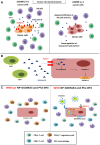
Correlation between pyroptosis and the tumor immune microenvironment. (A) Downregulation of perforin and granzyme B and reduced infiltration of NK cells and CTLs were observed in the GSDME-/- cancer cell line microenvironment. (B) Caspase-3-mediated pyroptosis in cancer cells is induced through perforin and granzyme B secreted by CAR-T cells. (C) The administration of NP-GSDMA3 and Phe-BF3 can induce GSMDA3-mediated pyroptosis in cancer cells and contribute to increased infiltration of immune cells, including CD4+ and CD8+ lymphocytes and M1 macrophages, and reduced infiltration of CD4+ regulatory T lymphocytes.

References

    1. Brill A, Torchinsky A, Carp H, Toder V. The role of apoptosis in normal and abnormal embryonic development. J Assist Reprod Genet. 1999;16(10):512–9. - PMC - PubMed
    1. Nainu F, Shiratsuchi A, Nakanishi Y. Induction of apoptosis and subsequent phagocytosis of virus-infected cells as an antiviral mechanism. Front Immunol. 2017;8:1220. - PMC - PubMed
    1. Reed JC. Mechanisms of apoptosis avoidance in cancer. Curr Opin Oncol. 1999;11(1):68–75. - "V体育ios版" PubMed
    1. Mortezaee K, Salehi E, Mirtavoos-Mahyari H, Motevaseli E, Najafi M, Farhood B. et al. Mechanisms of apoptosis modulation by curcumin: Implications for cancer therapy. J Cell Physiol. 2019;234(8):12537–50. - PubMed
    1. Su Z, Yang Z, Xie L, DeWitt JP, Chen Y. Cancer therapy in the necroptosis era. Cell Death Differ. 2016;23(5):748–56. - PMC - PubMed