Skip to main page content
U.S. flag

An official website of the United States government

Dot gov

The . gov means it’s official. Federal government websites often end in VSports app下载. gov or . mil. Before sharing sensitive information, make sure you’re on a federal government site. .

Https

The site is secure. The https:// ensures that you are connecting to the official website and that any information you provide is encrypted and transmitted securely V体育官网. .

. 2021 Sep 3;7(36):eabi9787.
doi: 10.1126/sciadv.abi9787. Epub 2021 Sep 3.

Single-cell transcriptome of early hematopoiesis guides arterial endothelial-enhanced functional T cell generation from human PSCs

Affiliations

Single-cell transcriptome of early hematopoiesis guides arterial endothelial-enhanced functional T cell generation from human PSCs

"V体育2025版" Jun Shen et al. Sci Adv. .

Abstract

Hematopoietic differentiation of human pluripotent stem cells (hPSCs) requires orchestration of dynamic cell and gene regulatory networks but often generates blood cells that lack natural function. Here, we performed extensive single-cell transcriptomic analyses to map fate choices and gene expression patterns during hematopoietic differentiation of hPSCs and showed that oxidative metabolism was dysregulated during in vitro directed differentiation. Applying hypoxic conditions at the stage of endothelial-to-hematopoietic transition in vitro effectively promoted the development of arterial specification programs that governed the generation of hematopoietic progenitor cells (HPCs) with functional T cell potential. Following engineered expression of the anti-CD19 chimeric antigen receptor, the T cells generated from arterial endothelium-primed HPCs inhibited tumor growth both in vitro and in vivo VSports手机版. Collectively, our study provides benchmark datasets as a resource to further understand the origins of human hematopoiesis and represents an advance in guiding in vitro generation of functional T cells for clinical applications. .


Figures (VSports手机版)

Fig. 1.
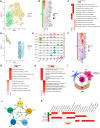
Fig. 1.. High-resolution dissection of hematopoietic differentiation of hPSCs using scRNA-seq.
(A) Schematic diagram of hematopoietic differentiation from H1 hESCs. Cells were collected on days 0, 2, 4, and 6 for scRNA-seq. Meso, mesoderm. (B) Identification of cell populations in the differentiation culture at days 0, 2, 4, and 6 visualized by uniform manifold approximation and projection (UMAP). Each dot represents one cell, and colors represent cell clusters as indicated. (C) UMAP visualization of the expression of typical feature genes for the identification of cell clusters. (D) Expression of genes in seven different categories of the indicated cell clusters. (E) Trajectory reconstruction of all single cells throughout hematopoietic differentiation (excluding day 0) reveals three branches: prebranch (before bifurcation), Mes lineage branch, and EC-HC lineage branch. (F) Gene expression heatmap of the top 3000 highly variable genes among cells used in the trajectory inference analysis presented in a pseudo-temporal order. (G) Expression dynamics of the top 3000 highly variable genes cataloged into four major clusters in a pseudo-temporal order shown as blue lines (Mes lineage) and red lines (EC-HC lineage). Thick lines indicate the average gene expression patterns in each cluster. Gene signatures and expression dynamics of representative genes in each gene cluster are shown. (H) Gene ontology (GO) analyses of each gene cluster identified in (F) and (G). ER, endoplasmic reticulum.
Fig. 2.
Fig. 2.. Heterologous cellular components and their interactions involved in EHT.
(A) UMAP visualization of EH-Mes, EPC, and D4-HE clusters based on subdivision of cells in the D4-EC cluster described in Fig. 1B. (B) Heatmap showing the average expression of the top seven cluster-specific TFs among the EH-Mes, EPC, and D4-HE clusters. (C) The major GO Biological Process (GO BP) terms in which cluster-specific genes are enriched for EH-Mes, EPC, and D4-HE clusters. (D) UMAP visualization of AE, VE, D6-HE, and D6-HPC clusters based on subdivision of cells in the D6-EC and HPC clusters described in Fig. 1B. (E) Violin plots showing the expression of feature genes in each cell cluster. (F) Heatmap showing the average expression of the top seven cluster-specific TFs among the AE, VE, D6-HE, and D6-HPC clusters. (G) The major GO:BP terms in which cluster-specific genes are enriched for D6-HE and D6-HPC clusters. (H) The major GO:BP terms in which cluster-specific genes are enriched for D4-HE and D6-HE clusters. ATP, adenosine triphosphate; SRP, signal recognition particle. (I) Schematic diagram showing the ligand-receptor interactions between HE or HPC and other niche clusters (AE, VE, and Mes). (J) Ligand-receptor interaction network showing the potential communications among niche cells (AE, VE, and Mes) and hematopoiesis-related cells (D6-HE and D6-HPC). (K) Heatmap showing the ligand-receptor pairs that exhibit different expression patterns among distinct niche clusters when coupled with D6-HE or D6-HPC.
Fig. 3.
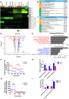
Fig. 3.. Down-regulation of oxygen metabolism at the stage of EHT.
(A) Classification heatmap showing that the identified subclusters differentiated in vitro are similar to those in human embryos. The data of Carnegie stage (CS) 10/11 and CS13 cells from human embryos were reanalyzed. Hema, hematopoietic cells; Epi, epithelial cells. (B) Enriched GO terms in D6-HE and HE in vivo, respectively. (C) Enriched GO terms in D6-HPC and hematopoietic cells in vivo, respectively. (D) Volcano plot displaying the differentially expressed genes (DEGs) between cells of the EC-HC lineage on days 4 and 6. Representative respiratory chain (red) and endothelial-hematopoietic (blue) genes are indicated. Gray dots represent non-DEGs [approximately <1.6 fold change (FC)]. (E) GO analysis of down-regulated and up-regulated genes in the EC-HC lineage comparing day 6 with day 4. ncRNA, noncoding RNA; rRNA, ribosomal RNA. (F) The raw data of oxygen consumption rate (OCR) on day 4 and day 6 H1-derived CD34+ cells determined using a Seahorse XF analyzer in the presence of the mitochondrial inhibitors (1 μM oligomycin, 5 μM carbonyl cyanide p-trifluoromethoxyphenylhydrazone, or 1.5 μM rotenone plus antimycin A). (G) Quantitation of basal, ATP-linked, and maximal OCR evaluated by two-way analysis of variance (ANOVA) (n = 5). (H) The raw data of glycolytic proton efflux on day 4 and day 6 H1-derived CD34+ cells following injection of rotenone plus antimycin A (0.5 μM) and 2-deoxy-d-glucose (50 mM); data are obtained using a Seahorse XF analyzer. (I) Quantitation of basal and compensatory glycolysis evaluated by two-way ANOVA (n = 5). Data are presented as means ± SEM. **P < 0.01 and ***P < 0.001.
Fig. 4.
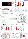
Fig. 4.. Hypoxia enhances hematopoietic differentiation by promoting the arterial specification program.
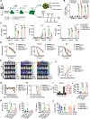
(A) Confocal microscopy visualization of emerging round hematopoietic cells on day 6 under the hypoxic (5% O2) or normoxic conditions. Scale bars, 40 μm. (B) The percentage of CD43+ hematopoietic cells on day 6 under the hypoxic (5% O2) or normoxic conditions. (n = 3; two-way ANOVA). (C and D) Diagram and representative flow cytometric analysis demonstrating the five conditions used to test the importance of hypoxia exposure (1% O2) in promoting hematopoiesis. (n = 3). (E) Mean gene expression of all cells at each time point. (F) HIF-1a expression measured by Western blot at each time point under the normoxic or hypoxic conditions (1% O2). (G and H) Representative flow cytometric and statistical analyses of the dynamic changes in the arterial cell population between days 4 and 6 under hypoxic (1% O2) or normoxic conditions. (n = 3; two-way ANOVA). (I) qRT-PCR analysis of the expression of DLL4 and NR2F2 from days 4 to 6 under the hypoxic (1% O2) or normoxic conditions. (n = 3; two-way ANOVA). (J) Representative flow cytometric analysis of the frequency of arterial cells in dimethyl sulfoxide (DMSO)– or U0126-treated day 5 cells. (n = 3). (K and L) Flow cytometric analysis of the frequency and number of CD43+ cells in DMSO- or U0126-treated day 6 cells. (n = 3; two-tailed Student’s t test). SSC, side scatter. For (J) to (L), U0126 or DMSO was added to the cultures from days 4 to 6 of differentiation. Experiments were performed on H1 unless otherwise indicated. Data are presented as means ± SEM. *P < 0.05 and ***P < 0.001.
Fig. 5.
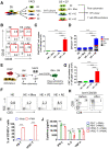
Fig. 5.. AE promote hematopoietic progenitor generation with T cell potential.
(A) Schematic of protocol for investigating the niche effect on hematopoiesis. (B) Representative flow cytometric analysis of the hematopoietic progenitors obtained from H1-derived HE, or HE cocultured with Mes (HE + Mes), VE (HE + VE), or AE (HE + AE) following 7 days of EHT culture (n = 3). (C) The fold change in CD43+CD45+ cell number compared to HE. The cells were generated under the conditions of HE, HE + Mes, HE + VE, or HE + AE. (n = 3; one-way ANOVA). (D) CFC assay of HPCs generated with or without coculture with AE, VE, or Mes. Colony-forming units (CFUs) per 2000 cells plated. (n = 3; two-way ANOVA). (E) Schematic diagram of the protocol for T cell differentiation. MACS, magnetic-activated cell sorting. (F and G) T cell potential of hematopoietic progenitors generated from D5 HE following 7 days of culture in the presence or absence of VE, AE, or Mes. (n = 3; one-way ANOVA). (H) Representative flow cytometric analysis of the expression of CD4 and CD8 by CD3+ cells before and after anti-CD3/CD28 stimulation (n = 3). (I) The proportion of CD107a in hPSC-derived T cells (hPSC-T) and peripheral blood T cells (PB-T) after treatment with phorbol 12-myristate 13-acetate (PMA)–ionomycin. (n = 3; two-way ANOVA). (J) Polyfunctional cytokine production by hPSC-T and PB-T after treatment with PMA-ionomycin. (n = 3; two-way ANOVA). Experiments were performed on H1 hESCs unless otherwise indicated. Data are presented as means ± SEM. *P < 0.05 and ***P < 0.001.
Fig. 6.
Fig. 6.. AE-primed hPSC-T showed therapeutic potential toward B-ALL following engineered expression of anti–CD19-CAR.
(A) Schematic diagram showing the protocol for generating hPSC-CAR-T for cytotoxicity assays both in vitro and in vivo. The AE used to prime T cells are generated under the hypoxic condition (1% O2). (B) The proportion of CD107a+ cells in effector cells after coincubation with target cells (E:T = 1:1) for 5 hours. (n = 3; two-way ANOVA). (C) ELISA data showing the release of IFN-γ, IL-2, and TNF-α after coincubation with target cells for 24 hours. (n = 3; two-way ANOVA). (D) Direct lysis of effector cells against target cells. Flow cytometry analysis of the percentage of CD19+CD3 cells. (n = 3; two-way ANOVA). (E and F) Representative bioluminescence imaging and statistical analysis of the bioluminescence intensity in nonobese diabetic/severe combined immunodeficient (NOD/SCID) mice. (n = 7; two-way ANOVA). (G) Body weight of the mice. (n = 7; two-way ANOVA). (H) Kaplan-Meier curve representing the percentage survival of the mice. (n = 7; log-rank test). (I) Specific fluorescence intensity (SFI) of CD19 on the bone marrow mononuclear cells (BMMNCs) in six patients with B acute lymphoblastic leukemia (B-ALL). (J) The proportion of CD107a+ cells in effector cells after coincubation with primary B-ALL target cells at E:T = 1:1 for 5 hours. (n = 6; one-way ANOVA). (K) The release of IFN-γ, IL-2, and TNF-α after coincubation with primary B-ALL target cells for 24 hours. (n = 6; one-way ANOVA). (L) Direct lysis of effector cells against primary B-ALL target cells. (n = 6; two-way ANOVA). All values are means ± SEM. *P < 0.05, **P < 0.01, and ***P < 0.001.

References

    1. Hansen M., von Lindern M., van den Akker E., Varga E., Human-induced pluripotent stem cell-derived blood products: State of the art and future directions. FEBS Lett. 593, 3288–3303 (2019). - PubMed
    1. Sugimura R., Jha D. K., Han A., Soria-Valles C., da Rocha E. L., Lu Y. F., Goettel J. A., Serrao E., Rowe R. G., Malleshaiah M., Wong I., Sousa P., Zhu T. N., Ditadi A., Keller G., Engelman A. N., Snapper S. B., Doulatov S., Daley G. Q., Haematopoietic stem and progenitor cells from human pluripotent stem cells. Nature 545, 432–438 (2017). - PMC - PubMed
    1. Gordon-Keylock S., Sobiesiak M., Rybtsov S., Moore K., Medvinsky A., Mouse extraembryonic arterial vessels harbor precursors capable of maturing into definitive HSCs. Blood 122, 2338–2345 (2013). - "VSports" PMC - PubMed
    1. Chen M. J., Yokomizo T., Zeigler B. M., Dzierzak E., Speck N. A., Runx1 is required for the endothelial to haematopoietic cell transition but not thereafter. Nature 457, 887–891 (2009). - PMC - PubMed
    1. Lancrin C., Mazan M., Stefanska M., Patel R., Lichtinger M., Costa G., Vargel O., Wilson N. K., Moroy T., Bonifer C., Gottgens B., Kouskoff V., Lacaud G., GFI1 and GFI1B control the loss of endothelial identity of hemogenic endothelium during hematopoietic commitment. Blood 120, 314–322 (2012). - PubMed