Skip to main page content
U.S. flag

An official website of the United States government

Dot gov

The . gov means it’s official. Federal government websites often end in . gov or VSports app下载. mil. Before sharing sensitive information, make sure you’re on a federal government site. .

Https

The site is secure. The https:// ensures that you are connecting to the official website and that any information you provide is encrypted and transmitted securely. V体育官网.

. 2021 Sep 8;13(610):eabd8995.
doi: 10.1126/scitranslmed.abd8995. Epub 2021 Sep 8.

scRNA-seq of human vitiligo reveals complex networks of subclinical immune activation and a role for CCR5 in Treg function

Affiliations

scRNA-seq of human vitiligo reveals complex networks of subclinical immune activation and a role for CCR5 in Treg function

Kyle J Gellatly et al. Sci Transl Med. .

"VSports" Abstract

Vitiligo is an autoimmune skin disease characterized by the targeted destruction of melanocytes by T cells. Cytokine signaling between keratinocytes and T cells results in CD8+ T cell infiltration of vitiligo lesions, but the full scope of signals required to coordinate autoimmune responses is not completely understood. We performed single-cell RNA sequencing on affected and unaffected skin from patients with vitiligo, as well as healthy controls, to define the role of each cell type in coordinating autoimmunity during disease progression. We confirmed that type 1 cytokine signaling occupied a central role in disease, but we also found that this pathway was used by regulatory T cells (Tregs) to restrain disease progression in nonlesional skin. We determined that CCL5-CCR5 signaling served as a chemokine circuit between effector CD8+ T cells and Tregs, and mechanistic studies in a mouse model of vitiligo revealed that CCR5 expression on Tregs was required to suppress disease in vivo but not in vitro. CCR5 was not required for Treg recruitment to skin but appeared to facilitate Treg function by properly positioning these cells within the skin. Our data provide critical insights into the pathogenesis of vitiligo and uncover potential opportunities for therapeutic interventions. VSports手机版.


Figures
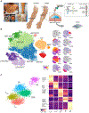
Fig. 1:
Fig. 1:. scRNA-seq of vitiligo skin reveals epidermal cell types and immune cells
Suction blister biopsies were obtained from healthy skin as well as non-lesional and active lesions from vitiligo subjects. The supernatant was analyzed by ELISA while the skin cells were analyzed by flow cytometry or single cell RNA-sequencing (A). Unbiased clustering of the scRNA-seq data resulted in 10 distinct clusters, consisting of 5 main cell types. Melanocytes (MEL), dendritic DCs (DC), and Macrophages were each identified as a single cluster. Keratinocytes (KRT) consisted of 5 clusters, Basal-1 (B1), Basal-2 (B2), Spinous (SP), Granular (GR), and Eccrine (ECR). Lymphocytes consisted of 2 clusters, IFNG− and IFNG+ (B). Refined clustering of the original TRAC+ cluster revealed 5 distinct cell types, Tregs (FOXP3+/CTLA4+), TConv (FOXP3−/IL13+/IL26+), CD8+ T cells (TRAC+/CD8+), γδ T cells (TRGC1+/TRDC+), and NK cells (FCER1G+/KIT+) (C).
Fig. 2:
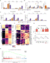
Fig. 2:. Activation of immune cells reveals subclinical inflammation in non-lesional vitiligo skin
Barplots reflecting the fraction of each cell type in each skin condition across all donors (healthy, non-lesional and lesional); error bars indicate standard error of the mean (A). Dotplot showing the paired proportions of lymphocytes and melanocytes within each patient, with an inset showing the ratio of Treg to CD8 T cells for patients where over 30 cells of these types were detected (B). Barplot of CPM (counts per million) values of IFNG in the T cell subsets and NK cells (C). Barplot of the weighted (CPM X Cell proportion) CPM values of IFNG in the T cell subsets and NK cells (D). K-means clustering of all differentially expressed genes within CD8+ T cells (E). K-means clustering of all genes found differentially expressed in more than two T Cell subsets (F). GSEA of the T cell activation pathway within Treg, TConv, and CD8+ T cells between all skin transitions. NES = normalized enrichment score, RES = running enrichment score (G). GSEA of the Th1 Activation pathways within Tregs in the transition from healthy to non-lesional skin (H). *FDR < 0.1, **FDR < 0.01
Fig. 3:
Fig. 3:. Response to IFNG in healthy and vitiligo skin
GSEA enrichment plot showing the IFNG response within all cell types detected within the epidermis and p value for the IFNG response between healthy to non-lesional, and non-lesional to lesional skin. NES = normalized enrichment score, RES = running enrichment score (A). Barplots showing the cell type specific expression of IFNG responsive genes, HLA-A, HLA-DPA1, IRF6, and IFITM3 (B). Barplot showing the weighted CPM expression levels of CXCL9/10/11 within APCs and keratinocytes (C). *p < 0.1, **p < 0.01
Fig. 4:
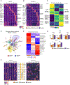
Fig. 4:. Ligand receptor mapping establishes communications between cells and their change in vitiligo
K-means clustering of all expressed ligands (A) and receptors (B) across each cell type and each skin condition, as annotated by a published database with representative GO terms for each k group. Each box represents the aggregate bulk CPM value for a given gene expressed in that cell type in each condition. K-means clustering of all ligand-receptor pairs in which at least one of the genes is significantly (p < 1.0E2) DE (C). Plot of the full receptor and ligand network showing the 18 identified clusters within the network, with the Chemotaxis, TGFB / Notch Signaling, and MHC-I clusters highlighted. Edge color denotes whether the given interaction is increased or decreased in signal between healthy and lesional skin (D). K-means clustering of the meta-edges of each cluster within the network across each of the skin conditions (E). Chemokine network heatmaps showing the expression of ligands and receptors, as well as their annotated interactions (F). Barplots showing CCL3/4/5 expression within CD8+ T cells and CCR5 expression on Tregs and CD8+ T cells (G)
Fig. 5:
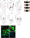
Fig. 5:. Treg CCR5 is required for optimal suppression of effector CD8+ T cells in the skin
Graph summarizing flow cytometry data comparing the average CCR5 expression (MFI) by CD4+FOXP3+ Tregs in blister fluid from the non-lesional and lesional skin of the same patient (A) (N=5, paired T-test). Quantification of CCR5 expression by Tregs in the skin draining lymph nodes (SDLN) and ear skin of uninduced FOXP3-GFP mice (No Vitiligo) and FOXP3-GFP with vitiligo (Vitiligo) (B) (3 experiments, N≥3, student T test). Ear vitiligo scores (C) and representative images (D) of ears from Rag−/− mice that received no Tregs, WT (WT→Rag) or CCR5−/− (CCR5−/−→Rag) Tregs 5 weeks post vitiligo induction (4 experiments, N≥2, student T test). Graphs depicts quantification of the average number of Tregs in the SDLNs and ear skin of Rag−/− mice that received WT or CCR5−/− Tregs 5 weeks post vitiligo induction (E) (3 experiments, N≥2, student T test). Quantification of WT and CCR5−/− Tregs in the SDLNs and ear skin of Rag−/− mice in a competitive Treg-adoptive transfer model of vitiligo (F). Graph depicts the proportion of WT or CCR5−/− Tregs in the indicated tissue relative to their proportion at input; (3 experiments, N≥3, student T test). Representative images of lesional vitiligo skin stained with CD8 (cell surface, AF488, green fluorescence), FOXP3 (nucleus, AF555, red fluorescence) and CCR5 (cell surface, AF647 cyan fluorescence); N=3 patients, 40x image, bar = 25μm (G). CCR5, FOXP3, and CD8 expression in the area defined by the white box is magnified in (H) and CCR5 expression in (I); yellow arrow highlights a FOXP3+ cell expressing CCR5, bar = 10μm. Bars depict the mean ± SD in all graphs.

References

    1. Rodrigues M, Ezzedine K, Hamzavi I, Pandya AG, Harris JE, Vitiligo Working Group, Current and emerging treatments for vitiligo. J. Am. Acad. Dermatol. 77, 17–29 (2017). - PubMed
    1. Frisoli ML, Harris JE, Vitiligo: Mechanistic insights lead to novel treatments. J. Allergy Clin. Immunol. 140, 654–662 (2017). - PubMed
    1. Klarquist J, Denman CJ, Hernandez C, Wainwright DA, Strickland FM, Overbeck A, Mehrotra S, Nishimura MI, Le Poole IC, Reduced skin homing by functional Treg in vitiligo. Pigment Cell Melanoma Res. 23, 276–286 (2010). - "V体育官网入口" PMC - PubMed
    1. Lili Y, Yi W, Ji Y, Yue S, Weimin S, Ming L, Global activation of CD8+ cytotoxic T lymphocytes correlates with an impairment in regulatory T cells in patients with generalized vitiligo. PLoS One. 7, e37513 (2012). - PMC - PubMed
    1. Dwivedi M, Laddha NC, Arora P, Marfatia YS, Begum R, Decreased regulatory T-cells and CD4(+) /CD8(+) ratio correlate with disease onset and progression in patients with generalized vitiligo. Pigment Cell Melanoma Res. 26, 586–591 (2013). - PubMed

Publication types