Skip to main page content
U.S. flag

An official website of the United States government

Dot gov

The VSports app下载. gov means it’s official. Federal government websites often end in . gov or . mil. Before sharing sensitive information, make sure you’re on a federal government site. .

Https

The site is secure. The https:// ensures that you are connecting to the official website and that any information you provide is encrypted and transmitted securely. V体育官网.

. 2021 Jan-Dec;13(1):1968257.
doi: 10.1080/19490976.2021.1968257.

Microbiota metabolite butyrate constrains neutrophil functions and ameliorates mucosal inflammation in inflammatory bowel disease

Affiliations

Microbiota metabolite butyrate constrains neutrophil functions and ameliorates mucosal inflammation in inflammatory bowel disease

Gengfeng Li et al. Gut Microbes. 2021 Jan-Dec.

Abstract

Host-microbial cross-talk plays a crucial role in maintenance of gut homeostasis. However, how microbiota-derived metabolites, e. g. , butyrate, regulate functions of neutrophils in the pathogenesis of inflammatory bowel disease (IBD) remains elusive. We sought to investigate the effects of butyrate on IBD neutrophils and elucidate the therapeutic potential in regulating mucosal inflammation. Peripheral neutrophils were isolated from IBD patients and healthy donors, and profiles of proinflammatory cytokines and chemokines were determined by qRT-PCR and ELISA, respectively. The migration and release of neutrophil extracellular traps (NETs) were studied by a Transwell model and immunofluorescence, respectively. The in vivo role of butyrate in regulating IBD neutrophils was evaluated in a DSS-induced colitis model in mice. We found that butyrate significantly inhibited IBD neutrophils to produce proinflammatory cytokines, chemokines, and calprotectins. Blockade of GPCR signaling with pertussis toxin (PTX) did not interfere the effects whereas pan-histone deacetylase (HDAC) inhibitor, trichostatin A (TSA) effectively mimicked the role of butyrate. Furthermore, in vitro studies confirmed that butyrate suppressed neutrophil migration and formation of NETs from both CD and UC patients. RNA sequencing analysis revealed that the immunomodulatory effects of butyrate on IBD neutrophils were involved in leukocyte activation, regulation of innate immune response and response to oxidative stress. Consistently, oral administration of butyrate markedly ameliorated mucosal inflammation in DSS-induced murine colitis through inhibition of neutrophil-associated immune responses such as proinflammatory mediators and NET formation. Our data thus reveal that butyrate constrains neutrophil functions and may serve as a novel therapeutic potential in the treatment of IBD VSports手机版. .

Keywords: Inflammatory bowel disease; butyrate; inflammatory mediators; neutrophil extracellular traps; neutrophils V体育安卓版. .


V体育2025版 - Conflict of interest statement

No potential conflict of interest was reported by the author(s).

Figures (VSports注册入口)
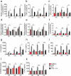
Figure 1.
Figure 1.
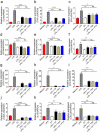
Butyrate inhibits neutrophils to produce proinflammatory cytokines, calprotectin and LCN2. Neutrophils were isolated from peripheral blood of healthy controls (HC, n = 8), patients with active CD (n = 10) and patients with active UC (n = 13) and were stimulated with LPS (300 ng/mL) in the presence or absence of C4 (0.5 mM) for 3 h. Cells were then collected and the levels of mRNA expression were analyzed for IL-6 (a), TNF-α (b), IFN-γ (c), S100A8 (d), S100A9 (e) and LCN2 (f), respectively, by qRT-PCR. Gene expression was normalized to GAPDH. Peripheral neutrophils (2 × 106) were isolated from healthy controls (HC, n = 7), patients with active CD (n = 7) and patients with active UC (n = 7) and were stimulated with LPS (300 ng/mL) in the presence or absence of C4 (0.5 mM) for 24 h, and the culture supernatants were then collected for detection of IL-6 (d), TNF-α (e), IFN-γ (f), S100A8/9 (j) and LCN2 (k) by ELISA. *p < .05, **p < .01, ***p < .001 and ****p < .0001; ns, not significant. Data are representative of 3 independent experiments
Figure 2.
Figure 2.
Butyrate inhibits production of chemokines and MPO by neutrophils. Neutrophils were isolated from peripheral blood of healthy controls (HC, n = 8), patients with active Crohn’s disease (CD, n = 10) and patients with active ulcerative colitis (UC, n = 13) and stimulated in vitro with LPS (300 ng/mL) in the presence or absence of C4 (0.5 mM) for 3 h. Cells then were collected and the levels of mRNA expression were analyzed for CCL3 (a), CCL4 (b), CXCL1 (c), IL-8 (d) and MPO (e), respectively, by qRT-PCR. Gene expression was normalized to GAPDH. Peripheral neutrophils (2 × 106) were isolated from healthy donors (HC, n = 7), patients with active CD (n = 7) and patients with active UC (n = 7) and stimulated in vitro with LPS (300 ng/mL) in the presence or absence of C4 (0.5 mM) for 24 h, and the culture supernatants were collected for detection of CCL3 (f), CCL4 (g), CXCL1 (h), IL-8 (i) by ELISA. *p < .05 and **p < .01; ns, not significant. Data are representative of 3 independent experiments
Figure 3.
Figure 3.
Butyrate inhibits neutrophil production of proinflammatory mediators in a HDACi-dependent manner. Neutrophils were isolated from peripheral blood of healthy donors (HC, n = 8) and stimulated in vitro with LPS (300 ng/mL) in the presence or absence of C4 (0.5 mM), TSA (10 μM) and PTX (100 ng/mL), respectively, for 3 h. The mRNA levels of proinflammatory mediators including IL-6 (a), TNF-α (b), IFN-γ (c), S100A8 (d), S100A9 (e), LCN2 (f), CCL3 (g), CCL4 (h), CCL20 (i), CXCL1 (j), IL-8 (k), and CXCL9 (l) were detected by qRT-PCR. Gene expression was normalized to GAPDH. *p < .05, **p < .01, ***p < .001 and ****p < .0001; ns, not significant. Data are representative of 3 independent experiments
Figure 4.
Figure 4.
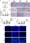
Butyrate suppresses migration and weakens the abilities of neutrophils to release ROS and NETs. Peripheral neutrophils (5 × 105) were isolated from peripheral blood of healthy donors (HC, n = 6), patients with active Crohn’s disease (CD, n = 10) and patients with active ulcerative colitis (UC, n = 10) and seeded on the upper chamber of a Transwell insert. IL-8 (20 ng/mL) was loaded into the lower chamber to attract neutrophils. (a) Schematic representation of neutrophil migration in vitro for 2 h at 37°C. (b) Representative images of migrated neutrophils on the lower membrane stained with 0.1% crystal violet and observed under optical microscopy. Original magnification: ×200. (c) The histogram represents the number of migrating neutrophils per high-power field (HPF). (d) Peripheral neutrophils (1.5 × 104) were isolated from peripheral blood of healthy donors (HC, n = 6), patients with active Crohn’s disease (CD, n = 10) and patients with active ulcerative colitis (UC, n = 10) and preincubated with or without C4 (0.5 mM) for 2 h, and then stimulated with PMA (100 ng/mL) to detect the production of ROS. The levels of ROS were measured by using Amplex Red Hydrogen Peroxide Assay Kit according to manufacturer’s instructions. (e, f) Peripheral neutrophils (5 × 105) were isolated from peripheral blood of healthy donors (HC, n = 6), patients with active Crohn’s disease (CD, n = 8) and patients with active ulcerative colitis (UC, n = 8) and seeded on coverslips which were coated with poly-L-lysine. Adherent neutrophils were stimulated with PMA (100 ng/mL) in the presence or absence of C4 (10 mM) for 3 h at 37°C. Histograms of fluorescence intensity of NETs detected by fluorometric plate reader (e). Representative images of NETs were obtained from a HC (upper panels), a patient with A-CD (middle panels) and a patient with A-UC (lower panels) (f). Original magnification: ×200. *p < .05, **p < .01 and ***p < .001; ns, not significant. Data are representative of 3 independent experiments
Figure 5.
Figure 5.
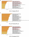
RNA sequencing analysis elicits the immunomodulatory effects of butyrate on IBD neutrophils. Neutrophils were isolated from healthy donors and patients with UC and treated in vitro with or without C4 (0.5 mM) for 3 h. The total RNA from four groups was extracted and performed with RNA sequencing to detect transcriptome differences. Differentially expressed genes (fold change > 2.0 or < 0.5 and p < .01) between two groups were enriched for GO functional analysis by Metascape. Top 10 pathways from the indicated comparisons are shown. (a-c) n = 3 biologically independent samples per group. HC_E, HC neutrophils treated without C4; HC_C4, HC neutrophils treated with C4; UC_E, UC neutrophils treated without C4; and UC_C4, UC neutrophils treated with C4
Figure 6.
Figure 6.
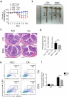
Oral administration of butyrate reduces neutrophil infiltration and protects from colitis induced by DSS. WT mice (n = 12 in each group) were administered 2% DSS in drinking water for 7 days followed by DSS free water for another 3 days. Mice from one of DSS-treated group were fed with C4 (200 mM) in drinking water from day 0 when DSS was given. (a) Changes of body weight during a period of 10-day observation. (b) Gross morphology of the colons on day 10 when mice were sacrificed. (c) Representative images of distal colonic sections after hematoxylin and eosin (H&E) staining. Scale bars: 200 μm. (d) Pathological scores of colon sections were calculated as indicated. (e) Lamina propria mononuclear cells (LPMC) were collected from colons of mice and flow cytometric analysis was performed to examine the percentages of CD11b+Ly6G+ neutrophils infiltrated in whole colon from each group. (f) Percentages of Ly6G+CD11b+ neutrophils in LPMC were shown in the bar chart. *p < .05 and **p < .01. Data are representative of 3 independent experiments
Figure 7.
Figure 7.
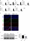
Butyrate inhibits NET formation and neutrophil-associated inflammatory mediators in DSS-induced murine colitis. Colon tissues were obtained from mice as described in Figure 6 on day 10 and total RNA was extracted to examine the mRNA levels of IL-6 (a), TNF-α (b), IFN-γ (c), CXCL1 (d), S100A8 (e), S100A9 (f) and LCN2 (g), respectively, by qRT-PCR. Gene expression was normalized to GAPDH. (h) Representative immunofluorescent images for detection of Citrullinated H3 (CitH3). Scale bars: 20 μm. (i, j) Lysates of the colon tissues were prepared and the protein levels of CitH3 were determined by Western blot with β-actin as a reference. *p < .05 and **p < .01. Data are representative of 3 independent experiments

References (V体育ios版)

    1. Maloy KJ, Powrie F.. Intestinal homeostasis and its breakdown in inflammatory bowel disease. Nature. 2011;474(7351):298–19. doi:10.1038/nature10208. - DOI - PubMed
    1. Rodríguez C, Romero E, Garrido-Sanchez L, Alcaín-Martínez G, Andrade RJ, Taminiau B, Daube G, García-Fuentes E. microbiota insights in clostridium difficile infection and inflammatory bowel disease. Gut Microbes. 2020;12(1):1725220. doi:10.1080/19490976.2020.1725220. - VSports最新版本 - DOI - PMC - PubMed
    1. Lavelle A, Sokol H. Gut microbiota-derived metabolites as key actors in inflammatory bowel disease. Nat Rev Gastroenterol Hepatol. 2020;17(4):223–237. doi:10.1038/s41575-019-0258-z. - DOI - PubMed
    1. Kelly CJ, Zheng L, Campbell EL, Saeedi B, Scholz CC, Bayless AJ, Wilson K, Glover L, Kominsky D, Magnuson A, et al. Crosstalk between Microbiota-Derived Short-Chain Fatty Acids And Intestinal Epithelial HIF augments tissue barrier function. Cell Host Microbe. 2015;17(5):662–671. doi:10.1016/j.chom.2015.03.005. - "V体育平台登录" DOI - PMC - PubMed
    1. Bilotta AJ, Ma C, Yang W, Yu Y, Yu Y, Zhao X, Zhou Z, Yao S, Dann SM, Cong Y, et al. Propionate enhances cell speed and persistence to promote intestinal epithelial turnover and repair. Cell Mol Gastroenterol Hepatol. 2021;11(4):1023–1044. doi:10.1016/j.jcmgh.2020.11.011. - DOI - PMC - PubMed

V体育官网入口 - Publication types

MeSH terms

LinkOut - more resources