Skip to main page content
U.S. flag

An official website of the United States government

Dot gov

The . gov means it’s official. Federal government websites often end in . gov or VSports app下载. mil. Before sharing sensitive information, make sure you’re on a federal government site. .

Https

The site is secure. The https:// ensures that you are connecting to the official website and that any information you provide is encrypted and transmitted securely. V体育官网.

. 2021 Aug 11:9:699263.
doi: 10.3389/fcell.2021.699263. eCollection 2021.

V体育ios版 - Spatiotemporal and Functional Heterogeneity of Hematopoietic Stem Cell-Competent Hemogenic Endothelial Cells in Mouse Embryos

Affiliations

Spatiotemporal and Functional Heterogeneity of Hematopoietic Stem Cell-Competent Hemogenic Endothelial Cells in Mouse Embryos

Yun-Qiao Li et al. Front Cell Dev Biol. .

Abstract

Hematopoietic stem cells (HSCs) are derived from hemogenic endothelial cells (HECs) during embryogenesis. The HSC-primed HECs increased to the peak at embryonic day (E) 10 and have been efficiently captured by the marker combination CD41-CD43-CD45-CD31+CD201+Kit+CD44+ (PK44) in the aorta-gonad-mesonephros (AGM) region of mouse embryos most recently. In the present study, we investigated the spatiotemporal and functional heterogeneity of PK44 cells around the time of emergence of HSCs. First, PK44 cells in the E10. 0 AGM region could be further divided into three molecularly different populations showing endothelial- or hematopoietic-biased characteristics. Specifically, with the combination of Kit, the expression of CD93 or CD146 could divide PK44 cells into endothelial- and hematopoietic-feature biased populations, which was further functionally validated at the single-cell level. Next, the PK44 population could also be detected in the yolk sac, showing similar developmental dynamics and functional diversification with those in the AGM region. Importantly, PK44 cells in the yolk sac demonstrated an unambiguous multilineage reconstitution capacity after in vitro incubation VSports手机版. Regardless of the functional similarity, PK44 cells in the yolk sac displayed transcriptional features different from those in the AGM region. Taken together, our work delineates the spatiotemporal characteristics of HECs represented by PK44 and reveals a previously unknown HSC competence of HECs in the yolk sac. These findings provide a fundamental basis for in-depth study of the different origins and molecular programs of HSC generation in the future. .

Keywords: developmental hematopoiesis; hematopoietic stem cells; hemogenic endothelial cells; heterogeneity; single-cell RNA-sequencing V体育安卓版. .


V体育2025版 - Conflict of interest statement

The authors declare that the research was conducted in the absence of any commercial or financial relationships that could be construed as a potential conflict of interest.

Figures

FIGURE 1
FIGURE 1
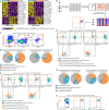
Transcriptomically different subpopulations in E10.0 AGM PK44 cells. (A) UMAP plots of the E10.0 AGM PK44 cells with clusters mapped onto it. (B) Heatmap showing top 10 DEGs of three subpopulations (endothelial-biased, transitional, and hematopoietic-biased population) in E10.0 AGM PK44 cells. DEGs were sorted by ROC power calculated through FindallMarkers function (test.use = “roc”) in Seurat2. (C) Dot plot showing the endothelial and hematopoietic scores of E10.0 AGM PK44 cells in three subpopulations to, respectively, represent the endothelial and hematopoietic characteristics molecularly. The statistical significance of differences was determined using Wilcox test. ∗∗∗P < 0.001. (D) Enriched GO terms of three subpopulations in E10.0 AGM PK44 cells and top 10 terms with P value < 0.05 are shown.
FIGURE 2
FIGURE 2
Functionally different subpopulations within E10.0 AGM PK44 cells. (A) Heatmap showing the differentially expressed surface marker coding genes of the subpopulations in E10.0 AGM PK44 cells arranged by ROC power (left) and fold change (right), respectively. For the convenience of FACS labeling and live cell sorting, only genes coding cell surface proteins whose antibodies for FACS labeling are also commercially available in China within the DEG list are displayed. (B) Schematic experimental design of screening candidate molecules by index sorting. (C) Flow cytometry gating strategy for isolating PK44 population (CD41 CD43 CD45 CD31+CD44+ Kit+CD201+) from the E10.0 AGM region. (D) FACS plots showing expression of Kit and CD93/CD146 on the index-sorted E10.0 AGM PK44 cells. Cells with different kinds of potentials based on in vitro functional evaluation (color dots) are mapped onto the reference FACS plots (gray dots). Data are from four independent experiments. The enlarged views of dashed black boxes are shown below. (E) Pie charts showing the proportions of four differentiation potentials in the putative subpopulations. (F) FACS plots showing expression of Kit and CD93/CD146 on the index-sorted candidate subpopulations of E10.0 AGM PK44 cells. Cells with different potentials based on in vitro functional evaluation (color dots) are mapped onto the reference FACS plots (gray dots). Data are from four independent experiments. The enlarged views of dashed black boxes are shown below. (G) Pie charts showing the proportions of four differentiation potentials in each of the isolated subpopulations. (H) FACS plots of Kit and CD146 expression with the indicated subpopulations mapped on. Orange boxes indicate the gates of the KithiCD146lo subpopulation; cyan boxes indicate the gates of the CD146hiKitlo subpopulation.
FIGURE 3
FIGURE 3
Developmental dynamics of the immunophenotypic PK44 cells in AGM and yolk sac. (A) Representative FACS plots for AGM/caudal half and yolk sac PK44 cells from E9.5 to E11.0. (B) Graphs showing the proportions of PK44 cells in endothelial cells (CD41 CD43 CD45 CD31+) (left) and in total cells of the corresponding tissues (middle). The statistical significance of differences was determined using Fisher’s exact test. ∗∗∗P < 0.001. NS, not significant. The average numbers of PK44 cells in corresponding tissues are shown to the right. Data are means ± SD. Data are from three independent experiments. The statistical significance of differences was determined using Mann–Whitney test. NS, not significant.
FIGURE 4
FIGURE 4
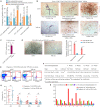
Functional evaluation of yolk sac PK44 cells. (A) Column charts showing the frequencies of potential-positive cells in the indicated populations for each kind of differentiation potentials. Data are from three independent experiments. (B) Representative CD31 and CD45 immunostaining on the cultures of single PK44 cells from E10.0 AGM and yolk sac, showing typical morphologies regarding distinct differentiation potentials. Cell frequencies of each kind of potentials are shown. Data are from three independent experiments. Scale bars, 100 μm. (C) Number of hematopoietic progenitors per embryo equivalent (ee) in the indicated populations derived from E10.5 yolk sac measured by the methylcellulose CFU-C assay. Data are means ± SD. The statistical significance of differences was determined using the Wilcox test. ∗∗P < 0.01. Data are from three independent experiments. (D) Morphology of hematopoietic progenies generated by AGM (left) and yolk sac (middle) PK44 cells after OP9-DL1 co-culture for 5 days. Scale bars, 100 μm. Column charts in the right showing the frequencies of AGM and yolk sac PK44 cells generating small or large hematopoietic clusters. Data are from three independent experiments. Data are means ± SD. The statistical significance of differences was determined using Pearson’s chi-squared test. P < 0.05; ∗∗P < 0.01. (E) Representative FACS plots of analyzing the progenies of E10.0 AGM and yolk sac PK44 cells following 7 days co-culture, showing a population of cells expressing putative HSC markers. (F) Detailed information of the transplantation assays performed with the progenies of E10.5 AGM and yolk sac PK44 cells. (G) Graphs showing the donor chimerism from 4 to 16 weeks after being transplanted with the progenies of the E10.5 AGM and yolk sac PK44 populations (three embryo equivalents per recipient), respectively. Data are means ± SD. The statistical significance of differences was determined using Mann–Whitney test. NS, not significant. (H) Bar plots showing the donor contribution to the granulocytes/monocytes (GM, red), B lymphocytes (green), and T lymphocytes (purple) in the peripheral blood of each of the primary recipients receiving the progenies of E10.5 AGM and yolk sac PK44 cells at 16 weeks post-transplantation.
FIGURE 5
FIGURE 5
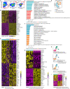
Transcriptomic characteristics of E10.0 yolk sac PK44 cells. (A) Flow cytometry gating strategy for isolating PK44 cells in E10.0 yolk sac. (B) Heatmap showing the top 15 DEGs between E10.0 yolk sac and AGM PK44 cells. (C) Enriched GO terms of E10.0 yolk sac and AGM PK44 populations. (D) UMAP plots of the E10.0 yolk sac PK44 cells with clusters mapped on. (E) Heatmap showing top 25 DEGs between two subpopulations (endothelial- and hematopoietic-biased) in E10.0 yolk sac PK44 cells. (F) Enriched GO terms of two subpopulations (endothelial- and hematopoietic-biased) in E10.0 yolk sac PK44 cells. (G) Heatmap showing top 20 DEGs between hematopoietic-biased subpopulation in E10.0 yolk sac PK44 cells and that in E10.0 AGM PK44 cells. (H) UMAP of integrated data of E10.0 AGM and yolk sac PK44 cells with subpopulations (endothelial-biased, transitional, and hematopoietic-biased population) mapped on. (I) Heatmap showing DEGs between the integrated endothelial- and hematopoietic-biased populations.

References

    1. Anders S., Pyl P. T., Huber W. (2015). HTSeq–a python framework to work with high-throughput sequencing data. Bioinformatics 31 166–169. 10.1093/bioinformatics/btu638 - DOI (V体育平台登录) - PMC - PubMed
    1. Chen M. J., Yokomizo T., Zeigler B. M., Dzierzak E., Speck N. A. (2009). Runx1 is required for the endothelial to haematopoietic cell transition but not thereafter. Nature 457 887–891. 10.1038/nature07619 - "V体育安卓版" DOI - PMC - PubMed
    1. Chong D. C., Koo Y., Xu K., Fu S., Cleaver O. (2011). Stepwise arteriovenous fate acquisition during mammalian vasculogenesis. Dev. Dyn. 240 2153–2165. 10.1002/dvdy.22706 - DOI - PMC - PubMed
    1. Corada M., Morini M. F., Dejana E. (2014). Signaling pathways in the specification of arteries and veins. Arterioscler. Thromb. Vasc. Biol. 34 2372–2377. 10.1161/ATVBAHA.114.303218 - DOI - PubMed
    1. Cumano A., Ferraz J. C., Klaine M., Di Santo J. P., Godin I. (2001). Intraembryonic, but not yolk sac hematopoietic precursors, isolated before circulation, provide long-term multilineage reconstitution. Immunity 15 477–485. 10.1016/s1074-7613(01)00190-x - DOI - PubMed