Skip to main page content
U.S. flag

An official website of the United States government

Dot gov

The . gov means it’s official. Federal government websites often end in . gov or . mil. Before sharing sensitive information, make sure you’re on a federal government site VSports app下载. .

Https

The site is secure V体育官网. The https:// ensures that you are connecting to the official website and that any information you provide is encrypted and transmitted securely. .

. 2021 Aug 28;27(1):96.
doi: 10.1186/s10020-021-00360-w.

G protein coupled estrogen receptor attenuates mechanical stress-mediated apoptosis of chondrocyte in osteoarthritis via suppression of Piezo1

Affiliations

G protein coupled estrogen receptor attenuates mechanical stress-mediated apoptosis of chondrocyte in osteoarthritis via suppression of Piezo1

Yi Sun et al. Mol Med. .

Abstract (V体育官网入口)

Background: Apoptosis of chondrocyte is involved in osteoarthritis (OA) pathogenesis, and mechanical stress plays a key role in this process by activation of Piezo1. However, the negative regulation of signal conduction mediated by mechanical stress is still unclear. Here, we elucidate that the critical role of G protein coupled estrogen receptor (GPER) in the regulation of mechanical stress-mediated signal transduction and chondrocyte apoptosis. VSports手机版.

Methods: The gene expression profile was detected by gene chip upon silencing Piezo1. The expression of GPER in cartilage tissue taken from the clinical patients was detected by RT-PCR and Western blot as well as immunohistochemistry, and the correlation between GPER expression and OA was also investigated. The chondrocytes exposed to mechanical stress were treated with estrogen, G-1, G15, GPER-siRNA and YAP (Yes-associated protein)-siRNA. The cell viability of chondrocytes was measured. The expression of polymerized actin and Piezo1 as well as the subcellular localization of YAP was observed under laser confocal microscope. Western blot confirmed the changes of YAP/ Rho GTPase activating protein 29 (ARHGAP29) /RhoA/LIMK /Cofilin pathway. The knee specimens of osteoarthritis model were stained with safranin and green. OARSI score was used to evaluate the joint lesions. The expressions of GPER and YAP were detected by immunochemistry. V体育安卓版.

Results: Expression profiles of Piezo1- silenced chondrocytes showed that GPER expression was significantly upregulated. Moreover, GPER was negatively correlated with cartilage degeneration during OA pathogenesis. In addition, we uncovered that GPER directly targeted YAP and broadly restrained mechanical stress-triggered actin polymerization. Mechanism studies revealed that GPER inhibited mechanical stress-mediated RhoA/LIMK/cofilin pathway, as well as the actin polymerization, by promoting expression of YAP and ARHGAP29, and the YAP nuclear localization, eventually causing the inhibition of Piezo1. YAP was obviously decreased in degenerated cartilage. Silencing YAP caused significantly increased actin polymerization and activation of Piezo1, and an increase of chondrocyte apoptosis. In addition, intra-articular injection of G-1 to OA rat effectively attenuated cartilage degeneration. V体育ios版.

Conclusion: We propose a novel regulatory mechanism underlying mechanical stress-mediated apoptosis of chondrocyte and elucidate the potential application value of GPER as therapy targets for OA VSports最新版本. .

Keywords: Chondrocyte; G protein coupled estrogen receptor; Mechanical stress; Osteoarthritis; Piezo1 V体育平台登录. .


Conflict of interest statement

The authors declare that they have no competing interests.

Figures

Fig. 1
Fig. 1
GPER is upregulated in chondrocytes exposed to mechanical stress after Piezo1 silencing. a Heatmap of differential gene expression values upon Piezo1 silencing in chondrocytes exposed to mechanical stress. b Disease and function analysis of differential gene expression values upon Piezo1 silencing in chondrocytes exposed to mechanical stress. c RT-qPCR analysis of GPER genes in Piezo1-silenced chondrocytes compared with non-silenced chondrocytes (n = 3). d Protein expression of GPER in Piezo1-silenced chondrocytes compared with non-silenced chondrocytes (n = 3). All data are presented in mean ± SD *P < 0.05, compared with non-silenced chondrocytes
Fig. 2
Fig. 2
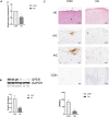
GPER expression is decreased in human osteoarthritis cartilage. a RT-qPCR analysis of GPER genes in degenerative cartilage compared with control cartilage (n = 10). b Protein expression of GPER in degenerative cartilage compared with control cartilage (n = 3). c Immunohistochemical staining of degenerative cartilage and control cartilage without (control) and with anti-GPER antibody (n = 10). All data are presented in mean ± SD *P < 0.05, compared with control cartilage. **P < 0.01, compared with control cartilage
Fig. 3
Fig. 3
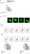
Activation of GPER reduces the apoptosis of chondrocytes induced by mechanical stress. a Cell viabilities for chondrocytes treated with mechanical stress, 8 μM estrogen, and 8 μM G15 (n = 3). b Protein expression of Bax and Bcl-2 for chondrocytes treated with mechanical stress, 8 μM estrogen, and 8 μM G15 (n = 3). c Cell viabilities for chondrocytes with GPER or scrambled siRNA after treatment with mechanical stress, and estrogen (n = 3). d Protein expression of Bax and Bcl-2 for chondrocytes with GPER or scrambled siRNA after treatment with mechanical stress, and estrogen (n = 3). e Cell viabilities for chondrocytes treated with mechanical stress, 10 μM G-1, and 8 μM G15. f Protein expression of Bax and Bcl-2 for chondrocytes treated with mechanical stress, 10 μM G-1, and 8 μM G15. g Cell viabilities for chondrocytes with GPER or scrambled siRNA after treatment with mechanical stress, and G-1 (n = 3). h Protein expression of Bax and Bcl-2 for chondrocytes with GPER or scrambled siRNA after treatment with mechanical stress, and G-1 (n = 3). All data are presented in mean ± SD (n = 3). *P < 0.05, compared with DMSO group. **P < 0.01, compared with DMSO group. #P < 0.05, compared with mechanical stress + DMSO group. ##P < 0.01, compared with mechanical stress + DMSO group
Fig. 3
Fig. 3
Activation of GPER reduces the apoptosis of chondrocytes induced by mechanical stress. a Cell viabilities for chondrocytes treated with mechanical stress, 8 μM estrogen, and 8 μM G15 (n = 3). b Protein expression of Bax and Bcl-2 for chondrocytes treated with mechanical stress, 8 μM estrogen, and 8 μM G15 (n = 3). c Cell viabilities for chondrocytes with GPER or scrambled siRNA after treatment with mechanical stress, and estrogen (n = 3). d Protein expression of Bax and Bcl-2 for chondrocytes with GPER or scrambled siRNA after treatment with mechanical stress, and estrogen (n = 3). e Cell viabilities for chondrocytes treated with mechanical stress, 10 μM G-1, and 8 μM G15. f Protein expression of Bax and Bcl-2 for chondrocytes treated with mechanical stress, 10 μM G-1, and 8 μM G15. g Cell viabilities for chondrocytes with GPER or scrambled siRNA after treatment with mechanical stress, and G-1 (n = 3). h Protein expression of Bax and Bcl-2 for chondrocytes with GPER or scrambled siRNA after treatment with mechanical stress, and G-1 (n = 3). All data are presented in mean ± SD (n = 3). *P < 0.05, compared with DMSO group. **P < 0.01, compared with DMSO group. #P < 0.05, compared with mechanical stress + DMSO group. ##P < 0.01, compared with mechanical stress + DMSO group
Fig. 4
Fig. 4
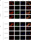
GPER inhibits mechanical stress-mediated actin polymerization and Piezo1 activation. a Immunofluorescence staining F-actin and Piezo1 (n = 3). b Cell viabilities for chondrocytes treated with mechanical stress, 10 μM G-1, and 8 μM cytoD (n = 3). c Protein expression of Bax and Bcl-2 for chondrocytes treated with mechanical stress, 10 μM G-1, and 8 μM cytoD (n = 3). All data are presented in mean ± SD
Fig. 4
Fig. 4
GPER inhibits mechanical stress-mediated actin polymerization and Piezo1 activation. a Immunofluorescence staining F-actin and Piezo1 (n = 3). b Cell viabilities for chondrocytes treated with mechanical stress, 10 μM G-1, and 8 μM cytoD (n = 3). c Protein expression of Bax and Bcl-2 for chondrocytes treated with mechanical stress, 10 μM G-1, and 8 μM cytoD (n = 3). All data are presented in mean ± SD
Fig. 5
Fig. 5
GPER regulates actin polymerization via YAP-ARHGAP29-RhoA-LIMK-Cofilin Signaling. a Protein expression of YAP in chondrocyte treated with G-1 or mechanical stress (n = 3), *P < 0.05, compared with control group. **P < 0.01, compared with control group. #P < 0.05, compared with mechanical stress group. ##P < 0.01, compared with mechanical stress group. b Immunofluorescence staining YAP (n = 3). (c) Cell viabilities for chondrocytes with YAP or scrambled siRNA after treatment with mechanical stress, and G-1 (n = 3), *P < 0.05, compared with DMSO group. **P < 0.01, compared with DMSO group. #P < 0.05, compared with mechanical stress + DMSO group. ##P < 0.01, compared with mechanical stress + DMSO group. d Protein expression of Bax and Bcl-2 for chondrocytes with YAP or scrambled siRNA after treatment with mechanical stress, and G-1 (n = 3), *P < 0.05, compared with DMSO group. **P < 0.01, compared with DMSO group. #P < 0.05, compared with mechanical stress + DMSO group. ##P < 0.01, compared with mechanical stress + DMSO group. e Immunofluorescence staining F-actin and Piezo1 (n = 3). f Protein expression of ARHGAP29-RhoA-LIMK-Cofilin Signaling in chondrocyte treated with G-1 or mechanical stress (n = 3) *P < 0.05, compared with control group. **P < 0.01, compared with control group. #P < 0.05, compared with mechanical stress group. ##P < 0.01, compared with mechanical stress group. All data are presented in mean ± SD
Fig. 5
Fig. 5
GPER regulates actin polymerization via YAP-ARHGAP29-RhoA-LIMK-Cofilin Signaling. a Protein expression of YAP in chondrocyte treated with G-1 or mechanical stress (n = 3), *P < 0.05, compared with control group. **P < 0.01, compared with control group. #P < 0.05, compared with mechanical stress group. ##P < 0.01, compared with mechanical stress group. b Immunofluorescence staining YAP (n = 3). (c) Cell viabilities for chondrocytes with YAP or scrambled siRNA after treatment with mechanical stress, and G-1 (n = 3), *P < 0.05, compared with DMSO group. **P < 0.01, compared with DMSO group. #P < 0.05, compared with mechanical stress + DMSO group. ##P < 0.01, compared with mechanical stress + DMSO group. d Protein expression of Bax and Bcl-2 for chondrocytes with YAP or scrambled siRNA after treatment with mechanical stress, and G-1 (n = 3), *P < 0.05, compared with DMSO group. **P < 0.01, compared with DMSO group. #P < 0.05, compared with mechanical stress + DMSO group. ##P < 0.01, compared with mechanical stress + DMSO group. e Immunofluorescence staining F-actin and Piezo1 (n = 3). f Protein expression of ARHGAP29-RhoA-LIMK-Cofilin Signaling in chondrocyte treated with G-1 or mechanical stress (n = 3) *P < 0.05, compared with control group. **P < 0.01, compared with control group. #P < 0.05, compared with mechanical stress group. ##P < 0.01, compared with mechanical stress group. All data are presented in mean ± SD
Fig. 6
Fig. 6
G-1 activated GPER inhibits articular cartilage degeneration in vivo. a Safranine and Fast Green double staining in each group and associated OARSI scores (n = 6). b Immunofluorescence staining GPER, and YAP (n = 6). All data are presented in mean ± SD *P < 0.05, compared with Sham + NC. **P < 0.01, compared with Sham + NC. #P < 0.05, compared with OA + NC. ##P < 0.01, compared with OA + NC

References

    1. Alexander A, Irving AJ, Harvey J. Emerging roles for the novel estrogen-sensing receptor GPER1 in the CNS. Neuropharmacology. 2017;113:652–660. doi: 10.1016/j.neuropharm.2016.07.003. - "VSports注册入口" DOI - PubMed
    1. Barton M, et al. Twenty years of the G protein-coupled estrogen receptor GPER: historical and personal perspectives. J Steroid Biochem Mol Biol. 2018;176:4–15. doi: 10.1016/j.jsbmb.2017.03.021. - DOI - PMC - PubMed
    1. Chagin AS, Chrysis D, Takigawa M, Ritzen EM, Savendahl L. Locally produced estrogen promotes fetal rat metatarsal bone growth; an effect mediated through increased chondrocyte proliferation and decreased apoptosis. J Endocrinol. 2006;188:193–203. doi: 10.1677/joe.1.06364. - DOI - PubMed
    1. Chang L, et al. The SWI/SNF complex is a mechanoregulated inhibitor of YAP and TAZ. Nature. 2018;563:265–269. doi: 10.1038/s41586-018-0658-1. - DOI - PMC - PubMed
    1. Delve E, et al. YAP/TAZ regulates the expression of proteoglycan 4 and tenascin C in superficial-zone chondrocytes. Eur Cell Mater. 2020;39:48–64. doi: 10.22203/eCM.v039a03. - DOI - PubMed

Publication types

MeSH terms

Substances

LinkOut - more resources