Skip to main page content
U.S. flag

An official website of the United States government

Dot gov

The . gov means it’s official. Federal government websites often end in . gov or . mil. Before sharing sensitive information, make sure you’re on a federal government site VSports app下载. .

Https

The site is secure. The https:// ensures that you are connecting to the official website and that any information you provide is encrypted and transmitted securely V体育官网. .

. 2021 Aug 16:14:3985-3999.
doi: 10.2147/JIR.S323054. eCollection 2021.

Human Amniotic Mesenchymal Stem Cells Inhibit aGVHD by Regulating Balance of Treg and T Effector Cells (V体育官网入口)

Affiliations

VSports注册入口 - Human Amniotic Mesenchymal Stem Cells Inhibit aGVHD by Regulating Balance of Treg and T Effector Cells

Ya Gao et al. J Inflamm Res. .

Abstract

Background: Acute graft versus host disease (aGVHD) remains a leading cause of transplant-related mortality following allogeneic haematopoietic cell transplantation (allo-HCT). Human amniotic mesenchymal stem cells (hAMSCs) are a novel mesenchymal stem cells (MSCs), which have stronger proliferation and immunomodulatory ability compared with bone marrow mesenchymal stem cells (BM-MSCs) VSports手机版. Besides, as the amniotic membrane is often treated as medical waste after delivery, hAMSCs can be obtained conveniently and noninvasively. The aim of this study was to explore the therapeutic efficacy and underlying mechanisms of hAMSCs transplantation for the humanized aGVHD mouse model. .

Methods: We established a humanized aGVHD mouse model by transplanting human peripheral blood mononuclear cells (PBMCs) into NOD-PrkdcscidIL2rγnull (NPG) mice, human amniotic membrane collected from discarded placenta of healthy pregnant women after delivery and hAMSCs were extracted from amniotic membrane and expanded in vitro. Mice were divided into untreated group (Control), aGVHD group (aGVHD), and hAMSCs treatment group (aGVHD+hAMSCs), the hAMSCs labeled with GFP were administered to aGVHD mice to explore the homing ability of hAMSCs. T effector and regulatory T cells (Tregs) levels and cytokines of each group in target organs were detected by flow cytometry and cytometric bead array (CBA), respectively V体育安卓版. .

Results: We successfully established a humanized aGVHD mouse model using NPG mice. The hAMSCs have the ability to inhibit aGVHD in this mouse model through reduced villous blunting and lymphocyte infiltration of the gut while reducing inflammatory edema, tissue destruction and lymphocyte infiltration into the parenchyma of the liver and lung. hAMSCs suppressed CD3+CD4+ T and CD3+CD8+ T cell expression and increased the proportion of Tregs, and besides, hAMSCs can reduce the levels of IL-17A, INF-γ, and TNF in aGVHD target organs V体育ios版. .

Conclusion: The NPG murine environment was capable of activating human T cells to produce aGVHD pathology to mimic aGVHD as in humans. The hAMSCs controlled aGVHD by decreasing inflammatory cytokine secretion within target organs by modulating the balance of Tregs and T effector cells in humanized mice. VSports最新版本.

Keywords: NPG mice; acute graft versus host disease; amniotic mesenchymal stem cells; humanized mouse model; immunomodulatory. V体育平台登录.


Conflict of interest statement (VSports注册入口)

The authors report no conflicts of interest in this work.

Figures (VSports)
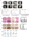
Figure 1
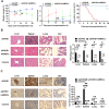
Figure 1
Induced aGVHD model in NPG mice. (A) Representative clinical manifestations including hunched posture, ruffed fur, damaged skin, and hematochezia in the aGVHD mouse model. (B) Measurement of weight change, clinical score, and survival time at different time points with those two groups. The weight loss rate in aGVHD group is faster than in the control group, the clinical score of aGVHD was higher than that of the control group, the survival time of the control group was also significantly longer than that of the aGVHD group. *p < 0.05 and **p < 0.01. (C) Representative histology of target organs, including the liver, spleen, lung and gut, showed inflammatory cell infiltration and tissue damage in aGVHD group. 400×. *p < 0.05 and **p < 0.01. (D) Representative immunohistochemistry of target organs. Human CD45+ cells were detected by immunohistochemistry in these target organs. 400×. *p < 0.05 and ***p < 0.001. (E) Human CD3+ T cells were not detected by flow cytometry in the control group. Human CD3+ T cells from each target organ were detected by flow cytometry in the aGVHD group.
Figure 2
Figure 2
Identification of human amnion mesenchymal stem cells. (A) Characteristics of passage three hAMSCs. (B) Osteogenic differentiation of hAMSCs indicated by Alizarin Red S. 100×. (C) Adipogenic differentiation of hAMSCs indicated by Oil Red O staining. 100×. (D) Surface antigens of hAMSCs were detected by flow cytometry. Cells were positive for CD90, CD105, and CD73 and negative for CD45, CD11b, CD34, and HLA-DR. (E) GFP-labeled hAMSCs and 293T cells were detected by fluorescence microscopy at day 3 following transfection with GFP-pseudovirion. 100×. ***p < 0.001.
Figure 3
Figure 3
Human amnion mesenchymal stem cells migrated to target organs. GFP-labeled hAMSCs detected by confocal microscopy in target organs at 24 after injection in vivo. At 72 h, GFP fluorescence in the spleen, liver, and gut was weakened while increased in the lung.
Figure 4
Figure 4
The capacity of hAMSCs inhibited the development of -aGVHD in vitro. (A) Measurement of weight change (aGVHD vs aGVHD+hAMSCs day 7 P=0.043, day 14 P=0.037), clinical score (aGVHD vs aGVHD+hAMSCs day 14 P=0.002), and survival in the different groups (aGVHD vs aGVHD+hAMSCs P<0.001). *p < 0.05 and **p < 0.01. (B) Representative histology of target organs, including the liver, spleen, lung, and gut. Compared with the hAMSCs treatment group, inflammatory cell infiltration and tissue damage in the aGVHD group were more severe. 400×. *p < 0.05 and **p < 0.01. (C) Representative immunohistochemistry of target organs, including the liver, spleen, lung, and gut. Infiltration and tissue damage in the aGVHD group were more severe than that of hAMSCs treatment group and Control group. 400×. **p < 0.01 and ***p < 0.001.
Figure 5
Figure 5
hAMSCs suppressed effector T cell and increased Tregs in target organs. (A) After hAMSCs treatment, the proportion of CD3+CD4+ T cells in liver, spleen and gut was significantly lower than that of aGVHD group. *p < 0.05. (B) After hAMSCs treatment, the proportion of CD3+CD8+ T in liver, spleen, lung and gut was significantly lower than that of aGVHD group. *p < 0.05 and ***p < 0.001. (C) After hAMSCs treatment, the proportion of CD4+CD25+Foxp3+ Tregs in liver, spleen, and gut was significantly increased compared with that of aGVHD group. *p < 0.05.
Figure 6
Figure 6
hAMSCs can inhibit the secretion of inflammatory cytokine in target organs. (A) IL-17A level in lung and gut were significantly decreased in aGVHD+hAMSCs group than that in aGVHD group. *p < 0.05 and **p < 0.01. (B) IFN-γ level in liver and gut were significantly decreased in aGVHD+hAMSCs group than that in aGVHD group. *p < 0.05. (C) TNF level in liver and blood were significantly decreased in aGVHD+hAMSCs group than that in aGVHD group. *p < 0.05 and **p < 0.01.

References

    1. Ghimire S, Weber D, Mavin E, Wang XN, Dickinson AM, Holler E. Pathophysiology of GvHD and other HSCT-related major complications. Front Immunol. 2017;8:79. doi:10.3389/fimmu.2017.00079 - DOI - PMC - PubMed
    1. Blazar BR, Murphy WJ, Abedi M. Advances in graft-versus-host disease biology and therapy. Nat Rev Immunol. 2012;12(6):443–458. doi:10.1038/nri3212 - DOI - PMC - PubMed
    1. Wolf D, von Lilienfeld-toal M, Wolf AM, et al. Novel treatment concepts for graft-versus-host disease. Blood. 2012;119(1):16–25. doi:10.1182/blood-2011-08-339465 - "V体育平台登录" DOI - PubMed
    1. Wu X, Jiang J, Gu Z, Zhang J, Chen Y, Liu X. Mesenchymal stromal cell therapies: immunomodulatory properties and clinical progress. Stem Cell Res Ther. 2020;11(1):345. doi:10.1186/s13287-020-01855-9 - DOI (V体育2025版) - PMC - PubMed
    1. Malard F, Huang XJ, Sim JPY. Treatment and unmet needs in steroid-refractory acute graft-versus-host disease. Leukemia. 2020;34(5):1229–1240. doi:10.1038/s41375-020-0804-2 - DOI - PMC - PubMed

LinkOut - more resources (VSports最新版本)