Skip to main page content
U.S. flag

An official website of the United States government

Dot gov

The . gov means it’s official. Federal government websites often end in . gov or . mil. Before sharing sensitive information, make sure you’re on a federal government site VSports app下载. .

Https

The site is secure. The https:// ensures that you are connecting to the official website and that any information you provide is encrypted and transmitted securely V体育官网. .

. 2021 Sep 8;29(9):1366-1377.e9.
doi: 10.1016/j.chom.2021.07.013. Epub 2021 Aug 19.

A bacterial bile acid metabolite modulates Treg activity through the nuclear hormone receptor NR4A1

Affiliations

A bacterial bile acid metabolite modulates Treg activity through the nuclear hormone receptor NR4A1

VSports手机版 - Wei Li et al. Cell Host Microbe. .

Abstract

Bile acids act as signaling molecules that regulate immune homeostasis, including the differentiation of CD4+ T cells into distinct T cell subsets. The bile acid metabolite isoallolithocholic acid (isoalloLCA) enhances the differentiation of anti-inflammatory regulatory T cells (Treg cells) by facilitating the formation of a permissive chromatin structure in the promoter region of the transcription factor forkhead box P3 (Foxp3) VSports手机版. Here, we identify gut bacteria that synthesize isoalloLCA from 3-oxolithocholic acid and uncover a gene cluster responsible for the conversion in members of the abundant human gut bacterial phylum Bacteroidetes. We also show that the nuclear hormone receptor NR4A1 is required for the effect of isoalloLCA on Treg cells. Moreover, the levels of isoalloLCA and its biosynthetic genes are significantly reduced in patients with inflammatory bowel diseases, suggesting that isoalloLCA and its bacterial producers may play a critical role in maintaining immune homeostasis in humans. .

Keywords: T cells; bile acids; human microbiome; inflammatory bowel disease V体育安卓版. .


Conflict of interest statement

Declaration of interests A. S. D. is a consultant for Takeda Pharmaceuticals and Axial Therapeutics. J. R. H. is a consultant for CJ Research Center and is on the scientific advisory board of ChunLab. C. H. is on the scientific advisory boards of Seres Therapeutics, Empress Therapeutics, and ZOE Nutrition VSports最新版本.

Figures

Figure 1.
Figure 1.. Identification of gut bacteria that produce isoalloLCA from the gut bacterial metabolite 3-oxoLCA.
(A) Proposed biosynthetic pathway for the conversion of the primary bile acid chenodeoxycholic acid (CDCA) (1) to isoalloLCA (4) by human gut bacteria. Bacteria that produce isoalloLCA have not yet been identified. (B) Representative UPLC-MS traces (left) and quantification of isoalloLCA production (right) by bacteria from a screen of human isolates following incubation for 96 hours with 3-oxoLCA (100 μM) (n = 3 biological replicates per group, data are shown as the mean ± SEM, N.D. = not detected). (C) Representative UPLC-MS traces (left) and quantification of isoalloLCA production (right) showing that a triculture of Clostridium scindens ATCC 35703, Eggerthella lenta DSM 2243 and Parabacteroides merdae ATCC 43184 converted CDCA to isoalloLCA while monocultures of these bacteria did not produce isoalloLCA. C. scindens ATCC 35703 contains bai operon genes that can transform CDCA to LCA (Ridlon et al., 2006), while E. lenta DSM 2243 contains a 3α-HSDH, Elen_0690, that transforms LCA into 3-oxoLCA (Paik et al., 2021) (n = 3 biological replicates per group, data are shown as the mean ± SEM, N.D. = not detected).
Figure 2.
Figure 2.. A gene cluster in Bacteroidetes converts 3-oxoLCA to isoalloLCA.
(A) Proposed biosynthetic pathways for the conversion of 3-oxoLCA (3) to isoalloLCA (4). Subsequent UPLC-MS analysis of Parabacteroides merdae ATCC 43184 cultures incubated with 3-oxoLCA (Figure 2B) detected the proposed intermediate 3-oxo-Δ4-LCA (5) as well as 3-oxoalloLCA (6) from the top (red) pathway, but not Δ4-isoLCA (7) from the bottom (blue) pathway, supporting the hypothesis that subsequent actions of a 5β-reductase, a 5α-reductase, and a 3β-HSDH are responsible for the conversion of 3-oxoLCA to isoalloLCA by Bacteroidetes species. (B) The bile acid metabolites 3-oxo-Δ4-LCA, 3-oxoalloLCA, and isoalloLCA were detected by UPLC-MS in cultures of P. merdae ATCC 43184 incubated with 100 μM 3-oxoLCA for 96 hrs, while Δ4-isoLCA was not detected (n = 3 biological replicates per group, data are shown as the mean ± SEM, N.D. = not detected). (C) BLASTP searches revealed a three-gene cluster in the isoalloLCA producers Bacteroides dorei DSM 17855, P. merdae ATCC 43184, and B. vulgatus ATCC 8489. Bacdor_03091, Parmer_04016 and Bvu_1964 encode putative a 5α-reductase, Bacdor_03090, Parmer_04017 and Bvu_1965 encode a putative 5β-reductase, and Bacdor_03089, Parmer_04018 and Bvu_1966 encode a putative 3β-HSDH (see Table S2). (D-E) Representative UPLC-MS traces (left) and quantification of bile acid product (right) showing conversion of 100 μM 3-oxoLCA to 3-oxo-Δ4-LCA and 100 μM 3-oxo-Δ4-LCA to 3-oxoalloLCA by cell lysates of E. coli heterologously expressing Bacdor_03090 (d) and Bacdor_03091 (e), respectively. Extracted ion chromatograms (EICs) for 3-oxo-Δ4-LCA (m/z 371) (d) and 3-oxoalloLCA (m/z 373) (e) are shown (n = 3 biological replicates per group, data are shown as the mean ± SEM, N.D. = not detected). (F) Representative UPLC-MS traces (left) and quantification of isoalloLCA (right) showing conversion of 20 μM 3-oxoalloLCA to isoalloLCA by purified Bacdor_03089. EICs for isoalloLCA (m/z 375) are shown (n = 3 biological replicates per group, data are shown as the mean ± SEM, N.D. = not detected). (G) Representative UPLC-MS traces (left) and quantification of bile acids (right) showing P. merdae ATCC 43184 wild-type (Pm WT) produces isoalloLCA as well as the intermediates 3-oxo-Δ4-LCA and 3-oxoalloLCA from 3-oxoLCA (100 μM), while the gene cluster knockout strain of P. merdae (PmΔ04016–18) does not produce detectable amounts of these metabolites (n = 3 biological replicates per group, data are shown as the mean ± SEM, N.D. = not detected). (H) In vitro testing of Bacteroidetes strains containing complete or partial gene clusters homologous to the cluster identified in Bacteroides dorei DSM 17855, P. merdae ATCC 43184, and B. vulgatus ATCC 8489 revealed additional isoalloLCA producers. The presence of a 5α-reductase can be seen as a predictor of overall function. Phylogenetic tree of gut Bacteroidetes strains was adapted from (Karlsson et al., 2011).
Figure 3.
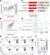
Figure 3.. ATAC-Seq analysis identifies NR4A1 as required for Treg induction by isoalloLCA.
(A) Two-way scatter plot comparing gene expression log2 scale fold changes under isoalloLCA or TGFβ treatment (all the expressed genes were used). Selected Treg signature genes are highlighted. R refers to the correlation coefficient. Naïve CD4+ T cells were purified from WT-B6 mice and culture under TH0 conditions (anti-CD3, anti-CD28, IL-2) in the presence of DMSO, isoalloLCA (20 µM), or TGFβ (0.25 ng/mL) for 48 hours. Cells were then collected for RNA-Seq analysis. Differentially expressed genes (DEGs) were determined by comparing DMSO- and isoalloLCA-treatments or DMSO- and TGFβ-treatments. (n = 3 biological replicates per group). (B) Scatter plot showing differentially enriched ATAC-Seq peaks between isoalloLCA and DMSO treatments. Naïve CD4+ T cells were purified from WT mice and culture under TH0 conditions (anti-CD3, anti-CD28, IL-2) in the presence of DMSO or isoalloLCA (20 µM) for 48 hours. Cells were then collected for the ATAC-Seq experiment (n = 2 biological replicates per group). (C) The top 4 transcription factor (TFs) binding motifs identified in differentially enriched ATAC-Seq peaks increased (Figure 3B, red), but not decreased (Figure 3B, blue), by isoalloLCA treatment. The specific TFs in the parenthese are the ones that have the smallest enrichment p values within the corresponding TF family. Data were analyzed using HOMER and the motifs were ranked by P value (see Method and Table S3). (D) Integrative Genomics Viewer (IGV) visualization of ATAC-Seq signals at the Foxp3 gene locus. Duplicated samples for naïve CD4+ T cells were purified from WT-B6 (CTL) or Nr4a1-deficient (KO) mice, cultured under TH0 condition (anti-CD3, anti-CD28, IL-2) in the presence of DMSO or isoalloLCA (20 µM) for 48 hours. Cells were then collected for the ATAC-Seq experiment. (n = 2 biological replicates per group, statistical tests were performed to compare the ATAC-Seq signals within the boxed region (unpaired t-test with 2-tailed p-value, ns, not significant, * P < 0.05). (E-F) Flow cytometry analysis and quantification of naïve CD4+ T cells purified from WT-B6 (CTL) or Nr4a1-deficient (KO) mice, cultured under TH0 conditions (anti-CD3, anti-CD28, IL-2) in the presence of DMSO, isoalloLCA (20 µM), TGFβ (0.25 ng/mL), or TGFβ + retinoic acid (RA, 1 nM) for 72 hours are shown. Cells were stained with FOXP3 as a marker for Treg cells. (n = 3 biological replicates per group, data are shown as the mean ± SEM, two-way ANOVA with Tukey’s multiple comparisons test, each compared to the DMSO group, ns, not significant, * P < 0.05, ** P < 0.01, *** P < 0.001).
Figure 4.
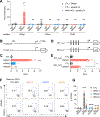
Figure 4.. NR4A1 activates Foxp3 gene transcription.
(A) Chromatin immunoprecipitation analysis of NR4A1 on the Foxp3 gene locus. Naïve CD4+ T cells purified from WT-B6 (CTL) or Nr4a1-deficient (KO) mice and cultured under TH0 conditions (anti-CD3, anti-CD28, IL-2) in the presence of DMSO or isoalloLCA (20 µM) for 48 hours. The obtained chromatin was immunoprecipitated with mouse IgG or anti-NR4A1 antibody and analyzed by real-time PCR analyses. Primers targeting a region 500bp or 150bp upstream of the FOXP3 transcription start site, or the CNS3 enhancer region, were used for qPCR quantification. Fold enrichment was presented relative to IgG (n = 2 for IgG group; n = 3 for NR4A1 group, biological replicates, data are shown as the mean ± SEM, one-way ANOVA with Dunnett’s multiple comparison test performed for NR4A1 group, compared to CTL-DMSO group, ns, not significant, **** P < 0.0001). (B) Schematics of a 2Kb (−2K to +170) Foxp3 promoter region used in Foxp3-firefly luciferase reporter constructs (Foxp3_Luc). (C) Luciferase reporter assays with the construct shown in (B). Human embryonic kidney (HEK) 293 cells were transfected with a Renilla luciferase construct along with control (CTL), NR4A1-, NR4A2-, or NR4A3- expressing vectors. The ratio of firefly to Renilla luciferase activity, indicating Foxp3-Luc reporter transcriptional activity, is presented (n = 3 biological replicates per group, data are shown as the mean ± SEM, one-way ANOVA with Dunnett’s multiple comparison test, compared to CTL group, ns, not significant, * P < 0.05, **** P < 0.0001). (D) Schematics of 1Kb (−800 to +170) Foxp3 promoter-firefly luciferase (WT_Luc) and the mutant constructs with deletions encompassing 4 putative NR4A1 binding sites mutant (Mut_Luc). (E) Luciferase reporter assays with constructs shown in (D) (n = 3 biological replicates per group), data are shown as the mean ± SEM by unpaired t-test with 2-tailed p-value, ** P < 0.01). (F-G) Flow cytometry analysis and quantification of naïve CD4+ T cells purified from WTmice (WT) or CNS3-deficient mice (ΔCNS3) cultured under TH0 conditions (anti-CD3, anti-CD28, IL-2) and transduced with retrovirus carrying GFP only (CTL), NR4A1, NR4A2, or NR4A3. Cells were stained with FOXP3 as a marker for Treg cells (n = 3 biological replicates per group, data are shown as the mean ± SEM, two-way ANOVA with Tukey’s multiple comparison test, compared to CTL group, ns, not significant, * P < 0.05, **** P < 0.0001).
Figure 5.
Figure 5.. IsoalloLCA is produced by gut bacteria in vivo.
(A) Design of bacterial colonization experiment. Germ-free B6 mice were gavaged with bacteria on day zero. Starting on day seven, mono-colonized mice were fed a control diet or a diet containing 3-oxoLCA (0.3% w/w) for seven days. (B) Bile acid quantification of germ-free B6 mice fed with 0.3% 3-oxoLCA in chow (w/w) and mono-colonized with bacteria producing isoalloLCA, showing in vivo production of isoalloLCA in the cecum (n = 2, 3, 3, 3 respectively, biologically independent samples, data are shown as the mean ± SEM, N.D. = not detected). (C) Quantification of isoalloLCA in the cecal contents of germ-free B6 mice colonized with either P. merdae WT (Pm WT) or P. merdaeΔ04016–18 (Pm KO) and Bacteroides sp. 3_1_19 (n = 10, 5 respectively, biological replicates pooled from two independent experiments, data are shown as the mean ± SEM by unpaired t-test with 2-tailed p-value, ** P < 0.01, N.D = not detected).
Figure 6.
Figure 6.. IsoalloLCA is negatively correlated with IBD in humans.
(A) Left: isoalloLCA abundance was significantly reduced (FDR q-values < 0.05) in longitudinal fecal samples from 67 Crohn’s disease (CD) subjects (n=265 samples) and 38 ulcerative colitis (UC) subjects (n=146 samples) compared to 27 non-IBD controls (n=135 samples). Right: isoalloLCA abundance was also significantly depleted in dysbiotic CD samples (n=48) relative to non-dysbiotic baselines (n=169). Zero values are plotted toward the y-axis minima and enumerated per-group as x-axis tick labels (in percent). Boxplot “boxes” indicate the first, second (median), and third quartiles of the data; points outside boxplot whiskers are outliers. Statistical significance was determined from linear mixed effects models (relevant q-values and coefficients are duplicated in the panel titles; see Table S4 for full details). (B) The normalized abundances of the 5α-reductase, 5β-reductase, and 3β-HSDH homologs were significantly depleted (FDR q-values < 0.05) in the dysbiotic states of both CD and UC subjects from the HMP2 cohort compared to their individual baselines (based on 1,298 metagenomes from 50 CD subjects, 30 UC subjects, and 26 non-IBD controls). Boxplot definitions and statistical modeling mirror the descriptions from 5D (see Table S4 for full results).

"VSports" References

    1. Adhikari AA, Seegar TCM, Ficarro SB, McCurry MD, Ramachandran D, Yao L, Chaudhari SN, Ndousse-Fetter S, Banks AS, Marto JA, Blacklow SC, Devlin AS (2020). Development of a covalent inhibitor of gut bacterial bile salt hydrolases. Nat. Chem. Biol 16, 318–326. - PMC - PubMed
    1. Anders S, Pyl PT, and Huber W (2015). HTSeq-a Python framework to work with high-throughput sequencing data. Bioinformatics 31, 166–169. - PMC - PubMed
    1. Andersson S, and Russell DW (1990). Structural and biochemical properties of cloned and expressed human and rat steroid 5 alpha-reductases. Proc Natl Acad Sci U S A 87, 3640–3644. - PMC (VSports app下载) - PubMed
    1. Arpaia N, Campbell C, Fan X, Dikiy S, van der Veeken J, deRoos P, Liu H, Cross JR, Pfeffer K, Coffer PJ, et al. (2013). Metabolites produced by commensal bacteria promote peripheral regulatory T-cell generation. Nature 504, 451–455. - V体育安卓版 - PMC - PubMed
    1. Arumugam M, Raes J, Pelletier E, Le Paslier D, Yamada T, Mende DR, Fernandes GR, Tap J, Bruls T, Batto JM, et al. (2011). Enterotypes of the human gut microbiome. Nature 473, 174–180. - PMC - PubMed

Publication types

MeSH terms

Substances

LinkOut - more resources