Skip to main page content
U.S. flag

An official website of the United States government

Dot gov

The . gov means it’s official. Federal government websites often end in . gov or VSports app下载. mil. Before sharing sensitive information, make sure you’re on a federal government site. .

Https

The site is secure. The https:// ensures that you are connecting to the official website and that any information you provide is encrypted and transmitted securely. V体育官网.

. 2021 Jun 5;12(1):325.
doi: 10.1186/s13287-021-02394-7.

MiR-375 reduces the stemness of gastric cancer cells through triggering ferroptosis

Affiliations

MiR-375 reduces the stemness of gastric cancer cells through triggering ferroptosis

Haiwei Ni et al. Stem Cell Res Ther. .

Abstract

Background: Gastric cancer stem cells (CSCs) are the main causes of metastasis and drug resistance. We previously indicated that miR-375 can inhibit Helicobacter pylori-induced gastric carcinogenesis; here, we aim to explore the effects and mechanisms of miR-375 on gastric cancer (GC) cell stemness VSports手机版. .

Methods: Lentivirus infection was used to construct GC cells with ectopic expression of miR-375. In vitro and in vivo experiments, including analysis of tumor spheroid formation, CD44+ sub-population with stemness, stemness marker expression, and tumor-initiating ability, were performed to evaluate the effects of miR-375 on the stemness of GC cells. Furthermore, microarray and bioinformatics analysis were performed to search the potential targets of miR-375 in GC cells. Luciferase reporter, RNA immunoprecipitation, and RNA-FISH assays were carried out to verify the targeting of miR-375. Subsequently, combined with tissue microarray analysis, erastin-resistant GC cells, transmission electron microscopy, a series of agonists and oxidative stress markers, the underlying mechanisms contributing to miR-375-mediated effects were explored V体育安卓版. .

Results: MiR-375 reduced the stemness of GC cells in vitro and in vivo. Mechanistically, SLC7A11 was identified as a direct target of miR-375 and miR-375 attenuated the stemness of GC cells mainly through triggering SLC7A11-dependent ferroptosis V体育ios版. .

Conclusion: MiR-375 can trigger the ferroptosis through targeting SLC7A11, which is essential for miR-375-mediated inhibition on GC cell stemness. These results suggest that the miR-375/SLC7A11 regulatory axis could serve as a potential target to provoke the ferroptosis and thus attenuate the stemness of GC cells VSports最新版本. .

Keywords: Ferroptosis; Gastric cancer; MiR-375; SLC7A11; Stemness V体育平台登录. .


Conflict of interest statement

The authors declare that they have no competing interests.

Figures
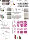
Fig. 1
Fig. 1
MiR-375 attenuates the stemness of GC cells. A, B The sphere size and number were examined in GC cells with or without miR-375 overexpression. C The protein expression of stemness markers was evaluated in GC cells with or without miR-375 overexpression. D CD44+ sub-population was determined in GC cells with or without miR-375 overexpression. E The images of tumors derived from different numbers of SGC7901 cells with or without miR-375 overexpression. F The tumor formation rate and confidence intervals for 1/(stem cell frequency) were calculated based on the results shown in E. G The images of tumors derived from different numbers of BGC-823 cells with or without miR-375 overexpression. H The tumor formation rate and confidence intervals for 1/(stem cell frequency) were calculated based on the result shown in G. I HE staining images of lung obtained from mice with injection of GC cells with or without miR-375 overexpression. J The correlation between miR-375 expression and the overall survival probability of GC patients was evaluated using the online analysis tool (http://kmplot.com). Data are presented as the mean ± SD, n ≥ 3, **p < 0.01 vs control, ##p < 0.01 vs LV3-NC
Fig. 2
Fig. 2
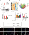
SLC7A11 is a direct target of miR-375 in GC cells. A The volcano map indicating the number of mRNAs in SGC-7901 cells with or without miR-375 overexpression. B The mRNA heatmap showing the change of mRNA expression in SGC-7901 cells with or without miR-375 overexpression. C The venn map showing the potential targets of miR-375. D SLC7A11 protein expression is detected in GC cells with or without miR-375 overexpression. Data are presented as the mean ± SD, n ≥ 3, *p < 0.05, **p < 0.001 vs control. E The diagram indicating the binding of miR-375 on SLC7A11. F Luciferase reporter analysis was performed to determine the effect of miR-375 on the activity of SLC7A11 3′UTR WT and SLC7A11 3′UTR MUT. Data are presented as the mean ± SD, ns means no significance, n ≥ 3, *p < 0.05 vs NC+vector. G RIP assay was constructed to measure miR-375 level in RNA pulled down by Anti-Ago2 in GC cells with SLC7A11 3′UTR WT or SLC7A11 3′UTR MUT overexpression. Data are presented as the mean ± SD, n ≥ 3, **p < 0.01 vs pMIR-report empty group. H RNA-FISH was performed to evaluate the co-localization of miR-375 and SLC7A11 in GC cells
Fig. 3
Fig. 3
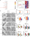
MiR-375 triggers ferroptosis through targeting SLC7A11. A KEGG pathway enrichment analysis in SGC-7901 cells with or without miR-375 overexpression. B GSEA analysis in SGC-7901 cells with or without miR-375 overexpression. C Transmission electron microscopy (TEM) assay on the mitochondria of GC cells with erastin treatment, miR-375 overexpression, SLC7A11 knockdown, or miR-375 overexpression as well as SLC7A11 overexpression. D Lipid peroxidation level was measured in GC cells with miR-375 overexpression as well as SLC7A11 overexpression or not. *p < 0.05 vs NC, ##p < 0.01 vs miR-375. E Cysteine concentration was examined in the cells as depicted in D. ***p < 0.001 vs si-NC, ##p < 0.01 vs NC+miR-375. F GSH concentration was detected in the cells described in D. **p < 0.01, ***p < 0.001 vs NC, ##p < 0.01 vs NC+miR-375. Data are presented as the mean ± SD, n ≥ 3
Fig. 4
Fig. 4
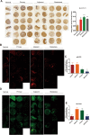
The expression levels of miR-375 and SLC7A11 in tissue chips. A Tissue chip containing different types of tissues as indicated was used to detect SLC7A11 protein level by IHC. **p < 0.01, ***p < 0.001 vs normal tissues. B Tissue chip was used to detect miR-375 level through RNA-FISH. C The fluorescence quantification was calculated based on the result. *p < 0.05, ***p < 0.001 vs normal tissues. D, E Tissue chip was used to detect SLC7A11 mRNA level through RNA-FISH and fluorescence quantification was shown
Fig. 5
Fig. 5
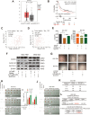
MiR-375 attenuates the stemness of GC cells by targeting SLC7A11. A Online dataset (http://gepia.cancer-pku.cn/index.html) was used to detect SLC7A11 mRNA level in GC (T) and normal (N) tissues. B The correlation between SLC7A11 mRNA expression and the overall survival probability of GC patients was evaluated using the online analysis tool (http://kmplot.com). C, D The correlation between SLC7A11 mRNA expression and ALDH1A1 (C) or Oct4 (D) mRNA expression was evaluated using the online analysis tool (R2: Genomics Analysis and Visualization Platform (https://hgserver1.amc.nl/cgi-bin/r2/main.cgi)). E CD44+ sub-population was determined in GC cells with miR-375 overexpression plus SLC7A11 overexpression or not. Data are presented as the mean ± SD, n ≥ 3, *p < 0.05, ***p < 0.001 vs LV3-NC+Vector, #p < 0.05, ##p < 0.01 vs Vector+LV3-miR-375. F The protein expression of stemness markers was examined in the GC cells. G The sphere size and number were evaluated in the GC cells. H Tumor images derived from BGC-823 cells described in H. I Tumor weight of tumors shown in I. J Tumor images derived from SCG7901 cells described in J. K, L Tumor formation rate (K) and confidence intervals for 1/(stem cell frequency) (L) were calculated based on the result shown in L. Data are presented as the mean ± SD, n ≥ 3, *p < 0.05, **p < 0.01 vs NC
Fig. 6
Fig. 6
Knockdown of SLC7A11 suppresses the tumor-initiating and metastatic ability of GC cells. A Tumor images and formation rate derived from SGC7901 cells with or without SLC7A11 knockdown. B The confidence intervals for 1/(stem cell frequency) was calculated based on the results shown in A. C Tumor images and formation rate derived from BGC823 cells with or without SLC7A11 knockdown. D The confidence intervals for 1/(stem cell frequency) was calculated based on the results shown in C. E HE staining images of lung obtained from mice with injection of GC cells with or without miR-375 overexpression
Fig. 7
Fig. 7
MiR-375 attenuates the stemness of GC cells mainly through inducing ferroptosis. A CD44+ sub-population was examined in GC cells with miR-375 overexpression plus Fer-1, Z-VAD-FMK, or Nec-1 treatment. Data are presented as the mean ± SD, n ≥ 3, *p < 0.05, **p < 0.01 vs LV3-NC+DMSO; ##p < 0.01, ###p < 0.001 vs LV3-miR-375+DMSO. B The expression of stemness markers was detected in the cells depicted in A. C The expression of stemness markers was determined in GC cells with SLC7A11 knockdown plus Fer-1 treatment or not. D The inhibition ratio of erastin was measured in WT and ER cells. Data are presented as the mean ±ean ar ≥ 3, **p < 0.01, ***p < 0.001 vs WT or ER. E CD44+ sub-population was assessed in WT and ER cells with miR-375 overexpression or miR-NC. Data are presented as the mean ±re pre ≥ 3, **p < 0.01 vs control+miR-NC; ns, no significance. F The expression of stemness markers was examined in the cells described in E. G Sphere-formation ability was evaluated in the cells depicted in E
Fig. 8
Fig. 8
Proposed model that miR-375 suppresses the stemness of GC cells. SLC7A11 is selective for uptaking cystine into the cell in exchange for glutamate. This system synthesizes glutathione (GSH) by taking extracellular cystine into the cell and thus represses lipid peroxidation in ferroptosis; this process is suppressed by miR-375 which can directly target SLC7A11. Additionally, miR-375 has no effect on iron concentration

References

    1. Li K, Dan Z, Nie YQ. Gastric cancer stem cells in gastric carcinogenesis, progression, prevention and treatment. World J Gastroenterol. 2014;20(18):5420–5426. doi: 10.3748/wjg.v20.i18.5420. - DOI - PMC - PubMed
    1. Gottesman MM. Mechanisms of cancer drug resistance. Annu Rev Med. 2002;53(1):615–627. doi: 10.1146/annurev.med.53.082901.103929. - DOI - PubMed
    1. Dixon SJ, Lemberg KM, Lamprecht MR, Skouta R, Zaitsev EM, Gleason CE, Patel DN, Bauer AJ, Cantley AM, Yang WS, Morrison B, III, Stockwell BR. Ferroptosis: an iron-dependent form of nonapoptotic cell death. Cell. 2012;149(5):1060–1072. doi: 10.1016/j.cell.2012.03.042. - DOI (V体育ios版) - PMC - PubMed
    1. Su Y, Zhao B, Zhou L, Zhang Z, Shen Y, Lv H, AlQudsy LHH, Shang P. Ferroptosis, a novel pharmacological mechanism of anti-cancer drugs. Cancer Lett. 2020;483:127–136. doi: 10.1016/j.canlet.2020.02.015. - DOI - PubMed
    1. Pervaiz S, Taneja R, Ghaffari S. Oxidative stress regulation of stem and progenitor cells. Antioxid Redox Signal. 2009;11(11):2777–2789. doi: 10.1089/ars.2009.2804. - DOI - PubMed

Publication types