Skip to main page content
U.S. flag

An official website of the United States government

Dot gov

The . gov means it’s official. Federal government websites often end in . gov or . mil. Before sharing sensitive information, make sure you’re on a federal government site. VSports app下载.

Https

The site is secure. The https:// ensures that you are connecting to the official website and that any information you provide is encrypted and transmitted securely V体育官网. .

. 2021 May 14:12:642856.
doi: 10.3389/fimmu.2021.642856. eCollection 2021.

"VSports在线直播" The Timing and Abundance of IL-2Rβ (CD122) Expression Control Thymic i NKT Cell Generation and NKT1 Subset Differentiation

Affiliations

The Timing and Abundance of IL-2Rβ (CD122) Expression Control Thymic i NKT Cell Generation and NKT1 Subset Differentiation

Hee Yeun Won (VSports手机版) et al. Front Immunol. .

VSports在线直播 - Abstract

Invariant NKT (iNKT) cells are thymus-generated innate-like T cells, comprised of three distinct subsets with divergent effector functions. The molecular mechanism that drives the lineage trifurcation of immature iNKT cells into the NKT1, NKT2, and NKT17 subsets remains a controversial issue that remains to be resolved. Because cytokine receptor signaling is necessary for iNKT cell generation, cytokines are proposed to contribute to iNKT subset differentiation also VSports手机版. However, the precise roles and requirements of cytokines in these processes are not fully understood. Here, we show that IL-2Rβ, a nonredundant component of the IL-15 receptor complex, plays a critical role in both the development and differentiation of thymic iNKT cells. While the induction of IL-2Rβ expression on postselection thymocytes is necessary to drive the generation of iNKT cells, surprisingly, premature IL-2Rβ expression on immature iNKT cells was detrimental to their development. Moreover, while IL-2Rβ is necessary for NKT1 generation, paradoxically, we found that the increased abundance of IL-2Rβ suppressed NKT1 generation without affecting NKT2 and NKT17 cell differentiation. Thus, the timing and abundance of IL-2Rβ expression control iNKT lineage fate and development, thereby establishing cytokine receptor expression as a critical regulator of thymic iNKT cell differentiation. .

Keywords: IL-15; IL-2 (interleukin-2); Tbet; cytokines; thymocytes V体育安卓版. .


Conflict of interest statement

The authors declare that the research was conducted in the absence of any commercial or financial relationships that could be construed as a potential conflict of interest.

Figures

Figure 1
Figure 1
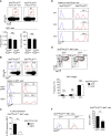
IL-2Rβ expression on thymic iNKT cells (A) C57BL/6 thymocytes were assessed for surface CD69 and CCR7 staining, which visualizes 5 distinct stages of differentiation (i.e., stages I–V), whereby thymocytes undergoing positive selection correspond to population III (top). iNKT cells were identified by PBS57-loaded CD1d tetramer (CD1dTet) staining among the 5 stages defined by CD69 and CCR7 (bottom). The results are representative of 5 independent experiments with a total of 5 WT C57BL/6 mice. (B) Surface cytokine receptor expression on thymic iNKT cell populations. iNKT cells in populations I, II, and III were assessed for γc and IL-2Rβ expression (red lines). Isotype control antibody staining are shown as gray lines. The results are representative of 3 independent experiments. (C) IL-2Rβ expression during iNKT cell differentiation. Thymic iNKT cells were divided into immature ST0 and mature stage 1–3 cells based on HSA expression (left, top). The abundance of IL-2Rβ was then assessed on ST0 (HSAhiCD44), ST1 (CD44NK1.1), ST2 (CD44+NK1.1), and ST3 (CD44+NK1.1+) iNKT cells (left, bottom). Histograms show surface IL-2Rβ expression for each subset (red line). Isotype control antibody staining are shown as gray lines. The results are representative of 5 independent experiments. (D) IL-2Rβ expression in thymic iNKT subsets. iNKT cells were assessed for intracellular PLZF and RORγt expression to identify NKT1, NKT2, and NK17 cells (left). Each iNKT subset was assessed for IL-2Rβ expression (right, red lines). Isotype control antibody staining are shown as gray lines. The results are representative of 3 independent experiments.
Figure 2
Figure 2
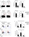
iNKT cell generation and differentiation in IL-2Rβ-floxed PLZF-Cre mice (A) Frequency and number of thymic iNKT cells in Il2rb fl/flPLZFCre and Il2rb fl/fl littermate control mice. The dot plots show thymic iNKT cells as identified by CD1dTet versus TCRβ staining (top). The bar graphs show the frequencies and numbers of iNKT cells from the indicated mice (bottom). The dot plot is representative, and the bar graphs are a summary of 4 independent experiments with a total of 11 Il2rb fl/flPLZFCre and 5 Il2rb fl/fl littermate control mice. (B) IL-2Rβ-deletion efficiency in Il2rb fl/flPLZFCre iNKT cells. The dot plots show thymic iNKT cells of the indicated mice CD1dTet versus HSA staining. HSAhiCD44 cells correspond to ST0 immature iNKT cells while HSAlo cells are mature ST1-3 iNKT cells. The histograms show GFP expression among the indicated population of thymic iNKT cells. (C) IL-2Rβ expression in GFP+ and GFP-negative mature iNKT cells of Il2rb fl/flPLZFCre iNKT mice. Bar graphs show the summary of analyzing 6 Il2rb fl/flPLZFCre mice. (D) GFP expression in ST1, ST2, and ST3 iNKT cells of Il2rb fl/flPLZFCre and littermate control thymocytes. The data are representative of 4 independent experiments. (E) Thymic iNKT cell differentiation in Il2rb fl/flPLZFCre mice. iNKT cells were stained for CD44 and NK1.1 to identify ST1, ST2, and ST3 cells as shown in the contour plots (top). The bar graph shows the frequencies of ST1, ST2, and ST3 cells in GFP+ and GFP thymic iNKT cells of Il2rb fl/flPLZFCre mice (bottom). Results show the summary of 6 independent experiments with a total of 14 Il2rb fl/flPLZFCre mice. (F) Bcl-2 expression in GFP+ and GFP iNKT cells of Il2rb fl/flPLZFCre thymocytes. Histograms are representative (left), and the bar graph (right) shows the summary of 3 independent experiments with a total of 8 Il2rb fl/flPLZFCre mice. **P < 0.01; ***P < 0.001. NS, Not Significant.
Figure 3
Figure 3
An IL-2Rβ requirement for thymic iNKT cell differentiation (A) Frequency and number of thymic iNKT cells in Il2rb –/– and WT littermate mice. Dot plots show thymic iNKT cells as identified by CD1dTet and TCRβ staining. Total thymocyte numbers are shown above the dot plots as the means ± SEM (left). Bar graphs show the frequencies and numbers of iNKT cells from the indicated mice (right). The dot plot is representative, and the bar graphs are a summary of 4 independent experiments with a total of 13 Il2rb –/– and 7 WT littermate mice. (B) Thymic iNKT cell differentiation in Il2rb –/– mice. HSAlo mature iNKT cells were stained for CD44 and NK1.1 to identify ST1, ST2, and ST3 cells in the indicated mice (left). The bar graph shows the frequencies of ST1, ST2, and ST3 cells in Il2rb –/– and WT littermate mice (right). The dot and contour plots are representative, and the bar graph shows the summary of 4 independent experiments. (C) Dot plots show the thymic iNKT subset composition in Il2rb –/– and WT littermate mice (left). The bar graph on top shows the frequency of thymic iNKT subsets as summary of 5 independent experiments with a total of 14 Il2rb –/– and 8 WT littermate mice. The bottom bar graph shows the cell numbers of thymic iNKT subsets as the summary of 4 independent experiments with a total of 9 Il2rb –/– and 4 WT littermate control mice. *P < 0.05; **P < 0.01; ***P < 0.001.
Figure 4
Figure 4
iNKT cell differentiation in IL-2Rβ transgenic mice (A) Frequency and number of thymic iNKT cells in IL-2RβTg and littermate C57BL/6 mice. The dot plots show thymic iNKT cells as identified by CD1dTet versus TCRβ staining (left). The total thymocyte numbers are shown above the dot plots as the means ± SEM. The bar graphs show the frequencies and numbers of iNKT cells from the indicated mice (right). The dot plot is representative, and the bar graphs are a summary of 4 independent experiments with a total of 7 IL-2RβTg and 6 littermate C57BL/6 mice. (B) Frequency and number of thymic iNKT cells in IL-2RβTg BALB/c and littermate WT mice. The dot plots show thymic iNKT cells as identified by CD1dTet versus TCRβ staining (left). The total thymocyte numbers are shown above the dot plots as the means ± SEM. The bar graphs show the frequencies and numbers of iNKT cells from the indicated mice (right). The dot plot is representative, and the bar graphs are a summary of 3 independent experiments with a total of 7 IL-2RβTg and 8 littermate WT mice. (C) The dot plots show the thymic iNKT subset composition in IL-2RβTg BALB/c and WT littermate mice based on PLZF versus T-bet (top) and PLZF versus RORγt analysis (bottom). Results are representative of 4 independent experiments. (D) The bar graphs show the summary of thymic iNKT subset distribution for the indicated mice. Subset frequencies are the summary of 5 independent experiments with a total of 10 IL-2RβTg and 10 littermate WT BALB/c mice (top). Cell numbers of each iNKT subset are the summary of 4 independent experiments with a total of 8 IL-2RβTg and 7 littermate WT BALB/c control mice (bottom). *P < 0.05; **P < 0.01; ***P < 0.001.
Figure 5
Figure 5
T-bet-ZsGreen reporter expression in iNKT cells of IL-2RβTg BALB/c mice (A) Thymic iNKT cells were identified in BALB/c and TBGRTg BALB/c mice (top) and assessed for T-bet-ZsGreen reporter expression (bottom). Results are representative of 5 independent experiments. (B) T-bet-ZsGreen reporter expression in thymic iNKT cells of TBGRTgIL-2RβTg BALB/c and TBGRTg littermate control mice. Dot plots show either PLZF versus T-bet-ZsGreen reporter expression (top) or T-bet protein versus T-bet-ZsGreen reporter expression (bottom) in HSAlo thymic iNKT cells of TBGRTg IL-2RβTg BALB/c and TBGRTg WT littermate mice. Bar graph shows the frequency of T-bet-ZsGreen+ iNKT cells (red box) of the indicated mice. Results are representative 3 independent experiments with 8 TBGRTgIL-2RβTg BALB/c and 5 TBGRTg littermate WT BALB/c mice. (C) T-bet-ZsGreen reporter expression upon NKT1 cell differentiation. Thymic iNKT cells of TBGRTg BALB/c mice were divided into three distinct populations, i.e. I, II, and III, based on the amount of T-bet-ZsGreen expression (dot plot, left). Surface expression of CD44, IL-2Rβ, and TCRβ was then assessed for each iNKT population (histograms, right). Results are representative of 2 independent experiments with a total of 6 TBGRTg BALB/c mice. (D) Subset compositions of thymic iNKT cells in TBGRTg BALB/c mice based on ZsGreen-reporter expression. Total iNKT cells, T-bet-ZsGreen-negative (population I), and T-bet-ZsGreen-intermediate (population II) iNKT cells were identified in TBGRTg WT BALB/c (top) and TBGRTgIL-2RβTg BALB/c thymocytes (bottom) and examined for their subset composition based on PLZF versus RORγt staining. Results are representative of 3 independent experiments with 8 TBGRTgIL-2RβTg BALB/c and 5 TBGRTg littermate WT BALB/c mice. **P < 0.01.
Figure 6
Figure 6
Thymic iNKT cell differentiation in IL-15-infused IL-2RβTg BALB/c mice (A) Effects of recombinant IL-15- or PBS-releasing Alzet osmotic pump installation on IL-2RβTg BALB/c mice. Spleen CD8 T cells of IL-2RβTg BALB/c mice that were infused with IL-15 or PBS for 10 days were assessed for the accumulation of memory-phenotype CD8 T cells by CD44 versus CXCR3 staining. Frequency and numbers of CD44hiCXCR3+ CD8 T cells are representative of 2 independent experiments with 4 IL-15 pump and 3 PBS pump implanted IL-2RβTg BALB/c mice (top). Picture shows spleens of IL-2RβTg BALB/c mice that were implanted with PBS or IL-15 Alzet pumps (bottom). Scale bar = 1 cm. (B) Frequencies and numbers of thymic iNKT cells of IL-2RβTg BALB/c mice implanted with IL-15- or PBS-releasing Alzet osmotic pumps. Dot plots are representative (top) and graphs are summary (bottom) of 3 independent experiments with a total of 5 IL-15 pump and 4 PBS pump implanted IL-2RβTg BALB/c mice. (C) Thymic iNKT subset composition IL-2RβTg BALB/c implanted with IL-15 or PBS-releasing Alzet osmotic pumps. NKT1 cells were identified by PLZF versus T-bet of HSAlo mature thymic iNKT cells. Results are representative of 3 independent experiments with 5 IL-15 pump and 4 PBS pump implanted IL-2RβTg BALB/c mice. (D) Frequencies and numbers of NKT1 cells in IL-2RβTg BALB/c implanted with IL-15 or PBS-releasing Alzet osmotic pumps. Results are summary of 3 independent experiments with 5 IL-15 pump and 4 PBS pump implanted IL-2RβTg BALB/c mice. (E) Frequency of thymic iNKT cells in CA-STAT5Tg and littermate C57BL/6 mice. The dot plots show thymic iNKT cells as identified by CD1dTet versus TCRβ staining (left). The total thymocyte numbers are shown above the dot plots as the means ± SEM (left). The bar graph shows the frequency of iNKT cells from the indicated mice (right). The dot plot is representative, and the bar graphs are a summary of 4 independent experiments with a total of 7 CA-STAT5Tg and 8 littermate WT mice (F) Cell numbers of DP thymocytes and thymic NKT1 cells from CA-STAT5Tg and littermate WT C57BL/6 mice. Bar graph shows the summary from a total of 7 CA-STAT5Tg and 5 littermate WT mice. *P < 0.05; **P < 0.01. NS, Not Significant.

References

    1. Etzensperger R, Kadakia T, Tai X, Alag A, Guinter TI, Egawa T, et al. . Identification of Lineage-Specifying Cytokines That Signal All CD8(+)-Cytotoxic-Lineage-Fate ‘Decisions’ in the Thymus. Nat Immunol (2017) 18:1218–27. 10.1038/ni.3847 - DOI - PMC - PubMed
    1. Park JH, Adoro S, Guinter T, Erman B, Alag AS, Catalfamo M, et al. . Signaling by Intrathymic Cytokines, Not T Cell Antigen Receptors, Specifies CD8 Lineage Choice and Promotes the Differentiation of Cytotoxic-Lineage T Cells. Nat Immunol (2010) 11:257–64. 10.1038/ni.1840 - DOI - PMC - PubMed
    1. Lin JX, Leonard WJ. The Common Cytokine Receptor Gamma Chain Family of Cytokines. Cold Spring Harb Perspect Biol (2018) 10:a028449. 10.1101/cshperspect.a028449 - DOI - PMC - PubMed
    1. Hong C, Luckey MA, Park JH. Intrathymic IL-7: The Where, When, and Why of IL-7 Signaling During T Cell Development. Semin Immunol (2012) 24:151–8. 10.1016/j.smim.2012.02.002 - "V体育平台登录" DOI - PMC - PubMed
    1. Weinreich MA, Odumade OA, Jameson SC, Hogquist KA. T Cells Expressing the Transcription Factor PLZF Regulate the Development of Memory-Like CD8+ T Cells. Nat Immunol (2010) 11:709–16. 10.1038/ni.1898 - DOI - PMC - PubMed

Publication types