Skip to main page content
U.S. flag

An official website of the United States government

Dot gov

The VSports app下载. gov means it’s official. Federal government websites often end in . gov or . mil. Before sharing sensitive information, make sure you’re on a federal government site. .

Https

The site is secure. The https:// ensures that you are connecting to the official website and that any information you provide is encrypted and transmitted securely. V体育官网.

. 2021 Mar 21;13(6):1434.
doi: 10.3390/cancers13061434.

A Cystine-Cysteine Intercellular Shuttle Prevents Ferroptosis in xCTKO Pancreatic Ductal Adenocarcinoma Cells

Affiliations

A Cystine-Cysteine Intercellular Shuttle Prevents Ferroptosis in xCTKO Pancreatic Ductal Adenocarcinoma Cells

Willian Meira et al. Cancers (Basel). .

V体育官网入口 - Abstract

In our previous study, we showed that a cystine transporter (xCT) plays a pivotal role in ferroptosis of pancreatic ductal adenocarcinoma (PDAC) cells in vitro. However, in vivo xCTKO cells grew normally indicating that a mechanism exists to drastically suppress the ferroptotic phenotype. We hypothesized that plasma and neighboring cells within the tumor mass provide a source of cysteine to confer full ferroptosis resistance to xCTKO PDAC cells. To evaluate this hypothesis, we (co-) cultured xCTKO PDAC cells with different xCT-proficient cells or with their conditioned media. Our data unequivocally showed that the presence of a cysteine/cystine shuttle between neighboring cells is the mechanism that provides redox and nutrient balance, and thus ferroptotic resistance in xCTKO cells. Interestingly, although a glutathione shuttle between cells represents a good alternative hypothesis as a "rescue-mechanism", our data clearly demonstrated that the xCTKO phenotype is suppressed even with conditioned media from cells lacking the glutathione biosynthesis enzyme. Furthermore, we demonstrated that prevention of lipid hydroperoxide accumulation in vivo is mediated by import of cysteine into xCTKO cells via several genetically and pharmacologically identified transporters (ASCT1, ASCT2, LAT1, SNATs). Collectively, these data highlight the importance of the tumor environment in the ferroptosis sensitivity of cancer cells VSports手机版. .

Keywords: cysteine transporters; cysteine-cystine shuttle; ferroptosis; resistance; tumor environment V体育安卓版. .


Conflict of interest statement

The authors declare no conflict of interest.

V体育官网入口 - Figures

Figure 1
Figure 1
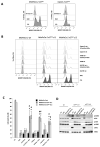
Fibroblasts reverse the characteristic ferroptosis phenotype of pancreatic ductal adenocarcinoma (PDAC) xCTKO cells. (A). The expression level of xCT in human dermal fibroblasts (HDF) under basal and amino acid (AA) starvation conditions (Western blot, left panel—ARD1 used as loading control), their sensitivity to xCT inhibition by erastin (ERA) seen through the lipid hydroperoxide accumulation after 24 h (BODIPY 591/581 C11 staining, middle panel), i.e., cell viability after 48 h (right bar graph). (B). Optimization of the co-culture conditions: accumulation of the lipid hydroperoxides (left) and cell viability (right) of MiaPaCa-2 xCTKO cells co-cultured with 3%, 6%, 12% or 25%, of HDF cells during 24 h and 48 h, respectively. (C). BODIPY 591/581 C11 staining of MiaPaCa-2 wild type (wt) and xCTKO cells cultured for 24 h in full or fractionated (lower (<) or higher (>) then 3 kDa, left and right panel, respectively) conditional media (CM) of HDF; dCM = dialyzed conditional media (>3 kDa). All experiments have been performed in triplicate and the representative blots and histograms are shown. Bar graphs show mean ± SEM; n = 3; *, p < 0.05, comparison with control group.
Figure 2
Figure 2
xCT-expressing cells export a redox-sensitive agent which prevents ferroptosis and restores amino acid imbalance in xCTKO cells. (A). Lipid hydroperoxide accumulation in xCTKO PDAC cells co-cultured with other PDAC wt cells or in media supplemented with an alternative cysteine-donor—N-acetylcysteine (NAC). (B,C). Phenotype of MiaPaCa-2 xCTKO cells co-cultured with different xCT-expressing cells (LS174T, A549 and Capan wt) in media +/− the low dose of pro-oxidant, glucose oxidase (0.2 mU/mL GOx): B. BODIPY 591/581 C11 staining; (C). Cell death. (D). MiaPaCa-2 wt and xCTKO cells were cultivated for 24 h in DMEM +/− 1 mM NAC, or in conditioned media (CM) of LS174T/A549 wt. Changes in phosphorylation status and protein abundance of members of the two major AA-sensing pathways GCN2 (p-GCN2/ATF4) and mTORC1 (p-S6K1 and p-RPS6) were analyzed by Western blot. All experiments have been performed in triplicate and the representative blots and histograms are shown. Bar graph shows mean ± SEM; n = 3; *, p < 0.05, comparison with WT control group; #, p < 0.05, comparison with the corresponding untreated group.
Figure 3
Figure 3
Cystine-cysteine shuttles fuel cooperation between WT and xCTKO cells. (A). Schematic representation of the cystine-cysteine (CySSCy, CySH) shuttle: xCT-expressing cells (“host”) are able to import the oxidized form of cysteine, reduce it and export it. Or, in the alternative scenario, the “host” cells can synthesize and export glutathione (GSH), which then will be cleaved outside the cell to the constituent amino acids. Cysteine provided in one or both ways is taken up by the xCTKO cells (“guests”) maintaining the amino acid and redox balance. (B). MiaPaCa-2 wt or xCTKO cells alone (full outer lines of the histograms) or in the co-culture with A549 wt (CC—dashed outer lines of the histograms) counterparts in DMEM media +/− 1 mM N-acetylcysteine (NAC), 100 μM inhibitor of GSH biosynthesis (buthionine sulphoximine, BSO) or 1 μM inhibitor of xCT, erastin. (C). Accumulation of lipid hydroperoxides and (D). Cell viability of MiaPaCa-2 xCTKO cells in co-culture (CC) or cultivated in the presence of conditional media (CM) of Capan-2 GCLcKO or xCTKO cells during (C) 24 h and (D) 48 h. All experiments have been performed in triplicate and the representative histograms are shown. Bar graph shows mean ± SEM; n = 3; *, p < 0.05, comparison with WT control group.
Figure 4
Figure 4
Genetic disruption of the ASCT2 cysteine transporter affects cooperation between xCT-expressing and xCTKO cells. (A). A variety of xCT wt cells (LS174T, A549, MiaPaCa-2, Capan-2 and HDF) were grown in DMEM media +/− amino acids during 24 h and protein content of different cysteine transporters (ASCT1 (SLC1A4), ASCT2 (SLC1A5), EAAT3 (SLC1A1), EAAT4 (SLC1A6) were analyzed by Western blotting. (B,C). Lipid hydroperoxide accumulation and cell viability of MiaPaCa-2 xCTKO and xCT-ASCT2DKO cells (guest cells—CySH import) in control conditions or co-cultured with 6% A549 wt, ASCT1KO, ASCT2KO, ASCT1-ASCT2DKO, or LAT1KO (host cells—CySH export). All experiments have been performed in triplicate and the representative blots and histograms are shown. Bar graph shows mean ± SEM; n = 3; *, p < 0.05, comparison with WT control group.
Figure 5
Figure 5
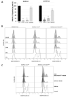
Multiple cysteine transporters are necessary for “guest–host” intercommunication. (A). Cysteine transport activity of A549 wt and LS174T wt cells was measured by the uptake of [14C]- 1, 2, 1′, 2′-cystine (14C-cystine) reduced by 100 μM β-mercaptoethanol in HBSS media containing 50 μM cold cystine and supplemented or not with 10 mM L-alanine, L-leucine or MeAIB. These results represent the average ± SEM, n = 2, comparison with WT control cells, * p < 0.05. (B). Accumulation of the lipid hydroperoxides in MiaPaCa-2 wt, xCTKO or xCT-ASCT2DKO (guest cells) co-cultured with A549 wt cells (host cells) in the presence or not of the 1 mM NAC, 3 mM L-alanine, 3 mM L-leucine or 3 mM MeAIB after 24 h. (C). Lipid hydroperoxides levels measured by BODIPY 591/581 C11 staining and FACS analysis of MiaPaCa-2 xCTKO and MiaPaCa-2 xCT-ASCT2DKO (guest cells) co-cultured for 24 h with A549 wt or A549 ASCT1-ASCT2DKO (host cells) in the presence or not of SNAT inhibitor, MeAIB; Control condition represents the phenotype of guest cells in the absence of host cells. Representative histograms of three independent experiments are shown.

References

    1. Siegel R.L., Miller K.D., Jemal A. Cancer statistics, 2019. CA Cancer J. Clin. 2019;69:7–34. doi: 10.3322/caac.21551. - DOI - PubMed
    1. Siegel R.L., Miller K.D., Jemal A. Cancer statistics, 2020. CA Cancer J. Clin. 2020;70:7–30. doi: 10.3322/caac.21590. - DOI (VSports在线直播) - PubMed
    1. Fox J.L., MacFarlane M. Targeting cell death signalling in cancer: Minimising ‘Collateral damage’. Br. J. Cancer. 2016;115:5–11. doi: 10.1038/bjc.2016.111. - DOI - PMC - PubMed
    1. Pfeffer C.M., Singh A.T.K. Apoptosis: A Target for Anticancer Therapy. Int. J. Mol. Sci. 2018;19:448. doi: 10.3390/ijms19020448. - DOI - PMC - PubMed
    1. Hanahan D., Weinberg R.A. Hallmarks of Cancer: The Next Generation. Cell. 2011;144:646–674. doi: 10.1016/j.cell.2011.02.013. - DOI - PubMed