Skip to main page content
U.S. flag

An official website of the United States government

Dot gov

The . gov means it’s official. Federal government websites often end in . gov or VSports app下载. mil. Before sharing sensitive information, make sure you’re on a federal government site. .

Https

The site is secure. The https:// ensures that you are connecting to the official website and that any information you provide is encrypted and transmitted securely V体育官网. .

. 2021 Apr;592(7856):778-783.
doi: 10.1038/s41586-021-03350-4. Epub 2021 Mar 17.

DPP9 sequesters the C terminus of NLRP1 to repress inflammasome activation (VSports手机版)

Affiliations

DPP9 sequesters the C terminus of NLRP1 to repress inflammasome activation

L Robert Hollingsworth et al. Nature. 2021 Apr.

Abstract

Nucleotide-binding domain and leucine-rich repeat pyrin-domain containing protein 1 (NLRP1) is an inflammasome sensor that mediates the activation of caspase-1 to induce cytokine maturation and pyroptosis1-4. Gain-of-function mutations of NLRP1 cause severe inflammatory diseases of the skin4-6. NLRP1 contains a function-to-find domain that auto-proteolyses into noncovalently associated subdomains7-9, and proteasomal degradation of the repressive N-terminal fragment of NLRP1 releases its inflammatory C-terminal fragment (NLRP1 CT)10,11. Cytosolic dipeptidyl peptidases 8 and 9 (hereafter, DPP8/DPP9) both interact with NLRP1, and small-molecule inhibitors of DPP8/DPP9 activate NLRP1 by mechanisms that are currently unclear10,12-14. Here we report cryo-electron microscopy structures of the human NLRP1-DPP9 complex alone and with Val-boroPro (VbP), an inhibitor of DPP8/DPP9. The structures reveal a ternary complex that comprises DPP9, full-length NLRP1 and the NLRPT CT. The binding of the NLRP1 CT to DPP9 requires full-length NLRP1, which suggests that NLRP1 activation is regulated by the ratio of NLRP1 CT to full-length NLRP1. Activation of the inflammasome by ectopic expression of the NLRP1 CT is consistently rescued by co-expression of autoproteolysis-deficient full-length NLRP1. The N terminus of the NLRP1 CT inserts into the DPP9 active site, and VbP disrupts this interaction. Thus, VbP weakens the NLRP1-DPP9 interaction and accelerates degradation of the N-terminal fragment10 to induce inflammasome activation. Overall, these data demonstrate that DPP9 quenches low levels of NLRP1 CT and thus serves as a checkpoint for activation of the NLRP1 inflammasome. VSports手机版.


VSports手机版 - Conflict of interest statement

Competing interests. H. W. is a co-founder of Ventus Therapeutics. The other authors declare no competing interests V体育安卓版.

Figures
Extended Data Fig. 1 |
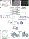
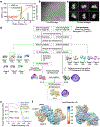
Extended Data Fig. 1 |. Structure determination of the NLRP1-DPP9 complex.
a, Purification of the NLRP1-DPP9 complex by ion exchange chromatography. The ternary complex peak is shaded in green and labelled with an arrow. b, A representative (>1000 images) cryo-EM micrograph. c, Representative 2D class averages. d, Workflow for the NLRP1-DPP9 complex structure determination. e, Map-map and map-model FSC curves. f, Local resolution distribution of the final map calculated with ResMap and coloured as indicated.
Extended Data Fig. 2 |
Extended Data Fig. 2 |. Crosslinking mass spectrometry analysis of the NLRP1-DPP9 complex.
a, Summary of BS3 crosslinking between DPP9 and NLRP1. High confidence crosslinked peptides are displayed, with residue ranges labelled and colours coded by domains. Crosslinked lysine pairs are indicated in red. Cα-Cα distances between these lysine residues interpreted by the final NLRP1-DPP9 model are shown, along with the figure panel names for their detailed depictions. All detected peptide pairs are tabulated in the Source Data file. b, Overview of BS3-mediated crosslinks. c-h, Zoom-ins highlighting crosslinked lysine pairs (red) interpreted by the final NLRP1-DPP9 model.
Extended Data Fig. 3 |
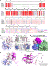
Extended Data Fig. 3 |. Sequence and structural analysis of FIIND.
a, ClustalW multiple sequence alignment between human NLRP1 (hNLRP1), mouse NLRP1 (mNLRP1, different isoforms), and rat NLRP1 (rNLRP1, different isoforms). COP: Copenhagen; ZUC: Zucker; LEW: Lewis; SD: Sprague Dawley; and CDF: Fischer. Secondary structures and residue numbers are denoted based on the human FIINDA structure in the NLRP1-DPP9 ternary complex. Interfacial residues in the NLRP1-DPP9 complex are annotated with asterisks, and residues in the catalytic triad (H1186, E1195, S1213) are boxed in black. b, FIINDA overview with ZU5 (blue) and UPA (light pink) subdomains. The catalytic triad residues (H1186, E1195, and S1213) are shown in sticks. c, Topology of the FIIND with secondary structures labelled. d, Superimposition of FIINDA onto the UPAB. NLRP1B must be free NLRP1-CT because a ZU5 subdomain at site B would have clashed with ZU5 and UPA at site A and with DPP9. e, The ZU5A-UPAA-UPAB module that binds DPP9. UPAA and UPAB interact with each other in a front-to-back manner with only a 9° rotation between them. f, Altered conformation of the UPAB N-terminus that binds in the DPP9 active site tunnel in comparison to UPAA in a complete FIINDA.
Extended Data Fig. 4 |
Extended Data Fig. 4 |. Structure determination of the NLRP1-DPP9 complex with VbP.
a, Purification of the NLRP1-DPP9 complex in the presence of VbP by ion exchange chromatography. The ternary complex peak is shaded in green and labelled with an arrow. b, A representative (>1000 images) cryo-EM micrograph. c, Representative 2D class averages. d, Workflow for the NLRP1-DPP9-VbP complex structure determination. e, Map-map and map-model FSC curves. f, Local resolution distribution of the final map calculated with ResMap and coloured as indicated.
Extended Data Fig. 5 |
Extended Data Fig. 5 |. VbP interactions in the DPP9 active site and comparison to a DPP substrate and NLRP1.
a, Schematic of covalent linkage between DPP9’s S730 and VbP. b, Fit of VbP into the cryo-EM density. VbP is shown in stick with carbon atoms in light brown. The charged amino group of VbP interacts with the DPP9 EE loop which also coordinates a substrate N-terminus, and the carbonyl oxygen of VbP interacts with R133 of the R-helix. The covalent linkage of VbP with S730, the catalytic serine, is displayed. c, Structural alignment of the VbP-bound DPP9 model (green) and the crystal structure of bacterial DPP4 bound to the substrate Ile-Pro (PDB ID: 5YP3, orange). VbP assumes a pose remarkably like a model substrate. d, NLRP1-CT-DPP9 complex in which DPP9 is coloured by Cα-Cα distances between NLRP1-bound and VbP-bound structures as indicated. A distance scale bar is shown, and VbP is displayed in sticks to mark the active site, with carbon atoms in green, oxygen atoms in red, nitrogen atoms in blue, and boron atoms in orange. NLRP1-CT (UPA) is shown in magenta.
Extended Data Fig. 6 |
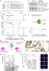
Extended Data Fig. 6 |. Lack of cleavage of intact NLRP1-CT but the cleavage of its isolated N-terminal peptide by DPP9.
a, N-terminal sequencing of the purified NLRP1-DPP9 complex showing that the NLRP1-CT is not cleaved by the co-expressed DPP9. b, Chemical enrichment of protease substrates (CHOPS) assay showing that DPP9 does not cleave NLRP1-CT. Briefly, WT NLRP1 expressed in DPP8/9 DKO HEK293T cells was incubated with PBS or recombinant DPP9 prior to labelling with a 2-pyridinecarboxaldehyde (2PCA)-biotin probe which selectively biotinylates free N-termini except those with a proline in the second position, followed by capture of biotinylated proteins. The inputs and the eluents were analysed by immunoblots using anti-NLRP1-CT (NLRP1-FL and NLRP1-CT), anti-GAPDH, and anti-Streptavidin (biotinylated proteins) antibodies. DPP9 treatment did not increase the biotinylation of NLRP1-CT, as would be expected after the removal of the N-terminal Ser-Pro dipeptide. c, Evidence of cleavage of the isolated 15 residue N-terminal peptide in NLRP1-CT by recombinant DPP9 from mass spectrometry analysis. d, Inhibition of DPP9 catalytic activity against AP-AMC by the isolated NLRP1-CT peptide. e, Schematic illustrates the ability of DPP9 to cleave an isolated UPA N-terminal peptide, but not dipeptides from an intact NLRP1-CT. f, Comparison of the binding modes of the UPAB N-terminal peptide in the NLRP1-DPP9 complex and the Ile-Pro dipeptide in an acyl-enzyme intermediate. g, Theoretical dipeptide cleavage does not dampen NLRP1 inflammasome activity by LDH release or inflammasome signalling. n=3 independent biological replicates. Data are mean ± SEM. Anti-FLAG (NLRP1-CT), anti-GSDMD, and anti-GAPDH antibodies were used in the immunoblots. p30: GSDMD N-terminal fragment from caspase-1 cleavage. h, Theoretical dipeptide cleavage does not dampen NLRP1 inflammasome activity by ASC speck formation. n = 10 quantified fields of view. Data are mean ± SEM. Right, representative superimposed images of nuclei (blue), RFP (red) and GFP-ASC (green). All data are representative of > 2 independent experiments. *: p < 0.05, **: p < 0.01, ***: p < 0.001 and ****: p < 0.0001 by unpaired two-sided t test, exact p-values are presented in the Source Data file.
Extended Data Fig. 7 |
Extended Data Fig. 7 |. Mutational analysis of the interactions in the NLRP1-DPP9 ternary complex.
a, Disorder to order transition of several DPP9 surface loops from the isolated DPP9 crystal structure (PDB ID: 6EOQ) to the NLRP1-bound DPP9 cryo-EM structure. b, Genomic confirmation of DPP8 KO generated in DPP9 KO HEK293T cells stably expressing Cas9 to create DPP8/9 DKO HEK293T cells. Single guide RNA (sgRNA) sequence is highlighted. c, Immunoblots of the input lysates for the FLAG co-immunoprecipitation with WT or mutant DPP9 and WT NLRP1-FLAG, related to Fig. 2h. Anti-DPP9, anti-NLRP1 (NLRP1-FL, NLRP1-CT), and anti-GAPDH antibodies were used in the immunoblots. d, Cleavage rate of a model DPP9 substrate, GP-AMC, by WT DPP9 and its structure-guided mutants. Only the catalytically dead mutant S730A disrupts catalytic activity and sensitivity to the DPP9 inhibitor, VbP. n=3 technical replicates. Data are mean ± SEM. e, Immunoblots of the input lysates for the FLAG co-immunoprecipitation with WT or mutant NLRP1-FLAG and WT DPP9, related to Fig. 2i. Anti-DPP9, anti-NLRP1 (NLRP1-FL, NLRP1-CT), and anti-GAPDH antibodies were used in the immunoblots. All data are representative of > 2 independent experiments.
Extended Data Fig. 8 |
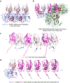
Extended Data Fig. 8 |. The ZU5 domain and DPP9 sterically hinder UPA polymerization.
a, Modelling of a FIIND polymer using the observed UPAA-UPAB relationship. Adjacent ZU5 molecules would clash, suggesting that UPA polymerization cannot occur in complete FIIND. b, Modelled recruitment of free UPA adjacent to UPAA and UPAB in the ternary complex with DPP9. The additional UPA subdomain next to FINDA clashes with the ZU5 subdomain, and the additional UPA next to UPAB clashes with both DPP9 monomers in the complex, suggesting that DPP9 inhibits UPA oligomerization. c, A modelled UPA oligomer based on the near front-to-back interaction in the NLRP1-DPP9 ternary complex. In the model, the N-terminal tails of free UPAs are shown in either the UPAA (pink) or UPAB (magenta) conformation in complex with DPP9, but in reality, this conformation is likely to be different. d, Modelling of a UPA oligomer formed on one side of a FIINDA-CTB complex. The NLRP1-FIINDA-NLRP1-CTB binary complex can polymerize with freed NLRP1-CT. a-d, DPP9 is coloured in green, and NLRP1 domains are coloured as indicated.
Extended Data Fig. 9 |
Extended Data Fig. 9 |. VbP displaces NLRP1 from DPP9 in vitro and in cells.
a, Schematic of the on-bead displacement experiment. The DPP9-ternary complex is expressed in 293T cells, which are then lysed and incubated with FLAG beads. Once bound, beads are split equally and washed with compounds or DMSO. The remainder of the protein is eluted off of the beads. MeBS, Bestatin methyl ester. b, Two structurally distinct VbP inhibitors, VbP and 8J, displace DPP9 from NLRP1-S1213A by the on-bead displacement assay. Anti-FLAG (S1213A-FL), anti-MYC (NLRP1-CT), and anti-V5 (DPP9) antibodies were used in the immunoblots. Representative of 2 independent experiments. c, Schematic of the dTAG experiment. FKBP12 with the F36V mutation (dTAG) is fused to the N-terminus of NLRP1-FL. The dTAG13 ligand recruits an E3-ligase to FKBP12-F36V, leading to its ubiquitination and N-terminal degradation of the fusion protein. NLRP1-CTs (UPA-CARDs) resulting from FIIND autoprocessing are released to assemble the inflammasome. d, NLRP1 FIIND-SA expression in reconstituted HEK293T inflammasome system rescues GSDMD cleavage resulting from dTAG13-induced NLRP1 degradation. VbP prevents GSDMD rescue without inducing additional NLRP1 degradation. Anti-HA (dTAG-NLRP1-FL, dTAG-NLRP1-NT), anti-FLAG (FIIND-SA), anti-GSDMD, and anti-GAPDH antibodies were used in the immunoblots. p30: GSDMD N-terminal fragment from caspase-1 cleavage. Representative of 2 independent experiments.
Fig. 1. |
Fig. 1. |. Structure of the NLRP1-DPP9 complex.
a, Domain organization. b, Representative (>3 independent experiments) SDS-PAGE of the purified NLRP1-DPP9 complex. HSP70 contamination is noted with an asterisk (*). c, Cryo-EM map (left) and the model (right) of the ternary NLRP1A-NLRP1B-DPP9 complex. The DPP9 dimer and the two copies of NLRP1 (A and B) are labelled with the colour scheme in (a). A schematic diagram (middle) denotes the entire NLRP1 and DPP9 molecules versus the ordered, resolved portions of the proteins (red circle).
Fig. 2. |
Fig. 2. |. Detailed interfaces in the NLRP1-DPP9 ternary complex and inhibition by the DPP9 inhibitor VbP.
a, Insertion of the N-terminal peptide of UPAB into the DPP9 active site. b, Cryo-EM map of the NLRP1-DPP9 complex in the presence of VbP. VbP binding reduces UPAB occupancy. c, Displacement of the UPAB N-terminal peptide from the DPP9 active site by VbP. d, Overview of three interfaces important for NLRP1-DPP9 association. Regions blocked in rectangles are shown in detail in (a) and (e-g). e-g, Zoom-ins of each NLRP1-DPP9 binding interface. h, FLAG co-immunoprecipitation using FLAG-tagged WT NLRP1 and the indicated His-tagged DPP9 constructs expressed in DPP8/9 DKO 293T cells. EV: empty vector. i, FLAG co-immunoprecipitation using FLAG-tagged NLRP1 expressed in HEK293T cells. FLAG-tagged GFP was used as a negative control. The Roman numerals in parentheses (h-i) represent the three interfaces in the NLRP1-DPP9 ternary complex. Each immunoblot is representative of > 2 independent experiments.
Fig. 3. |
Fig. 3. |. Functional consequences of interfacial mutations in the NLRP1-DPP9 ternary complex.
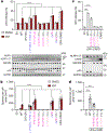
Interface I and II mutants cause NLRP1 autoactivation and UPA-UPA interactions are required for inflammasome activity. a, LDH release (top) and GSDMD processing (bottom) from transient expression of indicated constructs in a reconstituted HEK293T inflammasome system, with and without addition of VbP. n=3 independent biological replicates. *: non-specific bands; p30: GSDMD N-terminal fragment from caspase-1 cleavage. b, Quantification of speck formation induced by expression of indicated constructs in the presence and absence of VbP. n=15 quantified fields of view. c, LDH release (top) and GSDMD processing (bottom) by direct expression of WT or mutant NLRP1 UPA-CARDs. CARD alone was also included. n=3 independent biological replicates. d, Quantification of speck formation induced by expression of WT or mutant NLRP1 UPA-CARD. n=15 quantified fields of view. All data are representative of > 3 independent experiments. **** in (a) and (b): p < 0.0001 compared to EV by 2-way ANOVA with Tukey multiple comparison correction. *** in (c) and (d): p < 0.001 by unpaired two-sided t test, exact p-values are presented in the Source Data file. Data in (a-d) are mean ± SEM.
Fig. 4. |
Fig. 4. |. Repression of NLRP1-CT inflammasome activity by ternary complex formation.
a, Schematic representation of ternary complex formation between the autocatalysis-deficient NLRP1-S1213A-FL, Ub-NLRP1-CT, and DPP9. Ub-NLRP1-CT is co-translationally processed by endogenous deubiquitinases, resulting in NLRP1-CT with the native S1213 N-terminus. b, FLAG co-immunoprecipitation using FLAG-tagged WT NLRP1-FL or S1213A-FL and V5-tagged Ub-NLRP1-CT. S1213A-FL (site A) alone did not pull down endogenous DPP9. In contrast, co-expression of the two constructs formed a ternary complex with DPP9. c, Increasing amounts of S1213A-FL suppress NLRP1-CT-induced cell death measured by LDH release in a reconstituted HEK293 inflammasome system. n=3 independent biological replicates. d, NLRP1 FIIND-SA rescues ASC specks resulting from patient mutations except for P1214R, which reduces DPP9 binding. e, NLRP1 FIIND-SA rescues ASC specks resulting from dTAG13-induced NLRP1 degradation. f, ASC speck formation and FIIND-SA rescue for the NLRP1 P1214R patient mutation. FIIND-SA partially rescues dTAG-P1214R, but rescue of dTAG-P1214R is significantly less than for wild-type (WT) dTAG-NLRP1. g, DPP9 binding and catalytic activity contribute to repression of the NLRP1 inflammasome. ASC specks from NLRP1 and the indicated DPP9 constructs expressed in DPP8/9 DKO 293T cells. (d-g) n=10 quantified fields of view. Data in (c-g) are mean ± SEM. All data are representative of > 2 independent experiments (c-d, g) *: p < 0.05, **: p < 0.01 and ***: p < 0.001 by unpaired two-sided t test, (e-f) **: p < 0.01 and ***: p < 0.001 by 2-way ANOVA with Tukey multiple comparison correction, exact p-values are presented in the Source Data file. h, Schematic of NLRP1 activation and repression by DPP9. NLRP1-FL, together with DPP9, represses some threshold of free NLRP1-CTs. Enhanced degradation of NLRP1-FL, or displacement of the NLRP1-CT, leads to inflammasome signalling.

References

    1. Martinon F, Burns K & Tschopp J The Inflammasome: A Molecular Platform Triggering Activation of Inflammatory Caspases and Processing of proIL-β. Mol. Cell 10, 417–426 (2002). - PubMed
    1. Shen C, Sharif H, Xia S & Wu H Structural and mechanistic elucidation of inflammasome signaling by cryo-EM. Curr. Opin. Struct. Biol 58, 18–25 (2019). - PMC - PubMed
    1. Mitchell PS, Sandstrom A & Vance RE The NLRP1 inflammasome: new mechanistic insights and unresolved mysteries. Curr. Opin. Immunol 60, 37–45 (2019). - PMC - PubMed
    1. Taabazuing CY, Griswold AR & Bachovchin DA The NLRP1 and CARD8 inflammasomes. Immunol. Rev, 297, 1–13 (2020). - PMC - PubMed
    1. Soler VJ et al. Whole exome sequencing identifies a mutation for a novel form of corneal intraepithelial dyskeratosis. J. Med. Genet 50, 246–254 (2013). - PMC - PubMed

"VSports注册入口" Publication types